Noninvasive Assessment of Patients Undergoing Percutaneous
Intervention in Myocardial Infarction
Rica Dodo Delmar Buchler, Expedito E. Ribeiro, Antonio de Padua Mansur, Paola Smanio, Romeu Sergio Meneghelo,
William Azem Chalela, Carlos Alberto Buchpiguel, Jorge Roberto Buchler, Eric R. Bates, Eulogio E. Martinez
Instituto do Coração (InCor) da Faculdade de Medicina da Universidade de São Paulo, Instituto Dante Pazzanese de Cardiologia, University Of Michigan - AnnArbor - Michigan
Mailing address: Rica Dodo Delmar Buchler •
Rua Jose Maria Lisboa, 480/62 - Jardim Paulista - 01423-908 - São Paulo, SP - Brazil
E-mail: [email protected], [email protected]
Mnauscript received May 20, 2009; revised manuscript received April 04, 2010; accepted May 13, 2010.
Abstract
Background: Restenosis after primary percutaneous coronary intervention (PPCI) remains an important clinical problem, even with stent implantation. The ability of noninvasive testing to diagnose restenosis has had only inconsistent demonstration.
Objective: Our objective was to evaluate the ability of exercise treadmill testing (ETT) and myocardial perfusion imaging (MPI) to diagnose restenosis in patients treated by PPCI within 12 hours of ST-elevation myocardial infarction (STEMI).
Methods: From August 2003 to January 2006, 64 patients (mean age of 56.2 ± 10.2 years, 53 males) were enrolled
after PPCI. Only patients with left ventricular ejection fraction (LVEF) ≥ 40%, as assessed by resting transthoracic
echocardiography (TTE), were included. ETT with 12-lead ECG monitoring and right precordial leads, as also MPI were performed at 6 weeks, 6 months, and one year after intervention. Coronary angiography was performed at six months.
Results: Single-vessel disease was observed in 46.9% of the patients. The left anterior descending coronary artery was treated in 48.4% of the patients. Angiographic restenosis occurred in 28.8%. Sensitivity, specificity, positive predictive
value (PPV), negative predictive value (NPV), and accuracy of ETT in detecting restenosis were not significant. Right precordial leads did not add information. MPI sensitivity, specificity, PPV, NPV, and accuracy correlated with restenosis only in the 6-month follow-up, both when considering summed difference score >2 (p=0.006) and >4 (p=0.014).
Conclusion: ETT did not discriminate restenosis in this population. MPI performed at 6 months correlated with restenosis and proved useful during follow-up. (Arq Bras Cardiol 2010; 95(5): 555-562)
Keywords: Coronary restenosis; angioplasty; stent; myocardial infarction; critical pathways.
The purpose of the present study was to determine the prognostic value of ETT and MPI in the detection of in-stent restenosis during PPCI follow-up.
Patients and Methods
Primary balloon angioplasty and stenting during the acute phase of myocardial infarction was performed in two centers by experienced surgeons.
The patients underwent ETT, and MPI at 6 weeks, 6 months, and 1 year after intervention.
ETTs were performed with the patients using the medication prescribed, including betablockers. Follow-up coronary angiography was performed at six months. Resting TTE was performed prior to hospital discharge, to evaluate left ventricular ejection fraction (LVEF) and to include the patient in the protocol.
The study was approved by both institutional ethics committees and all patients gave written informed consent. The study was conducted in accordance with the clinical principles of the Declaration of Helsinki regarding research in humans.
Introduction
Primary percutaneous coronary intervention (PPCI) is superior to fibrinolytic therapy in the treatment of ST-elevation myocardial infarction (STEMI)1.Due to high rates of restenosis,
coronary stenting has emerged as a new strategy, but restenosis still remains a major limitation, with rates for bare-metal stenting ranging from 20% to 25%2,3.
Appropriate use of noninvasive testing following PPCI has not been systematically investigated. Among the noninvasive tests, exercise treadmill test (ETT) and myocardial perfusion imaging (MPI) have been used to detect restenosis. ETT is safe, simple, and easy to perform. Adding right precordial leads has been used to improve ETT sensitivity4. In contrast, the
Patients
From August 2003 to January 2006, 80 patients treated with PPCI and stenting within 12 hours of STEMI were prospectively selected at Instituto do Coração (InCor) and at Instituto Dante Pazzanesede Cardiologia in São Paulo, Brazil.
The inclusion criteria were: age ≤75 years, no prior
myocardial infarction, Killip class 16, resting LVEF ≥ 40% by
TTE, and ability to perform ETT. Bare metal stents were used in all patients. Sixteen patients (20%) were excluded: 5 did not meet the echocardiographic inclusion criteria and 11 withdrew before the final assessment. The remaining 64 patients were followed up for at least one year.
All interventions were performed according to current guidelines, including periprocedural glycoprotein IIb/IIIa inhibitors at the surgeon´s discretion7.
Angiographic success was defined as residual stenosis in
the target vessel ≤ 30% and presence of TIMI flow grade 38.
Only the infarct artery was treated.
Study protocol TTE
TTE studies were performed before hospital discharge with a 3-MHz Ultramark-9 HDI equipment (Advanced Technology Laboratories Inc., Cherry Hills, NJ). The methodology was focused on LVEF determined by Simpson´s method9. TTE
defined the inclusion and further maintenance of the patients inclusion in this protocol.
ETT
All patients underwent symptom-limited ETT using the Bruce protocol10 and gated-SPECT myocardial perfusion
imaging. The patients were monitored by a 12-lead ECG and right precordial leads V3R, V4R, and V5R in order to increase sensitivity and specificity. ETT was performed according to standard guidelines11.
Functional capacity was assessed by exercise duration in minutes and number of METS achieved, applying the Bruce equation12 (1 MET= 3.5 ml/O
2/kg/min).
Electrocardiographic criteria for ischemia were present
if ≥1 mm horizontal or upslopping ST-segment depression
80 miliseconds after the J point occurred in more than three consecutive QRS complexes with or without chest pain. Downslopping depression was measured at J point.
Typical angina was a clinical criterion for myocardial ischemia. The Duke treadmill score was calculated as previously described13.
At peak exercise, 444 to 555 mBQ of Technetium-99m sestamibi were administered intravenously in bolus. The patients continued to exercise for one additional minute before exercise termination. Before the exercise, sestamibi was injected at rest in the same dose. Gated-SPECT scyntigraphy was performed between 60 and 90 minutes after tracer injection.
Myocardial-SPECT perfusion image acquisition protocol All acquisitions were performed as described elsewhere14.
SPECT (Single Photon Emission Computed Tomography) was
performed in a rotating gamma camera (CardioMD, Philips Medical Systems, Milpitas, CA, USA).
Images were acquired using a 64 x 64 image matrix, and a 20% window centered in 140 keV peak. Qualitative and semiquantitative analyses were used to study myocardial perfusion.
For the quantitative analysis, perfusion was classified as normal; transient ischemia (low uptake during exercise only); fibrosis or fixed hypoperfusion (low uptake in both phases); and both fixed and transient hypoperfusion (coexistence of ischemic and fibrotic tissue, low uptake during the stress phase with partial improvement during the resting phase). Two independent observers analyzed the scintigraphic studies. For the semiquantitative analysis, the left ventricle was
divided into 17 anatomic segments and the perfusion
defects were analyzed using a score system ranging from zero to 4: 0- normal tracer uptake, 1- mild reduction, 2- moderate reduction, 3- severe reduction, and 4- absence of tracer uptake.
Summed stress scores were obtained by adding the scores
of the 17 segments of the stress images, and summed rest scores by adding the scores of the 17 segments at rest.
The summed difference score representing the amount of ischemic myocardium was defined as the difference between the stress and rest scores15. A summed difference score >2
was considered a reversible defect.
The analysis of all segments allowed us to identify ischemia related to other coronary lesions not treated by PPCI.
Follow-up coronary angiography
All angiographic procedures were performed according to the Judkins technique 6 months after PPCI, using 5 to 6 French guiding catheters.
In-stent restenosis was defined as a ≥50% diameter stenosis
on quantitative coronary angiography.
Endpoints
Major cardiac events were defined as death, nonfatal myocardial infarction, or target vessel revascularization.
Myocardial infarction was defined according to criteria described elsewhere16.
Target vessel revascularization was defined as a repeat intervention (either surgical or percutaneous) resulting from any lesion located in the vessel treated.
Statistical analysis
Continuous variables were expressed as mean and standard deviation and were compared using the Student’s t test.
Categorical variables were described as absolute and relative frequencies.
Sensitivity, specificity, positive predictive value, negative predictive value, and accuracy were calculated for ETT and MPI considering follow-up coronary angiography results.
The Student’s t test or the nonparametric Mann-Whitney test were used for non-normally distributed continuous variables.
The significance level was set at 0.05. Interobserver agreement for the interpretation of the myocardial perfusion images was evaluated using the Kappa statistics.
Results
The clinical characteristics of the patients are shown in Table 1.
The mean time from the onset of symptoms to treatment
was 4 hours and the mean door-to-balloon time was 75
minutes.
Coronary angiography
The left anterior descending artery was treated in 31 patients (48.4%), the right coronary artery in 22 (34.4%), the
left circumflex in 7 (10.9%), the left main coronary artery
in 2 (3.1%), the large diagonal branch in 1 (1.56%), and a saphenous vein bypass graft in 1 (1.56%).
Multivessel disease (other coronary lesions ≥ 50%) was
found in 34 patients (53.1%).
Complete revascularization was achieved in 30 patients with single vessel disease and in three with multivessel disease previously revascularized.
Mean pre- and post- procedural reference diameters were 2.9 ± 0.5 mm and 3.2 ± 0.4 mm, respectively. Mean pre- and post- procedural minimal luminal diameters were 0.34 ± 0.4 mm and 2.81± 0.4 mm, and mean pre- and post-procedural diameter
stenoses were 87.7% ± 14.6 % and 7.5 ± 4.2%. respectively.
Follow-up coronary angiography was performed in 59/64 patients, 3 before six months (one in the third month due to angina and 2 in the fourth month due to acute coronary syndrome).
Angiographic restenosis occurred in 17 patients (28.8%), 8 with 50.0%-70.0% stenosis and 9 with ≥ 70% stenosis. Repeat
target vessel revascularization was performed in 18.6% of the
cases. The final reference vessel diameter was 2.63 ± 0.72
mm and final minimal luminal diameter was 1.95 ± 0.90 mm. Final mean percent diameter stenosis was 39 ± 25.0%. No difference in restenosis occurred in patients with single or multivessel disease.
TTE
The mean LVEF found in the tests for patient inclusion in the protocol was 0.55.
ETT
The tests were performed with the patients receiving the medication prescribed after hospital discharge.
During the ETT performed at 6 weeks, 6 months, and
12 months, 85.0%, 79.0%, and 66.7% of the patients were using betablockers, and 45.9%, 7.5%, and 7.5% were using
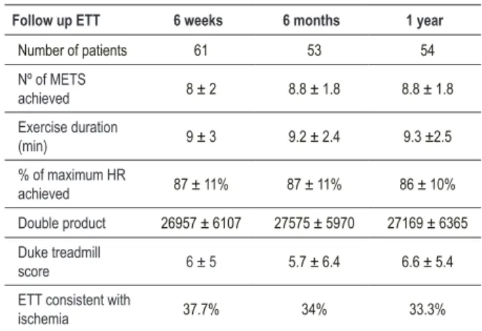
clopidogrel or ticlopidine, respectively. ETT data are shown in Table 2.
Sensitivity, specificity, PPV, NPV, and accuracy regarding results of ETT compared with follow-up angiography are seen in Table 3. When considering patients who reached 85.0% of maximum predicted heart rate, sensitivity, specificity, PPV, NPV, and accuracy
for the detection of restenosis were 63.6%, 67.7%, 41.2%, 84.0%, and 66,7%, respectively, at 6 weeks (p=0.086); 55.6%, 83.3%, 55.6%, 83.3%, and 75.7% at 6 months (p=0.073); and 40.0%, 66.7%, 30.8%, 75.0%, and 59.4% at 12 months (p= 0.716) after
treatment of myocardial infarction.
There was no correlation between restenosis and the Duke treadmill score, exercise duration, exercise angina, ST-segment depression, or right precordial lead abnormalities.
An increase in exercise duration was observed between the first and the second ETT (p=0.004) and between the first and the third ETT (p=0.004) in the 46 patients who performed all 3 tests. The same was observed for the number of METS achieved (p=0.002 and p=0.004, respectively).
Myocardial perfusion imaging
The results of MPI according to qualitative analysis are summarized in Table 4.
Table 1 - Clinical data
Demographic data n %
Men 53 82.8
Smokers 39 60.9
Systemic hypertension 39 60.9
Dyslipidemia 38 59.4
Diabetes mellitus 17 26.9
Previous CABG 6 9.4
Previous PCI* 5 7.8
Previous angina 29 45.3
Age (years) 56.2±10.6
CABG - coronary artery bypass graft; PCI - percutaneous coronary intervention; * - unrelated to the target vessel.
Table 2 - Follow-up ETT results
Follow up ETT 6 weeks 6 months 1 year
Number of patients 61 53 54
Nº of METS
achieved 8 ± 2 8.8 ± 1.8 8.8 ± 1.8
Exercise duration
(min) 9 ± 3 9.2 ± 2.4 9.3 ±2.5
% of maximum HR
achieved 87 ± 11% 87 ± 11% 86 ± 10%
Double product 26957 ± 6107 27575 ± 5970 27169 ± 6365
Duke treadmill
score 6 ± 5 5.7 ± 6.4 6.6 ± 5.4
ETT consistent with
ischemia 37.7% 34% 33.3%
Table 3 - Comparison between ETT and follow-up angiography results
Variable
ETT x Follow-up angiography
6 weeks ETT 1
6 months ETT 2
1 year ETT 3
Sensitivity (%) 57.3 54.5 38.5
Speciicity (%) 69 70.7 66.7
Positive predictive value (%) 38 33.3 27.8
Negative predictive value (%) 80.6 85.3 76.5
Accuracy (%) 64.9 67.3 54.9
p 0.123 0.159 0.747
Table 4 - Myocardial perfusion imaging (MPI). Qualitative analysis
Myocardial perfusion 6 weeks MPI
6 months MPI
1 year MPI
Number of patients 61 53 50
Normal 8 (13.1%) 6 (11.3%) 9 (18%)
Reversible defect 5 (8.1%) 6 (11.3%) 1 (2%)
Partially reversible defect 26 (34.1%) 11 (20.75%) 6 (12%)
Persistent defect 22 (30.1%) 30 (56.6%) 34 (64%)
LV ejection fraction 0.51 0.50 0.49
LV - left ventricular.
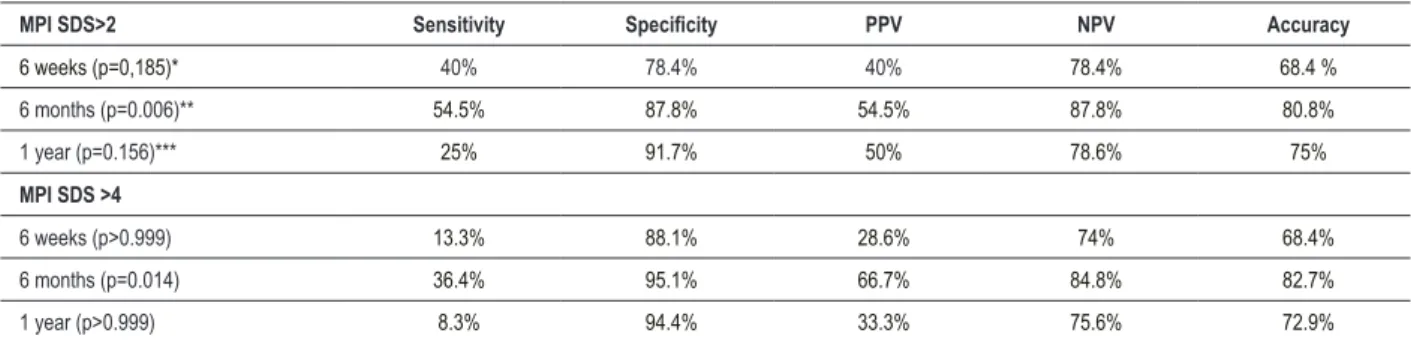
The semiquantitative analysis considered transient ischemia as a summed difference score > 2; for the same sample scores > 4 were also evaluated.
The results of this analysis are shown in Table 5. Interobserver reproducibility showed substantial agreement
with Kappa of 0.762 (95% CI 0.51-1.0), p < 0.00117. The summed difference score during the first year in 46 patients who underwent all sequential MPI did not show any statistical significance (p=0.194). The comparison between summed difference scores performed 6 weeks, 6 months, and one year after PPCI and restenosis was not statistically significant (p = 0.056).
Summed stress scores and summed rest scores were compared with restenosis in all MPI, and classified as 0 to
4, 4 to 8, and >8 according to the amount of damaged myocardium.
There was a correlation between the amount of damaged myocardium and restenosis at 1 year at rest (p=0.0019) and after exercise (p=0.004).
Follow-up
Angiographic restenosis occurred in 17/59 patients
(28.8%). Subacute stent thrombosis was observed 27 days
after PPCI in 1 patient. Non-ST-elevation acute coronary syndrome occurred in 2 patients during the fourth month (one of them treated by surgical revascularization and the other by balloon angioplasty).
Restenosis was treated in 11 patients (18.6%) by means of surgical revascularization in 3 (1 with restenosis > 50.0%
and < 70.0% plus angina) and angioplasty in 8 (2 with restenosis >50.0% and < 70.0% plus angina). In 6 patients
with restenosis, no procedure was performed.
Angioplasty was performed in 7 patients in non-infarct
arteries. No death was observed during follow-up.
Forty-four patients remained asymptomatic during the
first year, 11 patients had chest pain up to six months (7 with restenosis, of whom 5 had a lesion ≥70.0% ), and 9 reported
symptoms after 6 months of follow-up.
Discussion
Angiographic restenosis occurred in 17 patients (28.8%).
Restenosis rates in the PAMI-PILOT study18, STENT-PAMI
trial16, and CADILLAC study3 were 27.0%, 23.0% and 22.0%,
respectively.
Similar to Cutlip et al19, we followed up our patients for
one year to measure the real success rate19.
The STENT-PAMI16 and CADILLAC3 trials followed up their
patients for 6 months, while the GRAMI20 and STENTIM21 trials
reported a 12-month follow-up. Late repeat intervention after
PPCI was performed in 3.6% to 22.7% of the cases3,16,22. In our
study, the rate of target vessel revascularization was 18.6%. The appropriate use of noninvasive tests for the diagnosis of restenosis after PPCI remains controversial.
ETT is routinely used to detect residual ischemia, to assess exercise capacity, and for risk stratification after acute
Table 5 - MPI. Semiquantitative analysis of the left ventricle considering Summed Difference Score >2 and >4
MPI SDS>2 Sensitivity Speciicity PPV NPV Accuracy
6 weeks (p=0,185)* 40% 78.4% 40% 78.4% 68.4 %
6 months (p=0.006)** 54.5% 87.8% 54.5% 87.8% 80.8%
1 year (p=0.156)*** 25% 91.7% 50% 78.6% 75%
MPI SDS >4
6 weeks (p>0.999) 13.3% 88.1% 28.6% 74% 68.4%
6 months (p=0.014) 36.4% 95.1% 66.7% 84.8% 82.7%
1 year (p>0.999) 8.3% 94.4% 33.3% 75.6% 72.9%
myocardial infarction. It is the most commonly used test for evaluating patients after PCI23-25.
It is known that acute exercise can lead to a transient prothrombotic state in patients with coronary artery disease26.
Cases of in-stent thrombosis within hours after ETT have been previously reported27,28. In our study, ETT was safely performed.
The current ACC/AHA guidelines11 for ETT post-PCI do
not recommend routine screening for detection of restenosis, but there are insufficient data to determine the correct ETT strategy, especially in patients at high risk of restenosis such as those in the proximal LAD.
The rationale of the strategy of routine indication of functional tests after PCI is based on the theory that the diagnosis of silent ischemia can stratify patients at higher risk5. Previous studies demonstrated that ETT has low
sensitivity and ability to localize ischemia11,29. Chest pain is an
unreliable index of restenosis after PCI, since approximately 50.0% of patients with restenosis remain asymptomatic and, conversely, up to 45.0% of patients developing chest pain do not have angiographic restenosis5. In patients with incomplete
revascularization, the ischemic response may result from either stent restenosis or myocardial ischemia unrelated to the target vessel.
A meta-analysis showed ETT sensitivity and specificity to
be of 46.0% and 77.0%, respectively, for restenosis > 50.0%; and 50.0% and 84.0%, respectively, for restenosis > 70.0%29.
ETT performed 6 weeks after myocardial infarction showed low sensitivity and moderate specificity. Although ETT for the detection of restenosis had been performed 6 months after the procedure, we did not find improved sensitivity and specificity in relation to the earlier ETT; however, its high negative predictive value stresses the importance of a nonischemic test in the clinical approach to these patients.
Similar results regarding sensitivity and specificity, with lower negative predictive value (61.0%), were reported in symptomatic patients after PCI25.
ADORE was the first trial to compare the strategies of routine and selective ETT after PCI; this study evaluated patients undergoing complete revascularization with no angiographic follow-up, and found no benefits with routine ETT in relation to selective ETT, which was performed in symptomatic patients30.
In our study, no association with restenosis was found in the presence of complete or incomplete revascularization, even in patients who achieved more than 85.0% of maximum predicted heart rate. Our data confirm the low sensitivity of angina and electrocardiographic changes during exercise for the detection of restenosis31.
Improved ETT sensitivity was not observed when right precordial leads were included; this corroborates the findings from other studies32,33 but not Michaelides et al’s findings4.
The mean METS obtained was 8, which can be explained by our strict inclusion criteria, such as LVEF > 40.0%. In post-infarction patients, exercise capacity > 4 METS in association
with LVEF ≥ 40.0% was correlated with a favorable outcome34.
According to the DANAMI-2 trial, post-infarction patients with low exercise capacity had higher mortality rates35.
Exercise MPI has better diagnostic ability than ETT alone25.
MPI is frequently performed after PCI to detect restenosis and to identify other areas of myocardial ischemia unrelated to the target-vessel36. In our study, MPI performed at 6 weeks showed
low sensitivity, which could be explained by intermediary lesions or by the use of medications. Specificity and negative predictive value were moderate.
Given the high false positive rate, MPI early (<3 months)
after PCI should not be routinely indicated5. Rodes-Cabau et
al37 detected reversible defects in 17.0% of the patients who underwent MPI one week after PPCI, with 50.0% sensitivity and 54.0% specificity. Nevertheless, this finding may not exclude the possibility of restenosis. In 2 of our patients who presented with acute coronary syndrome during the fourth month of follow-up, no reversible ischemia was observed 6 weeks after PPCI.
In our study, MPI performed at 6 and 12 months showed low rates of sensitivity, but high rates of specificity, especially at 6 months, considering summed difference score > 2 and > 4. Sensitivity, specificity, and positive and negative predictive values may have been affected by drug therapy. The accuracy of MPI performed 6 months following PCI is usually very good5.
Galassi et al31 found similar accuracy (83.0%) in patients
who did or did not achieve maximum heart rate during exercise testing. The authors concluded that the most important characteristic is the homogeneity of the study population, like in our sample, in terms of type of stress and time interval between stenting and scintigraphy, and between scintigraphy and follow-up angiography. The inclusion of territories with previous myocardial infarction did not affect the accuracy of MPI. Unlike in our study, only complete revascularization was assessed.
Isaaz et al38 recently reported a poor correlation between
stress SPECT imaging and angiographic restenosis at 6 months in 149 patients treated by PPCI who remained asymptomatic during the follow-up. They showed sensitivity, specificity, positive predictive value, negative predictive value and
accuracy of 48.0%, 61.0%, 35.0%, 72.0% and 57.0%,
respectively, for the detection of restenosis. The authors
followed up 78 patients with single vessel disease for two
years and a half38.
Our results showed better specificity and negative predictive value.Kosa et al39 reported that preexisting
myocardial perfusion abnormalities in territories with previous infarction may result in lower sensitivity and specificity values. Population heterogeneity may account for some of the discrepancies observed in these studies.
The sensitivity and specificity of MPI performed at different times after PCI range from 39.0% to 100.0%, and from 46.0% to 100.0%, and improve with time after revascularization5.
The presence of reversible defects on MPI performed between 12 and 18 months after PCI is associated with an increased risk of events in the follow-up. The extent and severity of reversible perfusion defects on SPECT strongly predict event-free survival late after PCI15. This information
LVEF < 40.0% as determined by gated SPECT was
correlated with a poor prognosis during follow-up40. In our
case series, the good outcome may be explained by the LVEF > 45.0% observed on follow-up MPI. We know that patients with ejection fraction > 40.0% are not always found in the real world. However, in tertiary care centers, PPCI can be performed within the first few hours after myocardial infarction. Thus, it is possible to find patients with preserved ejection fraction and thereby obtain information from noninvasive tests similar to that obtained after elective PCI.
Clinical implications
The negative predictive value for MPI performed 6 months after the PPCI is high. The absence of reversible ischemia can be considered indicative of the absence of restenosis (or of the functional expression of restenosis) in these selected patient sample.
Study limitations
The limitations of this study include small sample size,
inclusion of only Killip class 1 patients with LVEF ≥ 40.0%, and
maintainence of usual medications during the entire protocol. The small sample was related to the occurrence of several trials running at the same time in both institutions, thus not allowing the same patient in more than one trial.
The selection of patients with LVEF > 40.0% was made
to ensure that they could undergo ETT.
Maintainance of drugs such as betablockers may affect the heart rate response to exercise, but our intention was to evaluate a real-world sample of patients after PPCI.
Conclusions
In conclusion, ETT showed low sensitivity to detect restenosis after PPCI during the follow-up. The inclusion of right precordial leads did not provide additional information. MPI performed 6 months after PPCI correlated with restenosis, showing high specificity and NPV in this patient population.
Potential Conflict of Interest
No potential conflict of interest relevant to this article was reported.
Sources of Funding
There were no external funding sources for this study.
Study Association
This article is part of the thesis of doctoral submitted by Rica Dodo Delmar Buchler, from Faculdade de Medicina da Universidade de São Paulo.
References
1. Keeley EC, Boura JA, Grines CL. Primary angioplasty versus intravenous thrombolytic therapy for acute myocardial infarction: a quantitative review of 23 randomised trials. Lancet. 2003; 361 (9351): 13-20.
2. Spaulding C, Henry P, Teiger E, Bramucci E, Carrié D, Slama MS, et al. Sirolimus-eluting versus uncoated stents in acute myocardial infarcyion. N Engl J Med. 2006; 355 (11): 1093-104.
3. Stone GW, Grines CL, Cox DA, Garcia E, Tcheng JE, Griffin JJ, et al. Controlled Abciximab and Device Investigation to Lower Late Angioplasty Complications (CADILLAC) Investigators. Comparison of angioplasty with stenting, with or without abciximab, in acute myocardial infarction. N Engl J Med. 2002; 346
(13): 957-66.
4. Michaelides AP, Psomadaki ZD, Dilaveris PE, Richter DJ, Andrikopoulos GK, Aggeli KD, et al. Improved detection of coronary artery disease by exercise electrocardiography with the use of right precordial leads. N Engl J Med. 1999; 340 (5): 340-5.
5. Giedd KN, Bergmann SR. Myocardial perfusion imaging following percutaneous coronary intervention: the importance of restenosis disease progression, and directed reintervention. J Am Coll Cardiol. 2004; 43 (3): 328-36.
6. Killip T 3rd, Kimball JT. Treatment of myocardial infarction in a coronary care
unit: a two year experience with 250 patients. Am J Cardiol. 1967; 20 (4): 457-64.
7. Smith SC Jr, Feldman TE, Hirshfeld JW Jr, Jacobs AK, Kern MJ, King SB 3rd, et
al. American College of Cardiology/American Heart Association Task Force on Practice Guidelines; ACC/AHA/SCAI Writing Committee to Update the 2001 Guidelines for Percutaneous Coronary Intervention. ACC/AHA/SCAI 2005 guideline update for percutaneous coronary intervention: a report of the American College of Cardiology/American Heart Association Task Force on Practice Guidelines (ACC/AHA/SCAI Writing Committee to Update the
2001 Guidelines for Percutaneous Coronary Intervention). J Am Coll Cardiol.
2006; 47 (1): e1-121.
8. The Thrombolysis in Myocardial Infarction (TIMI) trial. Phase I findings. TIMI Study Group. N Engl J Med. 1985; 312 (14): 932-6.
9. Lang RM, Bierig M, Devereux RB, Flachskampf FA, Foster E, Pellikka PA, et al. Chamber Quantification Writing Group; American Society of Echocardiography’s Guidelines and Standards Committee; European Association of Echocardiography. Recommendations for chamber quantification: a report from the American Society of Echocardiography’s Guidelines and Standards Committee and the Chamber Quantification Writing Group, developed in conjunction with the European Association of Echocardiography, a branch of the European Society of Cardiology. J Am Soc Echocardiogr. 2005;18 (12): 1440-63.
10. Balady GJ, Berra KA, Golding LA, Gordon NF, Mahler DA. ACSMs Guidelines for exercise testing and prescription. 6th ed. Baltimore, Maryland, U.S.A:
Lippincott, Williams and Wilkins; 2000. p. 97-8.
11. Gibbons RJ, Balady GJ, Bricker JT, Chaitman BR, Fletcher GF, Froelicher VF, et al. American College of Cardiology/American Heart Association Task
Force on Practice Guidelines (Committee to Update the 1997 Exercise
Testing Guidelines). ACC/AHA 2002 guideline update for exercise testing: summary article: a report of the American College of Cardiology/American Heart Association Task Force on Practice Guidelines (Committee to Update
the 1997 Exercise Testing Guidelines). Circulation. 2002; 106 (14): 1883-92.
12. Bruce RA, Kusumi F, Hosmer D. Maximal oxygen intake and normographic assessment of functional aerobic impairment in cardiovascular disease. Am
Heart J. 1973; 85 (4): 546-62.
14. Berman DS, Kiat H, Friedman JD, Wang FP, van Train K, Matzer L, et al. Separate acquisition rest thallium-201/stress technetium-99m sestamibi dual-isotope myocardial perfusion single photon emission computed tomography: a clinical validation study. J Am Coll Cardiol. 1993; 22 (5): 1455-64.
15. Acampa W, Petretta M, Florimonte L, Mattera A, Cuocolo A. Prognostic value of exercise cardiac tomography performed late after percutaneous coronary intervention in symptomatic and symptom free patients. Am J Cardiol. 2003; 91 (3): 259-63.
16. Grines CL, Cox DA, Stone GW, Garcia E, Mattos LA, Giambartolomei A, et al. Coronary angioplasty with or without stent implantation for acute myocardial infarction. Stent Primary Angioplasty in Myocardial Infarction Study Group. N Engl J Med. 1999; 341 (26): 1949-56.
17. Landis JR, Koch GG. The measurement of observer agreement for categorical data. Biometrics. 1977; 33 (1): 159-74.
18. Stone GW, Brodie BR, Griffin JJ, Costantini C, Morice MC, St Goar FG, et al. Clinical and angiographic follow-up after primary stenting in acute myocardial infarction: the Primary Angioplasty in Myocardial Infarction(PAMI) stent pilot trial. Circulation. 1999; 99 (12): 1548-54.
19. Cutlip DE, Chauhan MS, Baim DS, Ho KK, Popma JJ, Carrozza JP, et al. Clinical restenosis after coronary stenting: perspectives from multicenter clinical trials. J Am Coll Cardiol. 2002; 40 (12): 2082-9.
20. Rodriguez A, Bernardi V, Fernandez M, Mauvecin C, Ayala F, Santaera O, et al. In-hospital and late results of coronary stents versus conventional balloon angioplasty in acute myocardial infarction (GRAMI trial). Gianturco-Roubin in Acute Myocardial Infarction. Am J Cardiol. 1998; 81 (11): 1286-91.
21. Maillard L, Hamon M, Khalife K, Steg PG, Beygui F, Guermonprez JL, et al. A comparison of systematic stenting and conventional balloon angioplasty during primary percutaneous transluminal coronary angioplasty for acute myocardial
infarction. STENTIM-2 Investigators. J Am Coll Cardiol. 2000; 35 (7): 1729-36.
22. Zhu MM, Feit A, Chadow H, Alam M, Kwan T, Clark LT. Primary stent implantation compared with primary balloon angioplasty for acute myocardial infarction: a meta-analysis of randomized clinical trials. Am J
Cardiol. 2001; 88 (3): 297-301.
23. Rosanio S, Tocchi M, Stouffer GA. Use of stress testing to evaluate patients with recurrent chest pain after percutaneous coronary revacularization. Am J Med Sci. 1998; 316 (1): 46-52.
24. Laarman G, Luijten HE, van Zeyl LG, Beatt KJ, Tijssen JG, Serruys PW, et al. Assessment of “silent” restenosis and long term follow- up after successful angioplasty in single vessel coronary artery disease : the value of quantitative exercise electrocardiography and quantitative coronary angiography. J Am
Coll Cardiol. 1990; 16 (3): 578-85.
25. Dori G, Denekamp Y, Fishman S, Biherman H. Exercise stress testing, myocardial perfusion imaging and stress echocardiography for detecting restenosis after successful percutaneous transluminal coronary angioplasty: a review of performance. J Intern Med. 2003; 253 (3): 253-62.
26. Koenig W, Ernst E. Exercise and thrombosis. Coron Artery Dis. 2000; 11 (2):
123-7.
27. Samuels B, Schumann J, Kiat H, Friedman J, Berman DS. Acute stent
thrombosis associated with exercise testing after successful percutaneous
transluminal coronary angioplasty. Am Heart J. 1995; 130 (5): 1120-2.
28. MeurinP, DomniezT, Weber H, Tournadre P, Elhadad S, Bourmayan C, et al. Occlusion of a coronary endoprosthesis after a negative stress test. Arch Mal
Coeur Vaiss. 1999; 92 (3): 369-72.
29. Garzón P, Eisenberg MJ. Functional testing for the detection of restenosis after percutaneous transluminal coronary angioplasty: a meta-analysis. Can
J Cardiol. 2001; 17 (1): 41-8.
30. Eisenberg MJ, Blankenship JC, Huynh T, Azrin M, Pathan A, Sedlis S, et al. Evaluation of routine functional testing after percutaneous coronary
intervention. Am J Cardiol. 2004; 93 (6): 744-7.
31. Galassi AR, Foti R, Azzarelli S, Coco G, Condorelli G, Russo G, et al. Usefulness of exercise tomographic myocardial perfusion imaging for detection of restenosis after coronary stent implantation. Am J Cardiol. 2000; 85 (11): 1362-4.
3. Bokhari S, Blook DK, Bergmann SR. Failure of right precordial electrocardiography during stress testing to identify coronary artery disease. J Nucl Cardiol. 2001; 8 (3): 325-31.
33. Bainey KR, Kalia N, Carter D, Hrynchyshyn G, Kasza L, Lee TK, et al. Right precordial leads and lead aVR at exercise electrocardiography: does it change
test results? Ann Noninvasive Electrocardiol. 2006; 11 (3): 247-52.
34. Dutcher JR, Kahn J, Grines C, Franklin B. Comparison of left ventricular ejection fraction and exercise capacity as predictors of two- and five-year
mortality following acute myocardial infarction. Am J Cardiol. 2007; 99 (4):
436-41.
35. Valeur N, Clemmensen P, Saunamaki K, Grande P. (DANAMI-2 investigators). The prognostic value of pre-discharge exercise testing after myocardial infarction treated with either primary PCI or fibrinolysis: a DANAMI-2
sub-study. Eur Heart J. 2005; 26 (2): 119-27.
36. Ritchie J,Bateman TM, Bonow RO, Crawford MH, Gibbons RJ, Hall RJ, et al. Guidelines for clinical use cardiac radionuclide imaging. A report of the American Heart Association/American College of Cardiology Task Force on Assessment of Diagnostic and Therapeutic Cardiovascular Procedures, (Committee on Radionuclide Imaging), developed in collaboration with the
American Society of Nuclear Cardiology. Circulation. 1995; 91 (4): 1278-303. 37. Rodes-Cabau J, Candell-Riera J, Domingo E, Castell-Conesa J, Anivarro I,
Angel J, et al. Frequency and clinical significance of myocardial ischemia detected early after coronary stent implantation. J Nucl Med. 2001; 42 (12):
1768-72.
38. Isaaz K, Afif Z, Prevot N, Cerisier A, Lamaud M, Richard L, et al.The value of stress single-photon emission computed tomography imaging performed routinely at 6 months in asymptomatic patients for predicting angiographic restenosis after successful direct percutaneous intervention for acute ST
elevation myocardial infarction.Coron Artery Dis. 2008; 19 (2): 89-97.
39. Kosa I, Blasini R, Schneider-Eicke J, Neumann FJ, Matsunari I, Neverve J, et al. Myocardial perfusion scintigraphy to evaluate patients after coronary stent
implantation. J Nucl Med. 1998; 39 (8): 1307-11.
40. Beller GA, Zaret BL. Contributions of nuclear cardiology to diagnosis and prognosis of patients with coronary artery disease. Circulation. 2000; 101