Myocardial Viability in a Single-Vessel
Disease: The Role of a Dobutamine Stress
Echocardiography
Márcia Bueno Castier, José Raul Alves Carrinho, Márcia Alves Carrinho, Francisco Manes Albanesi Filho,
Álvaro Villela de Moraes, Fernando Portugal Morcerf - Brazil
Universidade do Estado do Rio de Janeiro e ECOR Serviço de Ecocardiografia - Rio de Janeiro, RJ - Brazil
Mailing Address: M á r c i a B u e n o C a s t i e r • Av. Epitácio Pessoa, 4254/201 – 22471-001 – Rio de Janeiro, RJ - Brazil E-mail: [email protected] Received on 01/22/04 • Accepted on 06/08/05
O
BJECTIVETo investigate a group of patients that have a signifi cant lesion in a single-vessel and to demonstrate whether or not the sensitivity and specifi city of a dobutamine stress echocardiography (DSE) was valuable in the evaluation of myocardial viability for these patients.
M
ETHODSTwenty patients who had undergone percutaneous transluminal coronary angioplasty (PTCA) were studied. This group was evaluated 2 to 7 days (3.65 ± 1.69) before the procedure and 2 to 5 days (4 ± 0.80) after the procedure with a DSE. Myocardial viability was assessed three months after the procedure using a two dimensional echocardiogram. Twelve patients underwent PTCA on the left anterior descending artery (LAD), seven on the right coronary artery (RC) and 1 on the circumfl ex artery (CX). Only one right coronary artery procedure was not 100% successful.
R
ESULTSFrom the 340 segments that were studied, 99 (29.18%) demonstrated contractile alterations of which 63 were hypokinetic (63.4%), 28 akinetic (28.28%) and 8 dyskinetic (8.08%). In reference to the segments involved, we obtained a sensitivity of 92.59%, specifi city of 84.45%, and accuracy of 88.88% for the DSE. The solitary case of PTCA for the circumfl ex artery demonstrated 100% sensitivity. The LAD demonstrated a sensitivity of 88.58%, specifi city of 95% and accuracy of 90.91%. For the RC segments, sensitivity was 91.30%, specifi city 83.33% and accuracy 88.71%. All dyskinetic segments were unviable. The DSE predicted a 91.48% recovery rate for the 63 hypokinetic segments.
C
ONCLUSIONThe DSE is an effective test for evaluating myocardial viability in patients with a significant single-vessel disease.
K
EY WORDSSince an echocardiogram is a non-invasive, reproducible, low cost procedure that can be performed at the side of the patient’s bed, it is now used extensively to detect coronary diseases. One of the techniques used during an echocardiography test is the infusion of stress producing agents such as dobutamine to evaluate the presence of ischemia and/or myocardial viability since coronary disease evaluation using an echocardiogram at rest is limited due to the necessity to have a critical reduction of coronary blood fl ow in order to have a detectible contractile alteration. Myocardial viability detection is crucial to defi ne the therapeutic strategy for patients with coronary disease and is directly related to the prognosis. The decision to conduct a revascularization procedure in patients with myocardial viability also involves prognosis modifications1. Since 1990, the dobutamine stress echocardiogram (DSE) has been used to detect myocardial viability as it can be used for both the acute and chronic phases of coronary disease2. The presence of a biphasic response, in which there is an increase of contractibility followed by a deterioration as the drug dosage is increased, is said to be too sensitive for the identifi cation of viable areas1.
Although the method is not new, clinical cardiologists have been showing a greater interest in the procedure because of the assistance it can offer when deciding the best therapeutic strategy for the patient. The studies published to date on the use and importance of DSE in the evaluation of myocardial viability combine various subgroups of patients (for example: those with and without previous intervention, single and multi-vessel diseases). Consequently, the objective of this study was to investigate a group of patients with a signifi cant lesion in a single-vessel and to demonstrate whether or not the sensitivity and specifi city of a dobutamine stress echocardiography was valuable in the evaluation of myocardial viability for these patients.
M
ETHODS
Twenty-two patients, scheduled consecutively for percutaneous transluminal coronary angioplasty (PTCA) for the treatment of an obstructive single coronary vessel desease were studied. The study inclusion criteria were: artery stenosis greater than 70% in a single-vessel, contractile alteration in the ventriculography in the corresponding region of the artery lesion, lack of obstructive damage in other coronary vessels greater than 50%. The exclusion criteria were: instable angina symptoms, myocardial infarction within six weeks of the start of the study, an inadequate echocardiography window to obtain images, valvular lesions, previous history of myocardial revascularization surgery and a deterioration of the clinical picture during follow-up.
From the initial 22 patients, two were excluded in the course of the study since after PTCA surgery they
presented angina symptoms and coronary restenosis was discovered. Consequently, twenty patients concluded the study. From these, twelve were male (60%), between the ages of 36 and 74 (55 ± 10 years). In relation to clinical characteristics, twelve (60%) had a previous history of myocardial infarction that had occurred more than eight weeks before the start of the study and eight (40%), had stable angina. The myocardial infarction patients had suffered the attack nine to 106 weeks before the start of the study (33 ± 29). Only one of the patients had previously undergone angioplasty surgery for another vessel. The coronary lesions varied in severity with obstructions between 70% and 95% (average of 84.75 ± 8.03).
This study consisted of three stages. The fi rst was conducted two to seven days (3.65 ± 1.69) before the PTCA procedure. On the same day, one and two dimensional echocardiograms were performed. The symptoms of all patients remained stable between the tests and the angioplasty revascularization procedure. The second stage was conducted two to five days (4.0 ± 0.8) after this intervention and both the basal echocardiogram and the DSE were repeated. During this interval, patient clinical follow-up was conducted by means of electrocardiograms and a series of stress tests. Three months after the procedure (third stage) myocardial viability was determined using one and two dimensional echocardiograms. Myocardial viability was evaluated by comparing the two dimensional tests that had been performed before the procedure, two to fi ve days after the procedure and three months after the procedure. The segments that presented an increase in contractibility in the echocardiogram performed three months after the procedure were considered viable.
The echocardiography tests were performed using an ATL instrument, model HDI 3000. All tests were recorded on video tapes for later analysis and fi led on optical disks.
The DES protocol consisted of three minute infusion stages in the dosages of 5, 10, 20, 30 e 40 µg/kg/minute3. When it was not possible to reach the sub-maximum frequency, up to 1 mg of atropine was administered via intravenous in the fi nal stage. The tests were reviewed later, evaluating the wall motion of the seventeen left ventricle segments4. In order to evaluate in a semi quantitative manner, the wall motion score index was used to evaluate contractibility in the left ventricle5.
Averages and standard deviations were used to study continuous variables. The Mann-Whitney non-parametric test was used to compare the averages of the scores. Sensitivity, specifi city, positive and negative predictive values and accuracy were used to evaluate the effi ciency of the diagnostic tests. A 5% alpha error was used for the tests.
R
ESULTS
92,59
84,45 87,72
88,88
50 60 70 80 90 100
S E PPV A
angioplasty (PTCA) on the left descending artery (LAD), seven (35%) on the right coronary artery (RC) and one (5%) on the circumfl ex artery (CX).
A total of 340 segments were analyzed during each stage of the study. In the echocardiogram performed before the PTCA, 99 segments (29.18%) presented contractile alterations. In this fi rst test, 63 segments (63.64%) were considered hypokinetic, 28 akinetic (28.28%) and eight dyskinetic (8.08%), corresponding to an average of 4.95 segments per patient.
In the second echocardiography that was performed during the week immediately following the procedure, it was observed that all of the 241 segments that had normal wall motion in the initial exam maintained the contractile standard. There was wall motion improvements in twenty (20.20%) of the 99 segments that had previous obstructions. Eighteen (28.57%) of the 63 hypokinetic segments were repaired and two (7.14%) of the 28 akinetic segments became hypokinetic. There was no change in the dyskinetic segments.
The average wall motion score in the fi rst test (pre-angioplasty) was 1.42 ± 0.31. The average score for the test performed three months after the procedure was 1.23 ± 0.33, which is a signifi cant statistical variation (z = -3.7, p < 0.001). These data are shown in fi gure 1. When compared with the initial exams and those conducted in the week following the procedure, no signifi cant variations in the wall motion score averages were observed (1.42 ± 0.31 vs. 1.36 ± 0.35, z = -1.13 , p = 0.255). Likewise, comparisons made during the administration of dobutamine in the dosage of 10 µg/kg/minute did not reveal any signifi cant variations (1.27 ± 0.38 vs 1.26 ± 0.38, z = -1.00, p = 0.317).
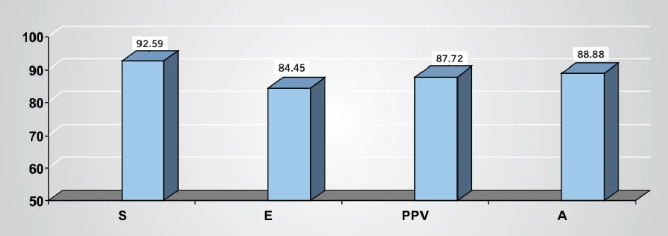
A total of 54 segments was considered viable in the test performed three months after the angioplasty. The DSE detected viability in fi fty (92.59%) of these segments. The test conducted three months after the procedure revealed a total of 45 unviable segment of which 38 had been correctly identifi ed by the DSE. The other seven unviable segments were identifi ed as viable (sensitivity 92.59%,
specifi city 84.45%, positive predictive value 87.72%, accuracy 88.88%). These data are shown in Figure 2.
Only one patient underwent CX angioplasty. The DSE for this patient demonstrated that the nine segments with contractile alterations were all unviable, which was confi rmed by the echocardiogram performed three months after the percutaneous intervention.
In relation to the twelve patients that underwent LAD angioplasty, 35 segments were considered viable (61.36%) and 20 unviable (38.64%). The DSE identifi ed 31 of these segments (88.58%) as viable.
In the seven patients that underwent RC angioplasty, 23 segments were considered viable (65.71%) and 12 unviable (34.29%). The DSE identifi ed 21 of these segments (91.30%) as viable (sensitivity 92.3%, specifi city 83.33%, positive predictive value 91.3%, negative predictive value 83.33% and accuracy 88.71%).
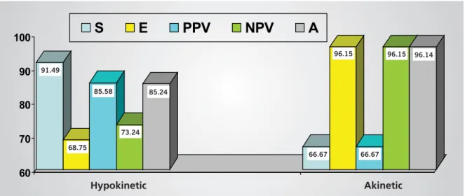
In regard to the type of segment alteration found, of the 63 hypokinetic segments, 47 were viable in the exam performed three months after the angioplasty (74.60%), and the DSE was able to identify 43 (91.48%) of these segments (sensitivity 91.49%, specificity 68.75%, positive predictive value 88.58%, negative predictive value 73.34% and accuracy 85.71%). Of the 28 akinetic segments, two (7.14%) became hypokinetic after the fi rst reevaluation and were correctly identifi ed
Fig. 2 – Sensitivity (S), Specifi city (E), Positive Predictive Value (PPV) and Accuracy (A). Indices obtained in the myocardial viability evaluation for patients with a single-vessel desease
1,42
1,23
0 0,2 0,4 0,6 0,8 1 1,2 1,4
Fig. 1- Wall motion index variations before and three months after angioplasty
Before After
1.4
1.2
1
0.8
0.6
0.4
0.2
0
1.42
1.23
92.59
84.45
by the DSE. Another akinetic segment was found to be hypokinetic in the study performed three months after the PTCA (sensitivity 66.67%, specificity 96.15%, positive predictive value 66.67%, negative predictive value 96.15% and accuracy 96.42%). All of the eight dyskinetic segments showed to be unviable after three months and were correctly evaluated by the DSE. These data are shown in Figure 3.
D
ISCUSSION
A DSE is a safe method with a low incidence of signifi cant events6, such as serious arrhythmias or acute myocardial infarction that occur in less than 0.5% of the patients6,7. In the twenty patients evaluated in our study, we did not fi nd any serious collateral effects due to the use of dobutamine. Despite the small sample, our data coincide with those of the Mertes study8, that also did not report any serious collateral effects in the 1,118 patients studied.
The detection of myocardial viability using a DSE could also be performed after an acute myocardial infarction, and was demonstrated in various case studies2,9,10,11,12,13, with sensitivity fi gures that vary from 77% to 89%, and specifi city fi gures between 68% and 93%.
In the chronic phase of coronary disease, studies have also demonstrated satisfactory results for the evaluation of myocardial viability with dobutamine. A study involving fourteen patients conducted by Marzullo et al14 revealed a sensitivity rate of 82% and a specifi city rate of 92%. Other authors have found, in a total of 224 patients, sensitivity rates from 78% to 92%, specifi city rates between 60% and 95%1,15-20 and excluding Afridi et al1, an accuracy higher than 83%.
In comparison with a myocardial scintillography, a dobutamine stress echocardiography has proven to provide equivalent information17,20.
The effi ciency index variations found in various studies
can be explained by differences in the populations evaluated, the protocols used and even the experience of observers, since this is one factor that interferes with DSE results. As demonstrated by Afridi et al.1 other factors can also affect method accuracy. In this case study, the presence of a biphasic response, characterized by improved contractibility at low dosages of dobutamine followed by a deterioration as the dosage is increased, revealed a sensitivity of 90% and a specifi city of 80%, suggesting that a biphasic response could be the best detection method for viability.
The sensitivity fi ndings in this study are similar to those found by Afridi et al1 and La Canna et al16, and higher than those found in other documented case studies17,19. This is in agreement with the concept that the DSE sensitivity for patients with a single-vessel desease is comparable with that found in multi-vessel cases which does not occur when a DSE is used to detect myocardial ischemia. This higher degree of sensitivity found in our study could also be partially explained by specifi c characteristics of our population, such as a single-vessel desease and the presence of contractile alterations at rest in the segments that it supplies blood to.
The comparison between the wall motion score averages before and in the week following the angioplasty surgery did not reveal any signifi cant differences (1.42 ± 0.31 before and 1.36 ± 0.35 after, p = 0.255). These fi ndings are comparable to those of McNeill et al21, but contrary to the fi ndings of other authors22,23, that noted improvements in the wall motion index scores for the tests performed in the fi rst few days after the procedure. Other studies24,25 demonstrate that early wall motion improvement occurs only in patients with hibernating myocardium. This improvement, in the majority of patients, occurs within three months but can take as long as four to six months for a complete recovery of contractile function. Based on these studies, we believe that even successful revascularization procedures require
Akinetic Hypokinetic
Fig. 3 – Sensitivity (S), Specifi city (E), Positive Predictive Value (PPV), Negative Predictive Value (NVP) and Accuracy (A) values found for the hypokinetic and akinetic segments
91,49
68,75 85,58
73,24 85,24
66,67 96,15
66,67
96,15 96,14
60
70
80
90
100
S
E
PPV
NPV
A
91.49
68.75 85.58
73.24 85.24
66.67 96.15
66.67
more time for a complete recovery of contractile function and this impression was confi rmed in our segmental wall motion score results in the test performed three months after the angioplasty.
Analysis of the types of arteries treated did not reveal any signifi cant sensitivity differences in a comparison between patients with LAD lesions and RC lesions. These fi ndings are contrary to evaluation studies for myocardial ischemia which demonstrate a lower sensitivity rate for patients with RC lesions2,26. From these data, we believe that in myocardial viability research for patients with a single-vessel desease, the type of artery treated has no infl uence on test sensitivity.
Analysis of the type of contractile alteration revealed a sensitivity of 91.49% for hypokinetic segments and
66.67% for akinetic segments. This lower sensitivity rating for akinetic segments is in agreement with other studies22,27. A lower dobutamine sensitivity to detect viability in akinetic areas is probably related to the higher quantity of fi brous tissue and consequently a lower quantity of viable myocytes in these areas. The DSE considered all dyskinetic segments as unviable, which was confi rmed in the test performed three months after the procedure and is in accordance with the fi ndings of De Filippi et al28.
In conclusion, we have verifi ed that a dobutamine stress echocardiogram is a valuable tool in the evaluation of myocardial viability, even in patients with single-vessel deseases, in which the test detection rates for coronary disease are lower than for other patient subgroups.
R
EFERENCES
1. Afridi A, Kleiman NS, Raizner AE, Zoghbi WA. Dobutamine echocardiography in myocardial hibernation. Optimal dose and accuracy in predicting recovery of ventricular function after coronary angioplasty. Circulation 1995; 91: 663-70.
2. Pierard LA, De Landsheere CM, Berthe C, Rigo P, Kulbertus HE. Identification of viable myocardium by echocardiography during dobutamine infusion in patients with myocardial infarction after thrombolytic therapy: comparison with positron emission tomography. J Am Coll Cardiol 1990; 15: 1021-31.
3. Armstrong WF, Pellikka PA, Ryan T, Crouse L, Zoghbi WA. Stress echocardiography: recommendations for per formance and interpretation of stress echocardiography. Stress Echocardiography Task Force of the Nomenclature and Standards Committee of the American Society of Echocardiography. J Am Soc Echocardiogr 1998; 11: 97-104.
4. Cerqueira MD, Weissman NJ, Dilsizian V et al. Standardized myocardial segmentation and nomenclature for tomographic imaging of the heart: a statement for healthcare professionals from the Cardiac Imaging Committee of the Council on Clinical Cardiology of the American Heart Association. Circulation 2002; 105: 539-42.
5. Bourdillon D, Broderick TM, Sawada SG et al. Regional wall motion index for infarct and noninfarct regions after reperfusion in acute myocardial infarction: comparison with global wall motion index. J Am Soc Echocardiogr 1989; 2: 398-407.
6. Picano E, Mathias W, Pingitore A, Bigi R, Previtali M. Safety and tolerability of dobutamine-atropine stress echocardiography: a prospective, multicenter study. Lancet 1994; 344: 1190-2.
7. Cortigiani L, Bigi R, Gigli G et al. Prognostic implications of intraventricular conduction defects in patients undergoing stress echocardiography for suspected coronary artery disease. Am J Med 2003; 115(1): 12-8.
8. Mertes H, Sawada SG, Ryan T et al. Symptoms, adverse effects, and complications associated with dobutamine stress echocardiography. Circulation 1993;88:15-9.
9. Smar t SC, Sawada S, Ryan T et al. Low-dose dobutamine echocardiography detects reversible dyfunction after thrombolytic therapy of acute myocardial infarction. Circulation 1993; 88: 405-15.
10. Previtali M, Poli A, Lanzarini L, Fetiveau R, Mussini A, Ferrario M. Dobutamine stress echocardiography for assessment of myocardial viability and ischaemia in acute myocardial infarction treated with thrombolysis. Am J Cardiol 1993; 72: 124-30G.
11. Watada H, Ito H, Oh H. Dobutamine stress echocardiography predicts reversible dysfunction after reperfusion of anterior myocardial infarction. J Am Coll Cardiol 1994; 24: 624-30.
12. Salustri A, Elhendy A, Garyfallidis P et al. Prediction of improvement of ventricular function after acute myocardial infarction using low-dose dobutamine stress echocardiography. Am J Cardiol 1994; 74: 853-6.
13. Elhendy A, Trocino G, Salustri A et al. Low-dose dobutamine echocardiography and rest-redistribution thallium-201 tomography in the assessment of spontaneous recovery of left ventricular function after recent myocardial infarction. Am Heart J 1996; 131: 1088-96.
14. Marzullo P, Parodi O, Reisenhofer B et al. Value of rest – thallium-201/technetium-99 sestamibi and dobutamine echocardiography for detecting myocardial viability. Am J Cardiol 1993; 71: 166-72.
15. Cigarroa CG, Defi lippi CR, Brickner ME, Alvarez LG, Wait MA, Grayburn A. Dobutamine stress echocardiography identifies hibernating myocardium and predicts recovery of left ventricular function after coronary revascularization. Circulation 1993; 88: 430-6.
16. La Canna G, Alfi eri A, Giubbibi R, Gargano M, Ferrari R, Visioli O. Echocardiography during infusion of dobutamine for identifi cation of reversible dysfunction in patients with chronic coronary artery disease. J Am Coll Cardiol 1994; 23: 617-26.
17. Charney R, Schwinger ME, Chun J et al. Dobutamine echocardiography and resting-redistribution thallium-201 scintigraphy predicts recovery of hibernating myocardium after coronary revascularization. Am. Heart J 1994; 128: 864-9.
18. Perrone-Filardi, Pace P, Prastaro M et al. Dobutamine echocardiography predicts improvement of hypoperfused dysfunctional myocardium after revascularization in patients with coronary artery disease. Circulation 1995; 91: 2556-65.
19. Arnese M, Cornel JH, Salustri A et al. Prediction of improvement of regional left ventricular function after surgical revascularization. Circulation 1995 ; 91: 2748-52.
20. Vanoverschelde J-LJ, D’Hondt A-M, Marwick T et al. Head-to-head comparison of exercise-redistribution-reinjection thallium single-photon emission computed tomography and low-dose dobutamine echocardiography for prediction of reversibility of chronic left ventricular ischemic dysfunction. J Am Coll Cardiol 1996; 28: 432.
22. Kao HL, Wu CC, Ho YL et al. Dobutamine stress echocardiography predicts early wall motion improvement after elective percutaneous transluminal coronary angioplasty. Am J Cardiol 1995; 76: 652-6.
23. Rambaldi R, Hambuerger JN, Geleijnse ML et al. Early recovery of wall motion abnormalities after recanalization of chronic totally occluded coronary arteries: a dobutamine echocardiographic, prospective, single-center experience. Am Heart J 1998; 136: 831-6.
24. Shivalkar B, Maes A, Borgers M et al. Only hibernating myocardium invariably shows early recovery after coronary revascularization. Circulation. 1996; 94(3): 308-15.
25. Vanoverschelde JL, D’Hondt AM, Marwick T et al. Head-to-head comparison of exercise-redistribution-reinjection thallium single-photon emission computed tomography and low-dose dobutamine echocardiography for prediction of reversibility of chronic left ventricular ischaemic dysfunction. J Am Coll Cardiol 1997; 28: 432-42.
26. Smart SC, Bhatia A, Hellman R et al. Dobutamine-atropine stress echocardiography and dipyridamole sestamibi scintigraphy for the detection of coronary artery disease: limitations and concordance. J Am Coll Cardiol 2000; 36(4): 1265-73.
27. Leclerq F, Messner-Pellen CP, Moragues C et al. Myocardial viability assessed by dobutamine echocardiography in acute myocardial infarction after successful primary coronary angioplasty. Am J Cardiol1997; 80: 6-10.