Case Report
Key words
Tomography; coronary disease; heart defects, congenital. Multidetector computed tomography (MDCT) with 64 channels provides to clinical practice an excellent method to detect coronary artery anomalies. The diagnosis of coronary anomalies consisting of origin of left coronary artery in the pulmonary trunk in adults with no history of congenital disease has few reports in literature. We report a case in a 30-year old female patient complaining of fatigue on major efforts and positive scintigraphy for ischemia. The diagnosis was made through 64-channel MDCT and thus it appears that the method can be used as baseline.
Anomalous Origin of Coronary Artery (ALCAPA) in 64-Channel TC
Scanner
Marcelo Souto Nacif
1,3,4, José Hugo Mendes Luz
3, Denise Madeira Moreira
2,3, Carlos Eduardo Rochitte
3,5, Amarino
Carvalho de Oliveira Júnior
3National Institutes of Health Clinical Center1, Bethesda, MD, EUA; Universidade Federal do Rio de Janeiro2; Hospital Pró-Cardíaco3, Rio de Janeiro,
RJ; Universidade Federal Fluminense4, Niterói, RJ; Instituto do Coração (InCor) do Hospital das Clínicas da Faculdade de Medicina da Universidade
de São Paulo5, São Paulo, SP - Brasil
Mailing address: Marcelo Souto Nacif •
4853 Cordell Avenue apt 419 - 20814 Bethesda, MD - EUA Site: www.msnacif.med.br
Manuscript received April 16, 2009; revised manuscript received November 10, 2009, accepted on December 11, 2009.
Case Report
Female patient, Caucasian, 30-year-old, reported that a couple of months before she had noticed the onset of fatigue related to major efforts, such as climbing stairs or physical activities she used to perform regularly. She also reported that when she was 2, she had a poorly-defined episode of “inflammation of the heart”, which was characterized as myocarditis. However, it was not investigated further. After consulting with a cardiologist, she underwent several tests, including echocardiography, which revealed a small left ventricular (LV) dilation and light dysfunction, and myocardial scintigraphy with pharmacological stress, which showed ischemia on the inferior wall.
The patient was referred to outpatient care to perform MDCT on a LightSpeed VCT 64 GE Healthcare device. She received 20 mg of intravenous metoprolol to control heart rate (HR), obtaining average HR of 70 bpm. Calcium scoring was analyzed through conventional acquisition with prospective synchronization and 3 mm collimation. Angiographic analysis used helical acquisition and retrospective electrocardiographic synchronization with 0.625 mm collimation during injection of 80 ml of nonionic contrast medium with a 5.0 ml/s flow. Twenty cardiac phases were reconstructed and the one with the lowest degree of cardiac motion at the level of the coronary arteries was selected. The best phase for coronary analysis in this case was that of 80%. For the analysis, we used axial imaging sources (Figure 1), multiplanar reformatting and three-dimensional curves and reconstructions per volume rendering with angiographic (Figure 2) and cardiac (Figure 3) reconstruction in ADW Workstation 4.3 (GE Healthcare).
Discussion
Anomalous Left Coronary Artery from Pulmonary Artery (ALCAPA) was first described in 1908 in a 60-year old female patient1. Subsequently, Bland et al1 described the same syndrome in 1933, studying the clinical and autopsy findings of a 3-month old child with dyspnea and myocardial infarction. The estimated incidence of ALCAPA is 1 for every 300,000 live births, representing a percentage between 0.24% and 0.46% of all congenital cardiac anomalies6-10. Some believe that this is significantly underestimated because an unknown portion of patients may spend a lifetime asymptomatically and remain undiagnosed7,8. The most common form of this syndrome is the
Introduction
Normal coronary anatomy is characterized by two ostia centrally located in the right and left Valsalva sinus. Anomalies stemming from coronary arteries are estimated to be about 1% of the general population according to studies deriving from coronary angiography1-4 performed in suspected obstructive diseases. In autopsy studies, we see a lower incidence, as in the study by Alexander and Griffiths5, in which 18,950 autopsies observed only 54 cases of origin anomalies (0.3%). These studies were limited due to selection of patient admissions and absence of clear diagnostic criteria. Congenital anomalies with hemodynamic consequences appear in isolation or as part of a secondary form associated with congenital heart disease. Moreover, suspicions that a particular clinical presentation is secondary to some anomalies deriving from coronary arteries, especially when it comes to young patients, remain a diagnostic challenge. This study describes a case of a young patient with dyspnea on major efforts and anomalies found in scintigraphy. The patient was referred to diagnostic evaluation with MDCT of coronary arteries.
Case Report
Nacif et al ALCAPA in a 64-channel CT scanner
Arq Bras Cardiol 2010;94(6) : e79-e82
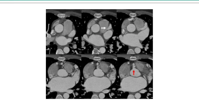
Figure 1 -Axial images of CT patterns that easily demonstrate the origins of coronary arteries. The right coronary artery originating in the right sinus (red arrow) and left coronary artery originating on the left side of the pulmonary artery (white arrow).
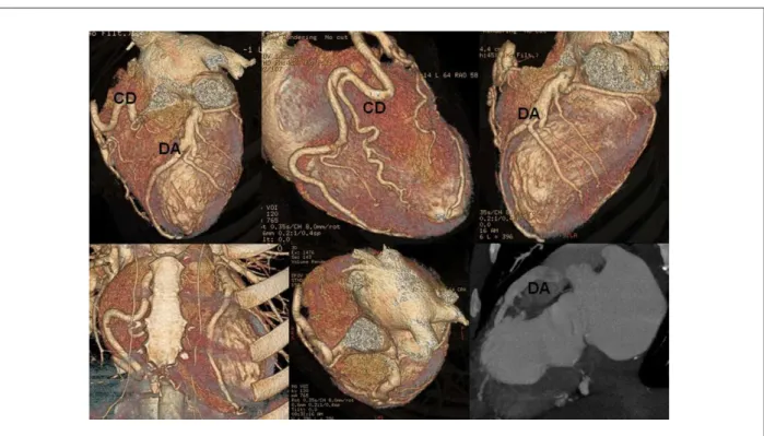
Figure 2 -Three-dimensional reconstruction by volume rendering of the coronary tree demonstrating dilatation and tortuosity of all vascular segments associated with septal defect of the origin of the left main coronary artery into the aorta.
left coronary artery coming out of the pulmonary artery and right coronary artery coming out of the aorta (Bland-White-Garland syndrome)1, which is exactly the type of syndrome reported in this study. ALCAPA develops before birth when the blood and lung pressure is equal and there is an antegrade flow in both coronary arteries. In the neonatal period, this flow changes gradually as the pulmonary pressure decreases and the ductus arteriosus closes, leading to reversal of flow in the left coronary artery6,9. The extent of myocardial ischemia in these patients is directly proportional to the development of collateral circulation between the right and left coronary arteries. If these patients with ALCAPA are not treated surgically,
they usually present a rather unfavorable development, with reports of 90% mortality early in childhood3,5. Such anomalous origin of coronary artery is extremely rare in adults and in the elderly7. The clinical features of this syndrome is generally nonspecific, with signs and symptoms such as syncope, arrhythmias, fatigue, nocturnal dyspnea and, less commonly, angina1-5. On examination we can find systolic murmur and ischemic changes (lateral wall) or adverse consequences from myocardial infarction on electrocardiogram (ECG)3,4,9. Chronic ischemia can be identified in tests with hemodynamic stress such as scintigraphy, echocardiography or magnetic magnetic resonance imaging7,10.
Case Report
Nacif et al
ALCAPA in a 64-channel CT scanner
Arq Bras Cardiol 2010;94(6) : e79-e82
Figure 3 -Three-dimensional reconstruction by volume rendering associating the cardiac image and the coronary tree. Note the tortuosity and dilation of the right coronary artery (aortic low). The combination of these images with the latter, a MIP (Maximum Intensity Projection) reconstruction, demonstrates the low concentration of contrast within the pulmonary artery and high density in the anterior descending coronary artery. These indings, combined with the low density contrast in the distal branches of the pulmonary arteries seen in axial planes (Figure 1) can provide hemodynamic data that the low is either too slow or reverse. It is also worth noting that the left ventricle muscle has a low concentration of contrast in cardiac reconstruction and this characterizes myocardial ischemia.
Until recently, such diagnosis has been performed with coronary catheterization. However, this method has limited use for this purpose due to its invasive characteristic and forecast analysis. With the onset of synchronized electrocardiogram MDCT, we can now, noninvasively and accurately, detect the origin, the course and termination of anomalous origin of coronary arteries. Some authors have shown the extent to which MDCT of coronary arteries is far more efficient as compared to conventional angiography in demonstrating the ostial origin and the proximal course of anomalous coronary arteries2,8,10.
The treatment of ALCAPA consists in the recreation of dual coronary perfusion. In children with this syndrome, you can directly deploy the anomalous coronary artery into the aorta or create an intrapulmonary conduit from the left coronary ostium into the aorta (Takeuchi procedure). In adults, the left coronary is usually tied to the pulmonary artery combined with grafting of the internal mammary artery or saphenous vein3,5,7,8.
We do not have image data through MDCT in medium and long term monitoring of patients after surgery, but as it is a non-invasive method and provides accurate anatomical
study, its use is well established9,11.
The success of surgical procedures will depend on the initial myocardial condition at the time of diagnosis and the impact on the patient’s clinical condition. Hence, the later such diagnosis is given, the greater will be the myocardial damage caused by ischemia and the greater will be the ventricular dysfunction and the degree of mitral regurgitation, which will impact the prognosis of these patients4,7,9,11.
Potential Conflict of Interest
No potential conflict of interest relevant to this article was reported.
Sources of Funding
There were no external funding sources for this study.
Study Association
This study is not associated with any post-graduation program.
Case Report
Nacif et al ALCAPA in a 64-channel CT scanner
Arq Bras Cardiol 2010;94(6) : e79-e82
References
1. Bland EF, White PD, Garland J. Congenital anomalies of the coronary arteries: report of an unusual case associated with cardiac hypertrophy. Am Heart J. 1933;8:787-801.
2. Baltaxe HA, Wixson D. The incidence of congenital anomalies of the coronary arteries in the adult population. Radiology. 1977;122 (1):47-52.
3. Yamanaka O, Hobbs RE. Coronary artery anomalies in 126,595 patients undergoing coronary angiography. Cathet Cardiovasc Diagn. 1990;21(1): 28-40.
4. Alexander RW, Griffith GC. Anomalies of the coronary arteries and their clinical significance. Circulation. 1956;14(5):800–5.
5. Cherian KM, Bharati S, Rao SG. Surgical correction of anomalous origin of the left coronary artery from the pulmonary artery. J Card Surg, 1994;9(4):386-91.
6. Schwartz ML, Jons RA, Colan SD. Anomalous origin of the left coronary artery from the pulmonary artery: recovery of left ventricular function after dual coronary repair. J Am Coll Cardiol. 1997;30(2):547-53.
7. Singh TP, Carli MF, Sullivan NM. Myocardial flow reserve in long-term survivors of repair of anomalous left coronary artery from pulmonary artery. J Am Coll Cardiol .1998;31(2):437-43.
8. Wollenek G, Damanig E, Salzer-Mufar U, Havel M, Wimmer M, Wolner E. Anomalous origin of the left coronary artery: a review of surgical management in 13 patients. J Cardiovasc Surg. 1993;34(5):399-405.
9. Shi H, Aschoff AJ, Brambs HJ, Hoffmann MH. Multislice CT imaging of anomalous coronary arteries. Eur Radiol. 2004;14(12):2172-81. 10. Wesselhoeft H, Fawcett JS, Johnson AL. Anomalous origin of the left coronary
artery from the pulmonary trunk: its clinical spectrum, pathology and pathophysiology, based on review of 140 cases with seven further cases. Circulation .1968;38(2):403-25
11. Brown JW, Ruzmetov M, Parent JJ, Rodelfed MD, Turrentine MW.. Does the degree of preoperative mitral regurgitation predict survival or the need for mitral valve repair or replacement in patients with anomalous origin of the left coronary artery from the pulmonary artery? J Thorac Cardiovasc Surg.2008; 136(3): 743-8.