Clinicoradiological Session
Case 6/2011 - 27-Year-Old Woman with Anomalous Origin of Left
Coronary Artery from the Pulmonary Trunk
Edmar Atik
Hospital Sírio-Libanês, São Paulo, SP, Brazil
Clinical aspects
A heart murmur had been auscultated in childhood and also at 18 years of age. For years, the patient remained with vague symptoms of dyspnea and chest pain irradiating to the left upper limb. Dilation of the right coronary artery, viewed by echocardiography, led to the initial presumptive diagnosis of Kawasaki disease, which was recently dismissed by angiography. The patient was taking acetylsalicylic acid.
On physical examination, the patient presented good general condition, was eupneic, with normal color, normal pulses, weighing 65 kg and height of 1.56 m. BP was at 110/70 mmHg and HR at 86 bpm. The aorta could not be felt at suprasternal notch. In the precordium, there were no deformities or impulsions and apical impulse was not palpated. Heart sounds were faint, and systolic murmur was auscultated, +/++ of intensity, soft timbre, of ejection, in the third, second and first left intercostal spaces at the sternal border. Liver could not be felt.
Complementary tests
Electrocardiogram
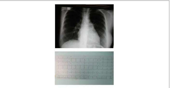
Showed sinus rhythm and signs of septal fibrosis in the absence of the first potential heart vector in V1 and V6. There were no signs of cavity overload, but left anterior hemiblock (Figure 1).
Chest radiography
Shows mild enlargement of the cardiac area at the expense of left ventricular arch. The middle arch was convex and the pulmonary vasculature was slightly prominent (Figure 1).
Echocardiography
Showed increased left cavities (AE = 37 mm, Ao = 30 mm, 69 mm = LVDD, LVSD = 47 mm) and dilatation of right
coronary artery (9 mm) compared to the left (4 mm). The left ventricular function was preserved with fractional shortening of myocardial fiber of 32.0%.
Cardiac catheterization and angiography and chest tomography
Established the diagnosis of anomalous origin of left coronary artery from the pulmonary trunk. The right coronary artery was dilated and nurtured the left coronary artery by strong collateral circulation providing flow from the left to the right at the level of the pulmonary trunk (Figure 2).
Diagnosis
Anomalous origin of the left coronary artery from the pulmonary trunk, with flow towards the pulmonary arterial tree in patients with few symptoms.
Clinical reasoning
The diagnosis of this defect is usually pointed out when there are clinical manifestations of heart failure due to ischemic cardiomyopathy that develops in the first months of age. However, in the presence of collateral circulation more pronounced between the two coronary arteries (if present), and with shunting of blood from the coronary artery to the pulmonary arterial tree, arteriovenous fistula simulating patent ductus arteriosus then develops. Therefore, we identified in this case the predominance of volume overload of left ventricle on myocardial ischemia (septal fibrosis on ECG). The vague symptoms, the soft murmur in the pulmonary area, septal fibrosis on electrocardiogram, increased pulmonary vasculature in retrospect could have led to the diagnosis of this anomaly. Echocardiography could also have advanced in the diagnosis if the clinical suspicion had prompted to do so. Hence the importance of examining all findings together.
Differential Diagnosis
Acyanogenic congenital heart disease with increased pulmonary flow should be evoked in the differentiation. From all of these, the ductus arteriosus, aortopulmonary window and arteriovenous fistulae in general are closer to the condition presented. The atrial septal defect is externalized by volume overload of the right chambers, ventricular septal defect in this age group is presented by left overload and slight dilation of the pulmonary trunk.
Mailing Address: Edmar Atik •
Rua Dona Adma Jafet, 74 conj. 73 - Bela Vista - 01308-050 - São Paulo, SP - Brazil
E-mail: [email protected], [email protected]
Manuscript received July 29, 2010; revised manuscript received January 13, 2011; accepted on January 13, 2011.
Keywords
Anomalous origin of left coronary artery, congenital heart defect, complications
Clinicoradiological Session
Atik Clinical-Radiographic Correlation
Arq Bras Cardiol 2011;97(5):e102-e104
Conduct
The surgical correction of the defect was considered in view of the impact of alterations resulting from increased pulmonary blood flow and the ischemic process.
During surgery, both coronary arteries were extremely dilated (10 mm diameter) as well as the pulmonary trunk. The left coronary artery emerging from the posterior wall of the pulmonary trunk in direct visualization of this structure
after incision. Since then, a tube was reconstructed with a patch from the pulmonary trunk which, containing the coronary ostium, was anastomosed with the lateral wall of the ascending aorta. The postoperative course, complicated by arrhythmia (supraventricular paroxysmal tachycardia) and anemia due to perioperative bleeding in the left ventricle outflow tract, required some cautions which postponed the discharge until the 12th day after the operation. On
Figure 2 - Coronary angiography shows dilated right coronary artery anastomosing by an intense collateral circulation (arrows) with the left coronary artery also dilated, and this in connection with the pulmonary trunk. This anastomosis has become sharper on the left chest tomography, which demonstrates the increased pulmonary arterial dilation in relation to the aorta. Abbreviations: RCA - right coronary artery; LCA - left coronary artery; AD - anterior descending; LMCA - left main coronary artery; and PT - pulmonary trunk.
Figure 1 -Chest X-ray shows increased left cavities, convex medial arch and increased pulmonary vasculature, suggestive of a malfunction with blood shunting from
left to right. Electrocardiogram reveals septal ibrosis and left anterior hemiblock.
Clinicoradiological Session
Atik
Clinical-Radiographic Correlation
the electrocardiogram, there were no alterations and the ventricular function was still preserved.
Considerations
The anomalous origin of the left coronary artery, usually from the pulmonary trunk, has recently benefited by the completion of an earlier diagnosis when, in a typical picture of dilated cardiomyopathy, there are alterations characteristic of myocardial ischemia on electrocardiogram. Such conditions manifests itself early in the first months of life predicted by persistent irritability,
uncontrollable crying, sweating and fatigue, which are early signs of myocardial ischemia. Undoubtedly, this initial condition should be paid closer attention for the completion of earlier diagnosis of this anomaly, so that surgical intervention can be done ideally before a stronger cardiac failure.
However, in the presence of a stronger collateral circulation between the two coronary arteries, this condition is milder and in the evolution, ischemia is discreet, as well as increased pulmonary flow. Even so, these patients must be corrected anatomically before more significant abnormalities appear.