Evaluation of Anomalous Coronary Arteries from
the Pulmonary Artery
Alper Guzeltas
1, MD; Erkut Ozturk
1, MD; Ibrahim Cansaran Tanidir
1, MD; Taner Kasar
1, MD; Sertac Haydin
2, MD
Abstract
Objective: This study evaluated clinical and diagnostic findings, treatment methods, and follow-up of cases of anomalous coronary arteries from the pulmonary artery.
Methods: The study included all cases diagnosed with anomalous coronary arteries from the pulmonary artery between January 2012 and January 2016. Data from patients’ demographic characteristics, electrocardiography, echocardiography, angiographic findings, operation, intensive care unit stay, and follow-up were evaluated.
Results: The study included 12 patients (8 male, 4 female), 10 with anomalous left coronary artery from the pulmonary artery (ALCAPA) and 2 with anomalous right coronary artery from the pulmonary artery (ARCAPA). Median age at diagnosis was 4 months (range, 1 month - 10 years old) and median weight was 5.5 kg (range, 3-30 kg). The most common complaints were murmur (n=7) and respiratory distress (n=5). In 4 cases, the initial diagnosis was dilated cardiomyopathy. Electrocardiographs were pathologic in all cases. Echocardiographic examination revealed medium to
severe mitral valve regurgitation in 4 cases and reduced (< 40%) ejection fraction in 6 patients. Of the 12 patients, 8 underwent direct implantation of the left coronary artery into the aorta, 2 underwent implantation of the right coronary artery into the aorta, and the remaining 2 underwent a Takeuchi procedure. There were no early mortalities. Median hospital stay was 20 days (range, 5-35 days). Median follow-up duration was 18 months (range, 5-36 months), and no cases required further surgery during follow-up.
Conclusions: Anomalous coronary arteries from the pulmonary artery can be successfully repaired providing there is early diagnosis and effective, appropriate intensive care unit follow-up. Therefore, coronary artery origins should be evaluated carefully, especially in cases with dilated cardiomyopathies.
Keywords: Coronary Vessel Anomalies. Bland White Garland Syndrome. Pulmonary Artery/Abnormalities. Cardiac Surgical Procedures.
DOI: 10.21470/1678-9741-2016-0082
1Department of Pediatric Cardiology, Istanbul Saglik Bilimleri University, Mehmet Akif Ersoy Thoracic and Cardiovascular Surgery Center, Istanbul, Turkey.
2Department of Pediatric Cardiovascular Surgery, Istanbul Saglik Bilimleri University, Mehmet Akif Ersoy Thoracic and Cardiovascular Surgery Center, Istanbul, Turkey.
This study was carried out at the Istanbul Saglik Bilimleri University, Mehmet Akif Ersoy Thoracic and Cardiovascular Surgery Center, Istanbul, Turkey.
No inancial support.
No conlict of interest.
Correspondence Address: Alper Guzeltas
İstanbul Mehmet Akif Ersoy Eğitim Araştırma Hastanesi, İstasyon Mahallesi İstanbul Caddesi Bezirganbahçe Mevki 34303 Küçükçekmece, Istanbul, Turkey.
E-mail: [email protected]
Article received on December 26th, 2016. Article accepted on January 5th, 2017. Abbreviations, acronyms & symbols
ALCAPA
ARCAPA
CVP
ECG
ECHO
ECMO
EF
= Anomalous left coronary artery from the pulmonary artery
= Anomalous right coronary artery from the pulmonary artery
= Central venous pressure
= Electrocardiography
= Echocardiography
= Extracorporeal membrane oxygenation
= Ejection fraction
ICU
LCAs
LV
NIRS
PICU
RCA
SD
TPN
= Intensive care unit
= Left coronary arteries
= Left ventricular
= Cranial near infrared spectroscopy
= Pediatric intensive care unit
= Right coronary artery
= Standard deviation
INTRODUCTION
Anomalous coronary arteries from the pulmonary artery are among the least-common congenital heart diseases. The majority of cases are of an anomalous left coronary artery from the pulmonary artery (ALCAPA), while cases of an anomalous right coronary artery from the pulmonary artery (ARCAPA) are rarely seen[1,2].
The indings and timeline for symptom development may vary depending on the efectiveness of the myocardial collateral branches concurrent with pulmonary artery pressure decrease. Some patients may be completely asymptomatic whereas others may experience death in the early period due to heart failure, valvular insuiciencies or myocardial infarction[1,2].
The deinitive treatment is surgery to normalize myocardial perfusion, after which, progressive remodeling and improvement in left ventricular (LV) function may be seen. Depending on the preference of the cardiac unit, either direct anastomosis may be performed on the anomalous coronary artery from the pulmonary artery to the aorta or, for patients in whom direct transfer of the coronary artery is not feasible, an intrapulmonary aortocoronary tunnel, as described by Takeuchi, may be implemented successfully[3-6].
This study evaluated presentation indings, diagnosis, treatment methods, and follow-up results of patients at a single tertiary center who were diagnosed with anomalous coronary arteries from the pulmonary artery.
METHODS
Study Design
For this retrospective study, the center’s computer database was searched for all patients diagnosed with anomalous coronary arteries from the pulmonary artery between January 1, 2012 and January 1, 2016. Cases with additional congenital heart diseases were excluded.
Patients’ demographic data, including age, gender, presentation type, initial diagnosis, and electrocardiography (ECG), telecardiography, echocardiography (ECHO), cardiac catheterization, and angiographic indings were evaluated. In addition, surgical methods, intensive care unit (ICU) follow-up data, observed complications, and outpatient-clinic follow-up results were assessed.
ECGs were evaluated according to the literature for sinus tachycardia, presence of pathological Q wave, ST elevation, ST depression, etc. ECHO indings before surgery, during ICU follow-up, and after hospital discharge were compared. Two-dimensional, M-mode, and Doppler ECHOs were performed using standard imaging techniques, in accordance with the recommendations of the American Society of Echocardiography.
Surgical techniques, namely the Takeuchi procedure and coronary implantation, were applied in accordance with the literature.
Postoperative Care
Patients were transferred to the ICU intubated, and mechanical ventilator support was initiated. In some patients, sternal closure was delayed.
In the ICU, all patients were monitored non-invasively by pulse oximetry, ECG, end-tidal capnography, and cranial near infrared spectroscopy (NIRS) and invasively by arterial blood pressure, central venous pressure (CVP), and left atrial pressure. Milrinone (0.5 microgram/kg/min) and low doses of epinephrine (0.05 microgram/kg/min) were the preferred inotropic support, beginning in the irst postoperative hours. Noradrenaline support was added if necessary. Fentanyl and midazolam were used for sedation and analgesia. All patients were started on total parenteral nutrition (TPN) in addition to minimal enteral feeding within the irst postoperative day.
Excessive volume loading was avoided. Vasodilator therapy, core cooling, neuromuscular blocking, and peritoneal dialysis were initiated in cases of low cardiac output that did not respond to inotropic support. Daily postoperative ECHO was performed in the pediatric intensive care unit (PICU). Tapering of postoperative medication, including inotropes, and weaning from the mechanical ventilator were decided on case by case, based on clinical evaluation and LV function on ECHO.
Statistical Analysis
Statistical analysis of the data was performed using SPSS for Windows, version 15.0 (SPSS Inc.; Chicago, IL, USA). Categorical variables were presented as absolute and percent frequencies, and quantitative variables were summarized as means and standard deviation (SD).
RESULTS
Demographic and Clinical Findings
This retrospective study included 12 patients, 10 with ALCAPA and 2 with ARCAPA. There were 8 males and 4 females. Median age at diagnosis was 4 months (range, 1 month to 10 years old), and median weight was 5.5 kg (range, 3-30 kg). The most common complaints were murmur (n=7) and respiratory distress (n=5). In 4 cases, the initial diagnosis was dilated cardiomyopathy. Table 1 shows the patients’ demographic data.
Electrocardiographic and Echocardiographic Findings
The ECGs of 7 patients showed a deep Q wave in the DI, aVL, and V6 derivations. It revealed sinus tachycardia in 5 cases, T wave inversion in the V5‒V6 derivation in 2 cases, and T wave inversion in the V1‒V3 derivation in 2 cases (Figure 1).
Of the 10 patients with ALCAPA, 9 had varying degrees of LV dilatation and dysfunction. ECHO revealed varying degrees of mitral valve regurgitation (Figure 2) and collateral blood low on the interventricular septum in all cases, papillary muscle hyperechogenicity in 9 cases, dilated right coronary artery (RCA) in 8 cases, and retrograde diastolic low toward the pulmonary artery in 4 cases. At the initial ECHO examination, coronary arteries of 4 patients were erroneously thought to originate from the aorta.
Table 1. Demographic and clinical indings of patients.
Patient Number
Sex/age
(months) Symptoms ECG Echocardiography
Catheter Angiography
1* F/20 Murmur Sinus tachycardia
EF = 30% LVEDd Z score: + 2
MR = mild IC = + PH = + RCA/AA = > 1.4
ALCAPA
2* M/120 Murmur V5-V6 T negativity
EF = 55% LVEDd Z score: 0-1
MR = severe IC = + PH = -RCA/AA = >1.4
ALCAPA
3* M/8 Shortness of breath,
DCM ALMI
EF = 30% LVEDd Z score = > +3
MR = severe IC = + PH = + RCA/AA > 1.4
ALCAPA
4* M/4 Restlessness, murmur ALMI
EF = 40% LVEDd Z score = > + 2
MR = mild IC = + PH = + RCA/AA= >1.4
ALCAPA
5* F/3 Shortness of breath,
DCM ALMI
EF = 35-40% LVEDd Z score = > + 3
MR = severe IC = + PH = + RCA/AA = >1.4
ALCAPA
6* F/2 Shortness of breath,DCM ALMI
EF: 25% LVEDd Z score = > +4
MR = mild IC = + PH = + RCA/AA = >1.4
ALCAPA
7* M/8 Murmur V5-V6 T negativity
EF: 30% LVEDd Z score = > +3
MR = mild IC = + PH = + RCA/AA = >1.4
ALCAPA
8* M/4 Shortness of breath,DCM ALMI
EF: 25% LVEDd Z score = >+4
MR = mild IC = + PH = + RCA/AA = >1.4
9* M/1 Restlessness, Murmur Sinus tachycardiaALMI
EF = 45% LVEDd Z score = > +2
MR = moderate IC = + PH = + RCA/AA = >1.4
ALCAPA
10* M/4 Shortness of breath ALMI
EF = 40% LVEDd Z score = > +2
MR = mild IC = + PH = + RCA/AA = > 1.4
ALCAPA
11 & F/1 Murmur V1-V3 T negativitySinus tachycardia
EF = 60 % LVEDd Z score = 0-1
MR = none IC = + PH = -LCA/AA = >1.3
ARCAPA
12 & M/18 Murmur V1-V3 T negativity
EF = 70 % LVEDd Z score = 0-1
MR = none IC = + PH = -LCA/AA = > 1.3
ARCAPA
*ALCAPA=anomalous left coronary artery from the pulmonary artery; &ARCAPA=anomalous right coronary artery from the pulmonary artery; ALMI=anterolateral myocardial infarction; DCM=dilated cardiomyopathy; ECG=electrocardiography; EF=ejection fraction; F=female; IC=collaterals; LCA/AA=left coronary artery/aorta artery; LVEDd= Left Ventricular End Diastolic Diameter; M=male; MR=mitral regurgitation; PA=pulmonary artery; PH=papillary hyperechogenicity; Pt=patient; RCA=right coronary artery
Catheterization and Angiography
All patients were evaluated with cardiac catheterization and angiography (Figure 3).
Surgical Treatment
Among the 10 patients with ALCAPA, 8 underwent direct implantation of the LCA into the aorta and 2 underwent the
Fig. 1 - Preoperative twelve lead electrocardiogram of patient 9. Electrocardiogram shows signs of acute anterolateral myocardial infarction; deep Q waves, ST segment elevation and T-wave inversion in leads I and aVL.
Takeuchi technique. In the 2 patients with ARCAPA, coronary reimplantation was applied. Median surgery time was 140 minutes (range, 76-375 minutes). In 3 cases, closure of the sternum was delayed and, in those cases, mean closure time was 96 hours.
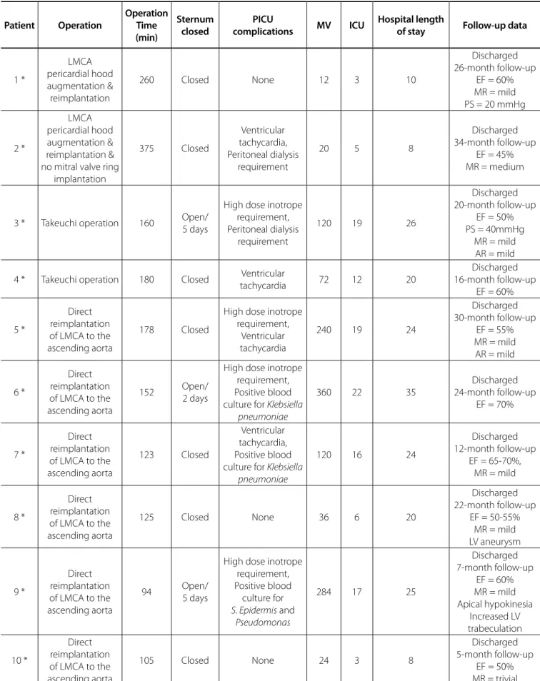
There were no early mortalities. Ventricular tachycardia occurred in 4 cases. In 3 cases, blood cultures were positive. In addition, 2 patients needed high-dose inotrope support, and another 2 patients needed peritoneal dialysis. Mean duration of mechanical ventilation was 54 hours (range, 12-360 hours), mean length of ICU stay was 9 days (range, 2-10 days), and mean length of hospital stay was 20 days (range, 5-35 days). Extracorporeal membrane oxygenation (ECMO) support was used for 1 patient. Table 2 shows surgical, ICU-stay, and follow-up data.
Follow-Up
Fig. 2 – A - Apical four chamber view echocardiogram showing dilated left ventricle, hyperechogenicity in papillary muscles, ventricular septal-defect-like appearance due to coronary collaterals and significant mitral regurgitation; B - Parasternal short axis view echocardiogram showing anomalous origin of the left coronary artery from pulmonary artery.
RV=right ventricle; LV=left ventricle; PA=pulmonary artery.
Table 2. Patients intensive care unit and follow-up data. Patient Operation Operation Time (min) Sternum closed PICU
complications MV ICU
Hospital length
of stay Follow-up data
1 *
LMCA pericardial hood augmentation & reimplantation
260 Closed None 12 3 10
Discharged 26-month follow-up
EF = 60% MR = mild PS = 20 mmHg
2 *
LMCA pericardial hood augmentation & reimplantation & no mitral valve ring
implantation 375 Closed Ventricular tachycardia, Peritoneal dialysis requirement
20 5 8
Discharged 34-month follow-up
EF = 45% MR = medium
3 * Takeuchi operation 160 Open/5 days
High dose inotrope requirement, Peritoneal dialysis
requirement
120 19 26
Discharged 20-month follow-up
EF = 50% PS = 40mmHg
MR = mild AR = mild
4 * Takeuchi operation 180 Closed tachycardiaVentricular 72 12 20
Discharged 16-month follow-up
EF = 60%
5 *
Direct reimplantation of LMCA to the ascending aorta
178 Closed
High dose inotrope requirement,
Ventricular tachycardia
240 19 24
Discharged 30-month follow-up
EF = 55% MR = mild
AR = mild
6 *
Direct reimplantation of LMCA to the ascending aorta
152 Open/2 days
High dose inotrope requirement, Positive blood culture for Klebsiella
pneumoniae
360 22 35
Discharged 24-month follow-up
EF = 70%
7 *
Direct reimplantation of LMCA to the ascending aorta
123 Closed
Ventricular tachycardia, Positive blood culture for Klebsiella
pneumoniae
120 16 24
Discharged 12-month follow-up
EF = 65-70%, MR = mild
8 *
Direct reimplantation of LMCA to the ascending aorta
125 Closed None 36 6 20
Discharged 22-month follow-up
EF = 50-55% MR = mild LV aneurysm
9 *
Direct reimplantation of LMCA to the ascending aorta
94 Open/5 days
High dose inotrope requirement, Positive blood
culture for
S. Epidermis and
Pseudomonas
284 17 25
Discharged 7-month follow-up
EF = 60% MR = mild Apical hypokinesia Increased LV trabeculation 10 * Direct reimplantation of LMCA to the ascending aorta
105 Closed None 24 3 8
Discharged 5-month follow-up
11 &
RCA ostium transfer to the
aorta
91 Closed None 12 3 7
Discharged 15-month follow-up
No problem
12 &
RCA ostium transfer to the
aorta
76 Closed None 16 2 5
Discharged 7-month follow-up
No problem *ALCAPA=anomalous left coronary artery from the pulmonary artery; & ARCAPA=anomalous right coronary artery from the pulmonary artery; AR=aortic regurgitation; EF=ejection fraction; ICU=intensive care unit; LMCA=left main coronary artery; LV=left ventricle; MR= mitral regurgitation; MV=mechanical ventilation; PICU=pediatric intensive care unit; PS=pulmonary stenosis; RCA=right coronary artery;
DISCUSSION
ALCAPA is a rare disorder, afecting 1 in 300,000 live births and representing approximately 0.5% of congenital heart defects[6,7]. Presentation and diagnosis timelines for ALCAPA vary depending on pulmonary vascular resistance and the presence of collateral vessels between the right and left coronary artery systems. A wide range of symptoms has been reported in the literature, including an one month-old baby with heart failure symptoms and an asymptomatic 53-year-old who was diagnosed incidentally[8]. A Danish study of 9 patients with ALCAPA reported that 77% had heart failure symptoms, including shortness of breath and wheezing, while the remaining 23% were asymptomatic at presentation[9]. A Chinese study of 27 cases reported acute heart failure (n=15), pneumonia (n=7), and cardiac murmurs (n=5) as presenting symptoms[10]. In the present study, 60% of patients presented with murmur, 40% with shortness of breath, and 30% were asymptomatic.
Because myocardial ischemia develops over time in an anterolateral distribution, pathological Q waves can be seen in derivations representing the region, namely the DI, aVL, and V4-V6. In patients with extensive collateral circulation, there might be nonspeciic ECG changes[5]. In patients with impaired LV functions, typical ECG changes should indicate the presence of an anomalous coronary artery[5,6].
Rodriguez-Gonzalez et al.[1] conducted a study of 12 patients and reported that 6 had speciic ECG changes compatible with acute lateral myocardial infarction. In the present study, the ECGs of 7 patients revealed pathological Q waves in the DI, aVL, and V6. The ECGs of the other 5 patients showed nonspeciic changes.
ECHO indings included the following: dilatation of the coronary artery with normal origin, increased ratio of the diameter of the coronary artery originating from the normal sinus to the aortic annulus (> 0.14), increased echogenicity of the papillary muscles, increased low toward the pulmonary artery, and either identiication of an inappropriate origin of the anomalous coronary artery from the pulmonary artery or non-identiication of the origin of the coronary artery. However, initial ECHOs have shown that 50-70% of patients had their coronary arteries arising from the aorta[7,11]. In the present study, 4 patients with ALCAPA and 1 with ARCAPA were diagnosed with normal coronary-artery patterns, and the ratio of false negative misdiagnoses was 43%.
Anomalous coronary arteries from the pulmonary artery
might be falsely diagnosed as idiopathic dilated cardiomyopathy or endocardial ibroelastosis. Zheng et al.[5] reported that 18 of 21 cases (78%) in their study were initially misdiagnosed. In the present study, 4 patients had initially been diagnosed with idiopathic dilated cardiomyopathy.
Various surgical methods can be used to repair an anomalous coronary artery from the pulmonary artery, all of them aiming to establish a system with two coronary arteries. One of the most common methods is direct reimplantation of the coronary artery into the aorta. The other is creating an aortopulmonary window that directs blood low from the aorta to the LCA (Takeuchi). After those procedures, no matter how impaired the ventricular functions had been, myocardial function can quickly heal[12,13].
Jin et al.[14] reported that 11 patients treated by direct reimplantation experienced improved LV function. In addition, Sarioglu et al.[3] reported the recovery of LV function in all patients, except for 1 mortality (5 coronary reimplantations, 2 Takeuchi). Ayik et al.[15] reported that 10 patients treated by the Takeuchi method had a 10% mortality rate. In the present study, coronary reimplantation was applied in 8 patients and the Takeuchi method in 2 patients. Regardless of the technique applied, LV function became normal in all patients.
In patients with ALCAPA, LV function is characterized by mitral valve dysfunction. Preoperative mitral valve regurgitation is a signiicant risk factor. However, mitral valve repair is controversial in ALCAPA patients. Vouhé et al.[16] suggested that resolution of myocardial ischemia leads to improved papillary muscle function. Therefore, they proposed that mitral valve repair should not be done at irst. In patients with ongoing mitral valve regurgitation, coronary artery restenosis should be investigated and, if necessary, the mitral valve should be repaired at this stage. In contrast, other researchers have argued that simultaneous mitral valve repair and anterolateral commissural annuloplasty during coronary artery reimplantation enhance early recovery of LV functions[17]. In the present study, only 1 patient needed mitral valve repair during the irst surgery. Mitral valve regurgitation improved in all patients except one, who required mitral valve repair during initial surgery.
chest pain, heart failure, arrhythmia, or sudden death. Time to diagnosis shows a wide distribution, from 1 month old to 90 years old. There is no consensus in the literature regarding treatment methods and diagnosis times in ARCAPA patients[18].
Although there are reports of various treatment methods, including surveillance, medical therapy, surgery, and surgical ligation, due to the 10-18% risk of sudden death, most authors advocate creating a dual coronary circulation by direct reimplantation, even in asymptomatic patients[18,19]. In the present study, both of the ARCAPA patients were asymptomatic, but underwent surgery, one at 9 months old and the other at 18 months old, in accordance with the literature.
Reported mortality rates range from 0-16%[20-22]. The literature suggests that the main reasons for mortality are low cardiac output and ventricular arrhythmias, and it advises using advanced life-support systems to reduce mortality. In the present study, 3 patients developed low cardiac output and 4 developed ventricular tachycardia. No ECMO was required, nor was there any early mortality.
Limitations
The most important limitations of the present study were the small number of patients included and its retrospective nature. In addition, the follow-up period was relatively short compared with those in the literature.
CONCLUSION
Anomalous coronary arteries from the pulmonary artery may present with various clinical, ECG, and ECHO indings. The condition can be successfully treated by surgery if accompanied by early diagnosis and efective, appropriate ICU follow-up. Coronary artery origins should be carefully evaluated, especially in patients with dilated cardiomyopathy.
Authors’ roles & responsibilities
AG
EO
ICT
TK
SH
Conception and study design; analysis and/or data interpretation; statistical analysis; final manuscript approval
Conception and study design; analysis and/or data interpretation; statistical analysis; final manuscript approval
Conception and study design; analysis and/or data interpretation; statistical analysis; final manuscript approval
Conception and study design; analysis and/or data interpretation; statistical analysis; final manuscript approval
Execution of operations and/or trials; manuscript writing or critical review of its content; final manuscript approval
REFERENCES
1. Rodriguez-Gonzalez M, Tirado AM, Hosseinpour R, Soto JS. Anomalous origin of the left coronary artery from the pulmonary artery: diagnoses and surgical results in 12 pediatric patients. Tex Heart Inst J. 2015;42(4):350-6.
2. Birk E, Stamler A, Katz J, Berant M, Dagan O, Matitiau A, et al. Anomalous origin of the left coronary artery from the pulmonary artery: diagnosis and postoperative follow up. Isr Med Assoc J. 2000;2(2):111-4. 3. Sarıoğlu T, Yalçınbaş YK, Erek E, Arnaz A, Türkekul Y, Avşar MK, et al.
Anomalous left coronary artery originating from pulmonary artery: recovery of left ventricular function after dual coronary system restoration and clinical results. Turkish J Thorac Cardiovasc Surg. 2013;21:1-6.
4. Dodge-Khatami A, Mavroudis C, Backer CL. Anomalous origin of the left coronary artery from the pulmonary artery: collective review of surgical therapy. Ann Thorac Surg. 2002;74(3):946-55.
5. Zheng JY, Han L, Ding WH, Jin M, Zhang GZ, Xiao YY, et al. Clinical features and long-term prognosis of patients with anomalous origin of the left coronary artery from the pulmonary artery. Chin Med J (Engl). 2010;123(20):2888-94.
6. Ceylan Ö, Örün UA, Koç M, Özgür S, Doğan V, Karademir S, et al. Anomalous coronary artery originating from the pulmonary artery: a report of four cases. Türk Göğüs Kalp Damar Cerrahisi Dergisi. 2013;21:122-6.
7. Karr SS, Parness IA, Spevak PJ, van der Velde ME, Colan SD, Sanders SP. Diagnosis of anomalous left coronary artery by Doppler color low mapping: distinction from other causes of dilated cardiomyopathy. J Am Coll Cardiol. 1992;19(6):1271-5.
8. Takemoto K, Hirata K, Tanimoto T, Matsuo Y, Ino Y, Kubo T, et al. Combined non-invasive Doppler echocardiography and coronary computed tomography lead to diagnosis of anomalous left coronary artery from the pulmonary artery (ALCAPA) syndrome. Circ J. 2015;79(5):1136-8. 9. Holst LM, Helvind M, Andersen HØ. Diagnosis and prognosis of
anomalous origin of the left coronary artery from the pulmonary artery. Dan Med J. 2015;62(9):A5125.
10. Chong M, Han L, Liu YL, Gu H, Jin M, Cheng P, et al. Diagnosis and surgery of anomalous origin of the left coronary artery from the pulmonary artery in children. Zhonghua Yi Xue Za Zhi. 2012;92(24):1673-6. 11. Chang RR, Allada V. Electrocardiographic and echocardiographic features
that distinguish anomalous origin of the left coronary artery from pulmonary artery from idiopathic dilated cardiomyopathy. Pediatr Cardiol. 2001;22(1):3-10.
12. Alsoui B, Sallehuddin A, Bulbul Z, Joufan M, Khouqeer F, Canver CC, et al. Surgical strategy to establish a dual-coronary system for the management of anomalous left coronary artery origin from the pulmonary artery. Ann Thorac Surg. 2008;86(1):170-6.
13. Alexi-Meskishvili V, Nasseri BA, Nordmeyer S, Schmitt B, Weng YG, Böttcher W, et al. Repair of anomalous origin of the left coronary artery from the pulmonary artery in infants and children. J Thorac Cardiovasc Surg. 2011;142(4):868-74.
14. Jin Z, Berger F, Uhlemann F, Schröder C, Hetzer R, Alexi-Meskhishvili V, et al. Improvement in left ventricular dysfunction after aortic reimplantation in 11 consecutive paediatric patients with anomalous origin of the left coronary artery from the pulmonary artery. Early results of a serial echocardiographic follow-up. Eur Heart J. 1994;15(8):1044-9. 15. Ayık MF, Oğuz E, Öztürk P, Atay Y, Ceylan N, Levent E, et al. Anomalous left coronary artery arising from the pulmonary artery repair with pulmonary artery reconstruction. Türk Göğüs Kalp Damar Cerrahisi Dergisi. 2012;20:735-40.
17. Huddleston CB, Balzer DT, Mendelof EN. Repair of anomalous left main coronary artery arising from the pulmonary artery in infants: long-term impact on the mitral valve. Ann Thorac Surg. 2001;71(6):1985-8. 18. Kühn A, Kasnar-Samprec J, Schreiber C, Vogt M. Anomalous origin of
the right coronary artery from the pulmonary artery (ARCAPA). Int J Cardiol. 2010;139(2):e27-8.
19. Williams IA, Gersony WM, Hellenbrand WE. Anomalous right coronary artery arising from the pulmonary artery: a report of 7 cases and a review of the literature. Am Heart J. 2006;152(5):e9-17.
20. Isomatsu Y, Imai Y, Shin’oka T, Aoki M, Iwata Y. Surgical intervention for anomalous origin of the left coronary artery from the pulmonary artery: the Tokyo experience. J Thorac Cardiovasc Surg. 2001;121(4):792-7. 21. Muzafar T, Ahmad Ganie F, Gpoal Swamy S, Wani NU. The surgical
outcome of anomalous origin of the left coronary artery from the pulmonary artery. Int Cardiovasc Res J. 2014;8(2):57-60.