https://doi.org/10.1590/0004-282X20170050
ARTICLE
Potential role of a cognitive rehabilitation program
following left temporal lobe epilepsy surgery
Reabilitação neuropsicológica em pacientes com epilepsia submetidos à lobectomia
temporal dominante
Camila de Vasconcelos Geraldi1,2, Sara Escorsi-Rosset1, Pamela Thompson2,3, Ana C. Gargaro Silva1,
Américo Ceiki Sakamoto1
Memory deicits in people with temporal lobe epilepsy (TLE) have been widely reported and memory decline is a well-established surgical outcome, particularly following dominant temporal lobe resections. Memory diiculties can have far ranging efects that afect academic and work per-formance but also can cause considerable personal distress. Research into memory and epilepsy has focused on measur-ing problems and explormeasur-ing causes with limited attention directed to the role of neuropsychological rehabilitation1
. Cognitive rehabilitation comprises a range of techniques, and
research in patients with various brain injuries and patholo-gies has provided some support for its eicacy2,3,4,5,6,7.
he evidence base for memory rehabilitation in epilepsy is limited particularly in temporal lobe surgical cases, a high-risk group. Well-controlled trials are few and data on generaliza-tion to daily activities and the persistence of efects are sparse1,8.
Existing reports have been encouraging8,9. Unfortunately few
stud-ies describe the rehabilitation process suiciently8, while for
oth-ers memory rehabilitation forms part of a more extensive set of interventions such that it is impossible to identify speciic efects10
.
1Universidade de São Paulo, Faculdade de Medicina de Ribeirão Preto, Departamento de Neurociências e Ciências do Comportamento, Ribeirão Preto SP, Brasil;
2UCL Institute of Neurology, Department of Clinical and Experimental Epilepsy, London, UK;
3Epilepsy Society Research Centre, Buckinghamshire, UK.
Correspondence: Camila de Vasconcelos Geraldi; Departamento de Neurociências e Ciências do Comportamento, FMRP / USP, Centro de Cirurgia de Epilepsia (CIREP) 4o andar; Av Bandeirantes, 3900; 14048-900 Ribeirão Preto SP, Brasil; E-mail: [email protected]
Conflict of interest: There is no conlict of interest to declare.
Received 15 September 2016; Received in inal form 01 February 2017; Accepted 14 February 2017. ABSTRACT
Research into memory and epilepsy has focused on measuring problems and exploring causes with limited attention directed at the role of neuropsychological rehabilitation in alleviating post-operative memory dificulties. Objectives: To assess the effects of a memory rehabilitation program in patients with left temporal lobe epilepsy following surgery. Methods: Twenty-four patients agreed to participate and 18 completed the study; nine received memory rehabilitation while nine had no input and were designated as controls. Verbal learning eficiency, naming abilities, memory subjective ratings, ecological activity measures and a language fMRI paradigm were used as outcome measures. Results: Improved verbal learning and naming test performance, increase in memory strategy use and improved self-perception were observed following the rehabilitation. Changes in fMRI activation patterns were seen in the rehabilitation group over the long term.
Conclusion: The indings support the potential role of a cognitive rehabilitation program following left temporal lobe surgery.
Keywords: epilepsy, temporal lobe; memory; neurologic rehabilitation; anterior temporal lobectomy.
RESUMO
As publicações na área de epilepsia e memória se focam em mensurar prejuízos e investigar causas, com poucos dados sobre reabilitação neuropsicológica em pacientes pós-cirúrgicos. Objetivos: Avaliar os efeitos da reabilitação neuropsicológica em pacientes submetidos a lobectomia temporal dominante. Métodos: Vinte e quatro pacientes iniciaram o estudo, apenas dezoito o concluíram, dos quais 9 foram participantes de sessões de reabilitação com enfoque em memória. Todos os participantes foram avaliados quanto a autopercepção de diiculdades de memória; ao uso de estratégias para minimizar tais diiculdades; a habilidade de nomeação e a aprendizagem verbal e foram submetidos à ressonância magnética funcional. Resultados: Foi encontrado efeito signiicativo da reabilitação neuropsicológica na autopercepção de diiculdades de memória; no uso de estratégias compensatórias; na aprendizagem verbal e na nomeação. Alterações no padrão de ativação na RMf foram observadas no grupo submetido a reabilitação. Conclusão: A reabilitação neuropsicológica pode beneiciar pacientes submetidos a lobectomia temporal antero-mesial dominante com prejuízos de memória.
Which features of the rehabilitation programs contribute to the cognitive gains reported and the mechanism of under-lying these changes is uncertain but available evidence sug-gests it can be efective. If memory rehabilitation has positive efects then this would provide support for inclusion in sur-gical programs and further research is of considerable clini-cal relevance. his study aimed to assess the efectiveness of memory rehabilitation for left TLE patients following surgery and utilized an fMRI paradigm to assess for changes in brain function in association with rehabilitation.
METHODS
Participants
Patients with epilepsy, who had undergone selec-tive left temporal lobe surgery at least six months previ-ously, were recruited. Participants were at least 16 years old and were right handed. No participant had a learning disability (IQ < 70) or a psychiatric history or any evidence of anti-epileptic drug toxicity.
Sixty-two patients met the study criteria but only 24 agreed to participate.
hirteen people were assigned to the active treatment program (the rehabilitation group) and 11 to the control group. Unfortunately, participant selection was not random, as those in the control group were people who were unable to commit to the time and travel requirements of the treat-ment program.
Written informed consent was obtained from all partici-pants. his study was approved by the ethics committee (pro-cess number 9968/2009).
Eighteen people completed the study, nine in the rehabili-tation group and nine in the control group. Six patients did not complete the entire program, four in the rehabilitation group and two in the control group. Five failed to attend one of the appointments for the fMRI scan and the cognitive re-assessment. he reasons for discontinuing the program pre-maturely were lack of time due to starting a new job (n = 1), other personal commitments (n = 2), depression (n = 1) and lack of resources for traveling to the hospital (n = 2).
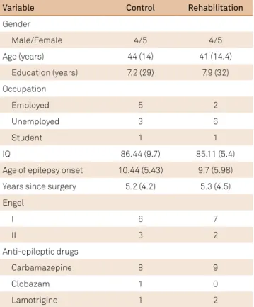
here were no signiicant diferences between the control and rehabilitation groups on demographic or epilepsy related variables (Table 1). Both groups experienced good surgery outcomes (Engel I and II).
Procedure
At the study onset, both groups completed tests of mem-ory, naming, a memory complaints questionnaire and an ecological memory measure. Each participant also under-went an fMRI study.
he rehabilitation group then received eight cognitive rehabilitation sessions, delivered on an individual basis, once a week for two months. hirty days following the completion
of the program, they had a second fMRI study and repeated the cognitive tests and questionnaire.
he control group underwent cognitive retesting and a second fMRI study at an interval of two months but received no rehabilitation. During these two months, they were not seen at the hospital.
A third fMRI was scheduled for both groups two months after the reassessment; however, this was only possible for the rehabilitation group.
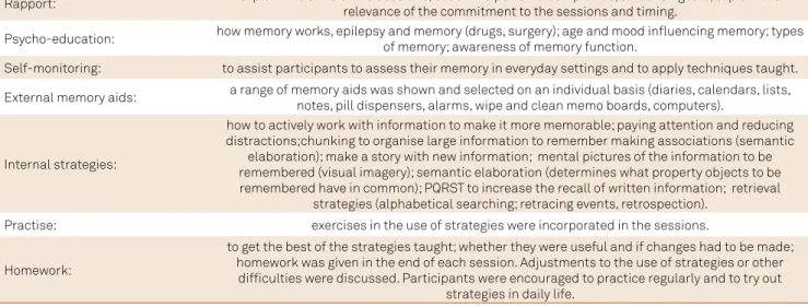
Cognitive rehabilitation sessions
Weekly sessions lasted one hour. Topics covered psy-cho-education about memory, and taught rehabilitation techniques focusing on attention, memory and word-finding problems. Cognitive strategies included self-mon-itoring and encoding level intervention, such as seman-tic elaboration, prospective memory training and visual imagery11
. Instruction was given in the use of external memory aids such as notes and diaries. Strategies and exercises were derived from those advocated by propo-nents of memory rehabilitation11
. Also, homework was given at the end of each session and checked at the begin-ning of the next session (Table 2).
At the beginning of the second and of the third sessions, a relative or living companion attended a session on the use of memory strategies and aids in order to provide support in the home setting when needed.
Table 1. Demographic and clinical characteristics of the rehabilitation and control groups.
Variable Control Rehabilitation
Gender
Male/Female 4/5 4/5
Age (years) 44 (14) 41 (14.4)
Education (years) 7.2 (29) 7.9 (32)
Occupation
Employed 5 2
Unemployed 3 6
Student 1 1
IQ 86.44 (9.7) 85.11 (5.4)
Age of epilepsy onset 10.44 (5.43) 9.7 (5.98)
Years since surgery 5.2 (4.2) 5.3 (4.5)
Engel
I 6 7
II 3 2
Anti-epileptic drugs
Carbamazepine 8 9
Clobazam 1 0
Lamotrigine 1 2
Outcome measures
Objective cognitive outcomes. he Boston Naming Test12
was used to assess retrieval from semantic memory. he subject is required to name 60 items of increasing diiculty, presented as line drawings. he score used was the total number of items named cor-rectly, considering spontaneous response plus semantic cues. he Rey Auditory Verbal Learning Test13 was used to
assess verbal learning and recall. he subject is read a list of 15 words over ive trials. On each trial, they have to recall as many words as they can. In this study, the total number of words recalled over the learning trials was used as the mea-sure of the memory performance (maximum score = 75).
he Ecological Activity was adapted from the Five Daily Living Test, part of the Neuropsychological Assessment Battery14. his test assesses memory in a context that
simu-lates performance in everyday life. he subject has to remem-ber what and where in the test room the examiner hides an object. he subject is also given a name to remember, e.g. Carolina Paiva. One point is given for each correct answer resulting in a maximum score of four points. he total score was used in the analysis.
Subjective memory outcomes.
he Self Report Memory Questionnaire2
was used to assess the self-perception of memory and the strategies used by the subject in everyday settings. It consists of 28 items; 24 items concern the experience of memory failures encoun-tered in daily life and the remaining four the use of memory support strategies. he questionnaire produces a maximum score of 96 points for the irst 24 questions – four points for each correct answer; the higher the score, the poorer the memory self-perception); and 20 points for the remaining four questions ( ive points for each answer; the higher the score, more strategies the subject uses). he scores were ana-lysed separately.
fMRI
A fluency paradigm to assess language networks pre- and post-rehabilitation provided a generic measure of brain activation involving the semantic processing sys-tem. Posterior temporal regions within a larger antero-posterior-basal temporal network have been identified in the process of word retrieval in TLE15 and word finding
difficulties have been found to correlate with resting-state metabolism of the postero-infero-temporo-basal region (BA 20-37), the posterior part of superior temporal gyrus (BA39) and inferior parietal lobule (BA40)15. Several
stud-ies have demonstrated that verbal fluency paradigms are sensitive to detecting impaired functioning of the prefron-tal cortex and brain interconnections, particularly of the left hemisphere16.
fMRI task
Participants underwent out-of-scanner training to max-imize task compliance. he task used followed a blocked experimental design and all the stimuli were presented via a digital sound recorder. Patients were asked to think silently of as many words as possible starting with a speciied letter (M, A, E, C and S- LET task)17. he whole paradigm consisted
of nine blocks: ive resting periods and four activations, in 30 second blocks. During the resting period the subjects were instructed to think of a “blank wall”.
fMRI acquisition
he MRI data acquisition was performed on a Philips Achieva 3T XSERIES scanner. Structural T1-weighted images were obtained using a standard gradient-echo sequence MPRAGE (TR = 6 ms, TE = 3 ms, matrix 256 x 256, FOV = 256mm, 1mm slice). One set of functional echo-planar images were acquired (TR/TE = 3000/30ms, FOV 230mm, voxel vol 1.79 x 1.79 x 4.00mm3, matrix dimension = 128 x 128).
Functional images were processed using the software Brain Table 2. Rehabilitation sessions content.
Rapport: explain the aims of the sessions; check the patients’ complaints; establish goals; explain the relevance of the commitment to the sessions and timing.
Psycho-education: how memory works, epilepsy and memory (drugs, surgery); age and mood inluencing memory; types of memory; awareness of memory function.
Self-monitoring: to assist participants to assess their memory in everyday settings and to apply techniques taught.
External memory aids: a range of memory aids was shown and selected on an individual basis (diaries, calendars, lists, notes, pill dispensers, alarms, wipe and clean memo boards, computers).
Internal strategies:
how to actively work with information to make it more memorable; paying attention and reducing distractions;chunking to organise large information to remember making associations (semantic
elaboration); make a story with new information; mental pictures of the information to be remembered (visual imagery); semantic elaboration (determines what property objects to be remembered have in common); PQRST to increase the recall of written information; retrieval
strategies (alphabetical searching; retracing events, retrospection).
Practise: exercises in the use of strategies were incorporated in the sessions.
Homework:
to get the best of the strategies taught; whether they were useful and if changes had to be made; homework was given in the end of each session. Adjustments to the use of strategies or other
Voyager™ QX (version 2.0) Brain Innovation, Maastricht, he Netherlands (Goebel et al., 2006)18. he fMRI data sets were
obtained in EPI-BOLD sequences and were co-registered with high-spatial resolution images (3D gradient-echo T1-weighted sequence) covering both brain hemispheres. Preprocessing consisted of 3D movement correction, a 4-mm FWHM Gaussian spatial ilter, and a temporal high-pass ilter.
Statistical analysis
All data were analysed using SPSS 15.0 version (SPSS Inc., Chicago, IL, USA). First the Kolmogorov-Smirnov, Shapiro-Wilk and c2
tests at a 5% signiicance level were per-formed and the normal distribution hypothesis was rejected. Due to the small sample size, data was then analysed using non-parametric statistics: Mann-Whitney U tests for com-parisons between groups and Wilcoxon tests for within group comparisons. Verbal learning and naming test scores and subjective memory questionnaire indices assessed one month after the completion of the rehabilitation program were the endpoints for analysis. he signiicance level was set at p < 0.05.
To determine the signiicance of changes in performance, each participant was classiied as improved or not based on reliable change indices derived from normative data avail-able from the Rey Auditory Verbal Learning Test and Boston Naming Test18,19,20.
he fMRI temporal series were analysed statistically using a general linear model with the evolution of each voxel being compared to a modelled reference function. In order to extend statistical inferences to the group level, the statistical maps of all subjects were registered to standard space and the signiicance of activation at each condition was nonpara-metrically evaluated using the paired Wilcoxon test for two related samples at 5% signiicance level for both voxels and cluster levels. he diference in the activation between the irst and the second scan, between the second and the third, and between the irst and the third scans were the indices used for the analysis. hreshold maps (p < 0.01) were super-imposed on the structural images for anatomic reference.
RESULTS
Objective cognitive outcomes
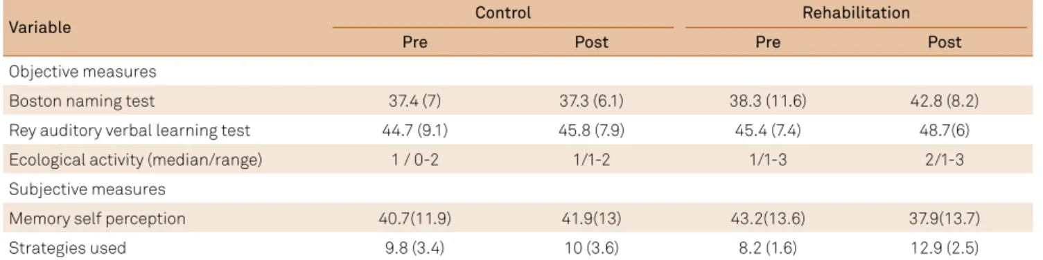
here were no statistically signiicant diferences between the groups on the irst assessment for the objec-tive cogniobjec-tive measures.
Signiicant improvements were recorded following the rehabilitation program for verbal learning (W = 2.38; p = 0.02); naming (W = 2.32; p = 0.02) and on the Ecological Activity measure (W = 2.34; p = 0.02). For the control group, improved performance was recorded only on the Ecological Activity measure (W = 2; p = 0.05) (Table 3).
hree participants in the rehabilitation group were classi-ied as having improved memory on a basis of reliable change indices for verbal learning versus one participant in the con-trol group. On the Boston Naming Test, two individuals were classiied as having a meaningful improvement in naming versus none in the control group.
Subjective memory outcomes
he two groups were matched at the initial assessment on measures of self-perception and in the use of support strate-gies. Both subjective memory measures indicated improve-ments in the rehabilitation group (self perception: W = 2.67; p = 0.008/strategies used: W = 2.67; p = 0.008). No changes were recorded for the control group (Table 3).
fMRI
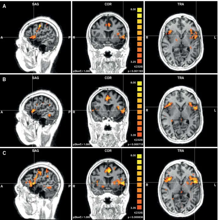
here were no signiicant diferences between the groups in activation patterns during the verbal luency paradigm for the irst fMRI scan. here were no changes between the irst and the second scans for the rehabilitation group, but changes from the irst to the third scan were observed in the right anterior prefrontal cortex (W = 1.96; p = 0.05), pars oper-cularis of Broca’s area in left hemisphere (W = 2.67; p = 0.01), and in the right inferior prefrontal gyrus (W = 2.43 p = 0.02). When the second scan was compared to the third scan there were changes in the right dorsal anterior cingulate cortex (W = 2.07; p = 0.04) and in the left angular gyrus (W = 2.21; p = 0.027) (Figure).
Table 3. Control and rehabilitation group outcome measures.
Variable Control Rehabilitation
Pre Post Pre Post
Objective measures
Boston naming test 37.4 (7) 37.3 (6.1) 38.3 (11.6) 42.8 (8.2)
Rey auditory verbal learning test 44.7 (9.1) 45.8 (7.9) 45.4 (7.4) 48.7(6)
Ecological activity (median/range) 1 / 0-2 1/1-2 1/1-3 2/1-3
Subjective measures
Memory self perception 40.7(11.9) 41.9(13) 43.2(13.6) 37.9(13.7)
Strategies used 9.8 (3.4) 10 (3.6) 8.2 (1.6) 12.9 (2.5)
Activation pattern changes for the control group were observed from the irst to the second scan in the primary somatosensory cortex (W = 2.03; p = 0.04) and the left fusi-form gyrus (W = 2.38; p = 0.02). Unfortunately, a third scan was not undertaken for technical reasons (Table 4).
DISCUSSION
Following a cognitive rehabilitation program, partici-pants who were all post left temporal lobe surgery, showed improvements in memory and naming test scores and in
Table 4. Brain area activation.
Group Brain area W p-value
Control group
scan 1x2 Primary somatosensory cortex -2.03 0.05*
Dorsal anterior cingulate cortex -2.38 0.02*
Rehab group
scan 1x3 Anterior prefrontal cortex -1.96 0.05*
1x3 Pars opercularis -2.7 0.01*
1x3 Inferior prefrontal gyrus -2.43 0.02*
2x3 Dorsal anterior cingulate cortex -2.07 0.04*
2x3 Superior temporal gyrus -2.21 0.03*
*p ≤ 0.05.
Figure. Activation pattern of language areas during fMRI word generation task in the rehabilitation group. A: before the intervention; B: one month after the program; C: three months after the program.
SAG COR TRA
SAG COR TRA
SAG COR TRA
A P R R L
A P R R L
A P R R L
3.25 t(2326) p < 0.001183 p(Bonf) < 1.000
8.00
3.39 t(2326) p < 0.000718 p(Bonf) < 1.000
8.00
3.35 t(2326) p < 0.000836 p(Bonf) < 1.000
8.00
A
B
subjective ratings which, with the exception of one test score but with a very small signiicance (p = 0.05), were not observed in the controls. All but one individual, classiied as demonstrating meaningful change based on reliable change indices, had received the memory training program. Changes in verbal luency fMRI activation patterns were recorded fol-lowing rehabilitation, but at the longer-term follow up.
Our indings support those reported by Jones et al.21
and Koorenhof et al.22
of improvements in verbal learning in left TLE surgery patients following strategy training. Bresson et al.23, in presurgical cases, demonstrated improved verbal
learning in association with training in the use of seman-tic strategies. Training-related improvements in objective measures of memory have been reported after group-based memory training8.
We also found gains on a naming test following rehabili-tation. Word-inding diiculties are a common complaint of people with TLE and naming decline has been observed in up to 60% of patients undergoing left anterior temporal lobe surgery24 and yet has rarely been the target of rehabilitation in
epilepsy. Gess et al.25 report on a patient who demonstrated
the beneit of errorless learning on naming performance after temporal surgery. Minkina et al.26 reported naming
improve-ments in a TLE patient after proper name retrieval training. Participants in the rehabilitation group reported an increased use of memory strategies and a more positive per-ception of their memory. his suggests possible generaliza-tion from the program to daily life. Increased use of com-pensation strategies has previously been reported23,24,27.
Improvements in subjective ratings of memory after cogni-tive training have been identiied in a few studies9,22,24. We did
not have any indication of the impact on quality of life and daily living skills and this is a shortcoming of the study.
Two months following the rehabilitation program, changes were recorded in left hemisphere networks involved in language, memory and attention and in right hemisphere networks involved in motivation28,29,30 that had not been
apparent a month after completion. hese changes may indi-cate a delayed positive impact of the rehabilitation program but this remains speculative due to the lack of a third scan in the controls. Changes in fMRI activation patterns observed in the controls were diferent and were apparent on the sec-ond scan and involved activation in the primary somatosen-sory cortex and the fusiform gyrus.
Our study has limitations. Participants were not ran-domly allocated: the control group consisted of patients who were unable to commit to the weekly rehabilitation sessions. We did not ind diferences in key demographic variables or initial cognitive performance levels but we cannot rule out that there were other possible diferences between the groups. Our sample size was small and fell below our target set based on efect size analysis. Larger sample sizes would be required to have adequate power to detect change and to enable parametric analyses and consideration of predic-tor variables. However, performance changes were observed with the smaller numbers and we were also able to consider individual diferences using reliable change indices. he lack of a third scan in the control group was a major limitation, which complicates the interpretation of the fMRI results.
However, this study provides support for the role of memory rehabilitation post-left temporal lobe surgery and the fMRI results are suggestive of accompanying changes in brain networks underlying the gains observed following reha-bilitation. Our indings, we believe, are encouraging and indi-cate that rehabilitation should be considered as part of the surgical evaluation and preparation.
References
1. Thompson PJ, Koorenhof L, Kapur N. Memory rehabilitation for people with epilepsy. In: Zeman A, Kapur N, Jones-Gotman M, editors. Epilepsy and memory. Oxford: Oxford Press; 2012. p. 425-39.
2. Ownsworth TL, Mcfarland K. Memory remediation in long-term acquired brain injury: two approaches in diary training. Brain Inj. 1999;13(8):605-26. https://doi.org/10.1080/026990599121340
3. Butler RW, Copeland DR, Fairclough DL, Mulhern RK, Katz ER, Kazak AE et al. A multicenter randomized clinical trial of a cognitive remediation program for childhood survivors of a pediatric malignancy. J Consult Clin Psychol. 2008;76(3):367-78. https://doi.org/10.1037/0022-006X.76.3.367
4. Berry E, Kapur N, Williams L, Hodges S, Watson P, Smyth G et al. The use of a wearable camera, SenseCam, as a pictorial diary to improve autobiographical memory in a patient with limbic encephalitis: a preliminary report. Neuropsychol Rehabil. 2007;17(4-5):582-601. https://doi.org/10.1080/09602010601029780
5. Dewar BK, Wilson BA. Cognitive recovery from encephalitis lethargica. Brain Inj. 2005;19(14):1285-91. https://doi.org/10.1080/02699050500309536
6. Fisher M, Holland C, Merzenich MM, Vinogradov S. Using neuroplasticity-based auditory training to improve verbal memory in schizophrenia. Am J Psychiatry. 2009;166(7):805-11. https://doi.org/10.1176/appi.ajp.2009.08050757
7. Franck N, Demily C. [Improving functional outcome of schizophrenia with cognitive remediation]. Presse Med. 2015;44(3):292-7. French. https://doi.org/10.1016/j.lpm.2014.06.031
8. Radford K, Lah S, Thayer Z, Miller LA. Effective group-based memory training for patients with epilepsy. Epilepsy Behav. 2011;22(2):272-8. https://doi.org/10.1016/j.yebeh.2011.06.021
9. Engelberts NH, Klein M, Adèr HJ, Heimans JJ, Trenité DG, Ploeg HM. The effectiveness of cognitive rehabilitation for attention deficits in focal seizures: a randomized controlled study. Epilepsia. 2002;43(6):587-95. https://doi.org/10.1046/j.1528-1157.2002.29401.x
10. Helmstaedter C, Loer B, Wohlfahrt R, Hammen A, Saar J,
Steinhoff BJ et al. The effects of cognitive rehabilitation on memory outcome after temporal lobe epilepsy surgery. Epilepsy Behav. 2008;12(3):402-9. https://doi.org/10.1016/j.yebeh.2007.11.010
12. Kaplan EF, Goodglass H, Weintraub S. The Boston naming test. Philadelphia: Lea & Febiger; 1983.
13. Schmidt M. Rey Auditory Verbal Learning Test (RAVLT): A Handbook. Los Angeles: Western Psychological Services; 1996.
14. Stern RA, White T. Neuropsychological Assessment Battery (NAB). Lutz: Psychological Assessment Resources; 2003.
15. Trebuchon-Da Fonseca A, Guedj E, Alario FX, Laguitton V, Mundler O, Chauvel P et al. Brain regions underlying word inding dificulties in temporal lobe epilepsy. Brain. 2009;132(10):2772-84. https://doi.org/10.1093/brain/awp083
16. Stuss DT, Alexander MP, Hamer L, Palumbo C, Dempster R,
Binns M et al. The effects of focal anterior and posterior brain lesions on verbal luency. J Int Neuropsychol Soc. 1998;4(3):265-78.
17. Wilke M, Lidzba K, Staudt M, Buchenau K, Grodd W,
Krägeloh-Mann I. An fMRI task battery for assessing hemispheric language dominance in children. Neuroimage. 2006;32(1):400-10. https://doi.org/10.1016/j.neuroimage.2006.03.012
18. Sawrie SM, Chelune GJ, Naugle RI, Lüders HO. Empirical methods for assessing meaningful neuropsychological change following epilepsy surgery. J Int Neuropsychol Soc. 1996;2(6):556-64. https://doi.org/10.1017/S1355617700001739
19. Bird CM, Papadopoulou K, Ricciardelli P, Rossor M,
Cipolotti L. Monitoring cognitive changes: psychometric properties of six cognitive tests. Br J Clin Psychol. 2004;43(2):197-210. https://doi.org/10.1348/014466504323088051
20. Strauss E, Sherman EMS, Spreen O. A compedium of neuropsychological tests. 3rd ed. New York: Oxford University Press; 2006.
21. Jones MK, Imagery as a mnemonic aid after left temporal lobectomy: contrast between material-speciic and generalized memory disorders Neuropsychologia. 1974;12(1):21-30. https://doi.org/10.1016/0028-3932(74)90023-2
22. Koorenhof L, Baxendale S, Smith N, Thompson P. Memory rehabilitation and brain training for surgical temporal lobe epilepsy patients: a preliminary report. Seizure. 2012;21(3):178-82. https://doi.org/10.1016/j.seizure.2011.12.001
23. Bresson C, Lespinet-Najib V, Rougier A, Claverie B, N’Kaoua B. Verbal memory compensation: application to left and right temporal lobe epileptic patients. Brain Lang. 2007;102(1):13-21.
https://doi.org/10.1016/j.bandl.2006.06.005
24. Aldekamp AP, Hendriks MPH. Managing cognitive and behavioural consequences of epilepsy. In: Baker GA, Jacoby A, editors. Quality of life in epilepsy. Amsterdam: Harwood Academic; 2000. p. 27-40.
25. Gess JL, Denham M, Pennell PB, Gross RE, Stringer AY. Remediation of a naming deicit following left temporal lobe epilepsy surgery. Appl Neuropsychol Adult. 2014;21(3):231-27. https://doi.org/10.1080/09084282.2013.791826
26. Minkina I, Ojemann JG, Grabowski TJ, Silkes JP, Phatak V, Kendall DL. Treatment of proper name retrieval deicits in an individual with temporal lobe epilepsy. Am J Speech Lang Pathol. 2013;22(2):S250-5. https://doi.org/10.1044/1058-0360(2012/12-0048)
27. Aldenkamp AP, Vermeulen J, Neuropsychological Rehabilitation of memory function in epilepsy. Neuropsychol Rehabil. 1991;1(3):199-214. https://doi.org/10.1080/09602019108520165
28. Badre D, D’Esposito M. Functional magnetic resonance imaging evidence for a hierarchical organization of the prefrontal cortex. J Cogn Neurosci. 2007;19(12):2082-99. https://doi.org/10.1162/jocn.2007.19.12.2082
29. De Baene W, Albers AM, Brass M. The what and how components of cognitive control. Neuroimage. 2012;63(1):203-11.
https://doi.org/10.1016/j.neuroimage.2012.06.050