DOI: 10.1590/0004-282X20160188 ARTICLE
Expression of NMDA receptor and
microRNA-219 in rats submitted to cerebral
ischemia associated with alcoholism
Expressão do receptor de NMDA e do microRNA-129 em ratos submetidos à isquemia
cerebral associada ao alcoolismo
Cristiane Iozzi Silva1, Paulo Cézar Novais2, Andressa Romualdo Rodrigues1, Camila A.M. Carvalho3,
Benedicto Oscar Colli1, Carlos Gilberto Carlotti Jr.1, Luís Fernando Tirapelli1, Daniela P.C. Tirapelli1
Cerebral ischemia is the third cause of death after cardiovascular diseases and cancer in Brazil, according to a survey of the Brazilian Society of Neurology in 2000, and the major cause of permanent sequelae that generate disability1.
Some studies show that the incidence of stroke in the United
States afects between 500,000 and 750,000 people, mainly over 65 years of age, annually2. here are several important
factors for cerebral ischemia risks, among them hyperten-sion, smoking, physical inactivity and obesity3.
Several studies have evaluated the role of neurotrans-mitters in experimental cerebral ischemia associated with harmful agents such as alcohol. It has long been accepted that excessive use of alcohol can cause struc-tural and functional abnormalities of the brain and oth-er organs. In the brain, these abnormalities have been demonstrated clinically, with imaging techniques and histopathology4. The role of alcohol as a risk factor for
stroke is not clear. A primary intracerebral hemorrhage is
1Universidade de São Paulo, Departamento de Cirurgia e Anatomia, Ribeirão Preto SP, Brasil; 2Universidade de Marília, Departamento de Odontologia, Marília SP, Brasil;
3Universidade Federal de Alagoas, Instituto de Ciências Biológicas e da Saúde, Maceió AL, Brasil.
Correspondence: Daniela P. C. Tirapelli; Faculdade de Medicina de Ribeirão Preto; Av dos Bandeirantes, 3900; 14049-900 Ribeirão Preto SP, Brasil;
E-mail:[email protected]
Conflict of interest: There is no conflict of interest to declare.
Received 22 July 2016; Accepted 10 October 2016.
ABSTRACT
Alcohol consumption aggravates injuries caused by ischemia. Many molecular mechanisms are involved in the pathophysiology of cerebral ischemia, including neurotransmitter expression, which is regulated by microRNAs. Objective: To evaluate the microRNA-219 and NMDA expression in brain tissue and blood of animals subjected to cerebral ischemia associated with alcoholism. Methods: Fifty Wistar rats were divided into groups: control, sham, ischemic, alcoholic, and ischemic plus alcoholic. The expression of microRNA-219 and NMDA were analyzed by real-time PCR. Results: When compared to the control group, the microRNA-219 in brain tissue was less expressed in the ischemic, alcoholic, and ischemic plus alcoholic groups. In the blood, this microRNA had lower expression in alcoholic and ischemic plus alcoholic groups. In the brain tissue the NMDA gene expression was greater in the ischemic, alcoholic, and ischemic plus alcoholic groups.
Conclusion: A possible modulation of NMDA by microRNA-219 was observed with an inverse correlation between them.
Keywords: brain ischemia; alcoholism; N-Methylaspartate; microRNAs.
RESUMO
Algumas condições podem agravar os danos causados pelo processo isquêmico, tais como o consumo de álcool, e diversos mecanismos moleculares que estão envolvidos na fisiopatologia da isquemia cerebral, incluindo a expressão de neurotransmissores, e estes podem estar regulados por microRNAs. Objetivo: Avaliar a expressão de NMDA e do microRNA-219 no tecido cerebral e no sangue de animais submetidos à isquemia cerebral associada ao alcoolismo. Métodos: 50 ratos Wistar foram divididos em: controle, sham, isquêmico, alcoólico e isquêmico mais alcoólico. A expressão de microRNA-219 e de NMDA foram analisadas por PCR em tempo real. Resultados: Quando comparado com o grupo controle, o microRNA-219 no tecido cerebral foi menos expresso nos grupos isquêmico, alcoólico e associado. No sangue, este microRNA teve menor expressão no grupo alcoólico e no associado. Em relação à expressão do gene do NMDA, em tecido cerebral foi maior nos grupos isquêmico, alcoólico e no associado. Conclusão: Uma possível modulação de NMDA pelo microRNA-219 foi observada, com uma correlação inversa entre eles.
strongly related to alcohol abuse, which is also involved in subarachnoid hemorrhage5.
More recently, many morphological, biochemical and molecular studies have aimed at examining the change that alcohol promotes in the nervous system. Sathyan, Miranda and Golden have shown that high doses of ethanol suppress
the expression of speciic microRNAs (miRNA) during cor
-tical neurogenesis (miR-21, miR-335, miR-9 and miR-153)6. Data using microarrays indicated that of these, miR-21 and miR-335 were the most highly suppressed, as miR-21 oc -curs through mechanisms of neurotransmitter action via, for example, a GABAA receptor-dependent mechanism. he
miRNA sensitivity to ethanol behaves agonistically or an -tagonistically towards the survival and growth of the
neuro-epithelium, and the coordinated suppression of miR-21 and miR-335 explains why ethanol leads to the proliferation rath -er than the death of precursors of the fetal c-erebral cortex.
hese data indicate that the miRNA sensitivity to ethanol controls important genes during development. MicroRNAs are a class of small RNA that negatively regulate gene expres
-sion at a post-transcriptional level. he discovery of small
noncoding molecules has improved our understanding of the mechanism of gene expression regulation7. MicroRNAs con -tain 18–25 nucleotides that regulate the stability or eiciency of the RNA transduction messenger (mRNA)8. Approximately 40% to 50% of mammalian mRNA can be regulated at the lev
-el of miRNA translation. In mammals, speciic miRNAs con -trol development, neuronal cell fate and apoptosis9.
Some studies have shown the potential of miRNAs as novel biomarkers of vascular injury and disease. he expression pro
-ile of miRNAs in rats with cerebral ischemia shows signiicant changes in several miRNAs that were highly expressed in isch -emic brains, which were detected in blood samples10.
Little is known about the role of miRNAs in the case of
cerebral ischemia especially as regards the glutamate
neu-rotransmitter. he changes in the expression proile of this
neurotransmitter in alcoholism are also relevant, as there is a public health concern of 20% of the population with somat-ic and psychsomat-ic complsomat-ications, with profound social
repercus-sions. herefore, not only it is our goal in this study, but it is of paramount importance that we clarify the role of miRNAs in
the mechanisms involved in the imbalance of the glutamater-gic neurotransmission in brain ischemia as well as its associa-tion with the possible damage caused by chronic alcohol con-sumption, which generates a broad impact on society globally.
METHODS
General procedures
he experiments were carried out according to the Ethical Principles for Experimental Animals and the study was ap
-proved by the Animal Experimentation Committee of the Medical School of Ribeirão Preto - University of São Paulo. Fifty
adult male rats (Rattusnorvegicus) weighing 280–310g were used. he animals were randomly divided into ive experimen
-tal groups: control: 10 animals sacriiced without being sub -mitted to the surgical procedure; sham: 10 control animals submitted to complete simulation of the surgical procedure
but without obstruction of the middle cerebral artery (MCA) and then sacriiced; ischemic: 10 animals submitted to focal ischemia by occlusion of the MCA for 90 minutes followed by reperfusion for 48 hours, and then sacriiced; alcoholic: 10 ani -mals received ethanol diluted to 20% in water for four weeks
and then they were sacriiced; and, ischemic and alcoholic:
10 animals subjected to the same treatment as the alcoholic group and after four weeks, were submitted to focal cerebral
ischemia for 90 minutes followed by reperfusion for 48 hours.
Weekly measurements of the weight of the animals were taken
in the diferent study groups.
he preparation of these experimental groups included
conditioning them to a brief period of gradual adaptation to the consumption of ethanol, which consisted of supplying ethanol diluted in water, changing its concentration
gradu-ally in weekly increasing doses of 5%, 10% and 20%. he ini -tiation of the experimental phase was at the beginning of the third week of treatment.
All animals were partially anesthetized by halothane in-halation and intubated with an orotracheal cannula. On two occasions during the ischemic period, arterial blood samples were collected for the determination of glycemia, paCO2, paO2 and pH. An MCA occlusion was created through the external carotid artery, which was ligated cranially and
sec-tioned for the retrograde introduction of a 2.5 cm long ob -structive 4-0 mononylon suture with one end thickened with
silicone over a section of 5 mm. he suture was introduced
until it reached the common carotid artery and then crani-ally progressed through the internal carotid artery to reach and obstruct the MCA.
After the period of ischemia, we removed the obstruct-ing thread, replacobstruct-ing the temporary clampon in the common
carotid artery to prevent the low of blood and in the inter
-nal carotid artery to prevent the blood relux. he proximal
stump of the external carotid artery was permanently
con-nected and the temporary clamps were removed. hen, the
skin and subcutaneous tissues were closed in animals from the ischemic, and ischemic and alcoholic groups.
Analysis of microRNA-219 and NMDA gene expression (tissue and serum)
For the analysis of gene expression, 10 animals were used per
group. A fragment was obtained from each animal and 1 ml of blood was collected at a single moment in the four study groups.
Total RNA was extracted with Trizol reagent (Applied Biossystems, USA) according to the manufacture’s instruc
-tions. To verify the integrity of the RNA obtained, each sam
-ple was subjected to electrophoresis on agarose gel 1% RNA
RNA concentration in a sample of 1 to 2 μl. In addition to the concentration, this device provides us with values relating to
the integrity of the samples (260/280 ratio). he ideal range to be obtained is 1.7 to 1.9.
To prepare the real-time polymerase chain reaction
(PCR), reverse transcription of RNA samples was performed using the High-Capacity cDNA kit (Applied Biossystems, USA). he cDNA was ampliied with quantitative real-time PCR using the TaqMan Master Mix (Applied Biosystems) for miRNA reaction.
Endogenous control for the reaction of the miRNA and
the gene, was through the use of U6 and GAPDH, respec-tively. All reactions were carried out in duplicate and
ana-lyzed with the 7500 Sequence Detection System apparatus (Applied Biosystems).
Immunohistochemical analysis
he brain tissue samples collected were stained for histo -pathological and morphometric analyses, both by Trichrome
Masson’s stain, and immunohistochemistry for protein ex -pression of NMDA.
he coronal sections of brain tissue (3µm) were ixed by
immersion in 4% paraformaldehyde and embedded in
paraf-in and subjected to immunohistochemical analysis by the
avidin-biotin-peroxidase method, and the brain tissue was incubated with NMDA antibodies.
he reading of the NMDA protein expression was per
-formed in ive animals of each group and two parame
-ters were analyzed: a) Percentage of positively stained cells (neurons and glia cells) in area 1 (dorsolateral cortex) and area 2 (lateral cortex), both of the left cerebral hemisphere; b) Percentage of positively stained cells (neurons and glia cells) in the striatum (striatum - area 3) of the left cerebral
hemisphere. In both parameters, the percentage of positive cells was calculated, from the count of the total number of positive and negative cells.
Statistical analysis
Data concerning the miRNA, gene and protein expres -sion and morphometric analysis in the various groups were analyzed statistically by Kruskal-Wallis test followed by the Bonferroni post-test using the GraphPad Prism software
(GraphPad Software, San Diego, CA, USA). he level of sig
-niicance was set at p < 0.05 for two-tailed tests.
RESULTS
he miR-219 in brain tissue was less expressed in the groups
submitted to cerebral ischemia, alcoholism, and the alcohol-ic and cerebral ischemia groups compared to the control group
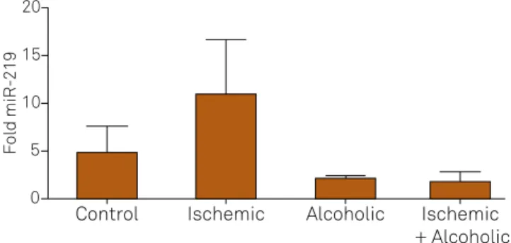
(Figure 1). However, the expression of miR-219 in the blood was
lower only in the alcoholic group and in the alcoholic and cerebral
ischemia group when compared to the control group (Figure 2).
The gene expression of NMDA neurotransmitter was greater in the brain tissue of animals with cerebral isch-emia, alcoholism, and the alcoholic and cerebral ischemia
groups compared to the control group (Figure 3). The
protein expression of the NMDA neurotransmitter in the brain tissue was greater in the mice in the cerebral isch-emia, alcoholism, and the alcoholic and cerebral ischemia
groups when compared to the control group (Figure 4).
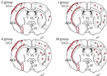
Cytoplasmic positive staining for NMDA protein was
ob-served in the three regions analyzed (dorsolateral cortex,
striatum and lateral cortex of the ischemic focus in the
nerve tissue of the left cerebral hemisphere) in all groups. Figure 5 shows the comparison of the expression of this
protein among the four groups.
Figure 2. Representation average (± standard deviation) of miR-219 in brain tissue among the groups (p = 0.0373, Kruskal-Wallis).
5
4
3
2
1
0
Control Ischemic Alcoholic Ischemic
+ Alcoholic
Fo
ld
miR-219
Figure 1. Representation average (± standard deviation) of miR-219 in the blood among the groups (p = 0.0461, Kruskal-Wallis).
20
15
10
5
0
Control Ischemic Alcoholic Ischemic + Alcoholic
Fo
ld
miR-219
Figure 3. Representation average (± standard deviation) of gene expression of NMDA receptor NR1 in the groups studied (p = 0.0211, Kruskal-Wallis test).
10
8
6
4
2
0
Control Ischemic Alcoholic Ischemic
+ Alcoholic
Fo
DISCUSSION
his work aimed at evaluating the possible modulation of the NMDA receptor NR1 by the action of miR-219 in animals
subjected to focal cerebral ischemia with or without an alco-hol abuse model.
Seeking to clarify the molecular mechanisms involved in controlling the expression of NMDA receptors, the literature
has highlighted the characterization of the expression proile of miRNAs in the modulation of neurotransmitters. In a study
with rats subjected to cerebral ischemia by occlusion of the MCA and reperfusion for 24 and 48 hours, Jeyaseelan, Lim, Armugam performed a comprehensive analysis of the
ex-pression proile of the miRNA technique for large-scale anal -ysis of microarrays, followed by the validation of the selected
real-time PCR in the blood of animals as well as in ischemic tissue miRNAs9.hree groups of miRNAs were identiied in the blood of animals. he irst miRNAs were diferentially ex -pressed only in the 24-hour reperfusion brain samples, and
some others overexpressed and underexpressed. he second group was comprised of miRNAs that changed their expres
-sion levels only after 48 hours of reperfu-sion. he third group was comprised of miRNAs that had diferent levels of expres -sion in both periods of reperfu-sion10.
Dharap et al. analyzed the role of miRNAs in the modula -tion of reperfusion in animals subjected to cerebral ischemia
by occlusion of the MCA. he authors analyzed 238 miRNAs,
of which eight overexpressed and 12 underexpressed11. It was observed that 20% of the miRNAs analyzed (47 of 238) show al
-terations over a period of reperfusion. herefore, the study in
-dicated that cerebral ischemia in the expression of miRNAs var -ies according to the time of reperfusion11. Kye et al.12 analyzed the expression of 187 miRNAs by real-time PCR in embryonic cultures of hippocampal neurons. he authors evaluated the mechanisms involved in the regulation of miRNAs after the in -traperitoneal administration of an antagonist of NMDA acid
receptors (CPP) and found 32 miRNAs had changed.
In our work we analyzed the expression of miR-219 in
ischemic brain tissue, which underexpressed in the group subjected to cerebral ischemia, the alcoholism group, and the alcoholic and cerebral ischemia group. We also observed an inverse correlation between the pattern of protein expression
of NMDA receptor NR1 and tissue expression of miR-219. Due to the speciicity of miR-219 in brain tissue, oth -er studies in the current lit-erature have demonstrated the
role of this miRNA in cerebral ischemia. Among them, Pai et al., who, after silencing Ago2 protein and RISC complex in rat long-term potentiation found by real-time PCR, dis -covered that the regulation of expression levels of several
miRNAs, including the miR-219, was totally dependent on the
NMDA receptor activation during long-term potentiation13.
In another study, Kocerba et al. analyzed the expression of
miR-219, a brain-speciic miRNA, after pharmacological in
-tervention (administration of dizocilpine) or genetic disrup
-tion (NR1 hypomorphism). he authors found that miR-219
quickly responded to changes in NMDA receptor signaling by two types of interventions14. Winbrad et al. have also demon-strated the regulation of the NMDA receptor by miR-219. he
authors found that the activation of NMDA receptors dur-ing long-term potentiation leads to a decreased expression of
miR-219, among other miRNAs15.
MicroRNAs have also been highlighted as biomarkers as -sociated with various diseases, among which are cerebral ischemia and alcoholism. In this regard, Liu et al.16 analyzed the expression proile of miRNAs in global cerebral tissue
and in the blood of animals undergoing the following mod-els of brain diseases: cerebral ischemia, cerebral hemorrhage induced by kainite, and epilepsy. Many overexpressed or
underexpressed miRNAs were found in each experimental condition. he authors describe functions associated with
common molecular mechanisms in brain tissue and blood as cell cycle, cell death, cell morphology and development of the organism, and they even suggested, according to the results,
Figure 5. Protein expression NMDA. Illustration of coronal sections of the brain the four groups showing in the left cerebral hemisphere, and from the three regions evaluated, diffuse distribution in protein expression of NMDA (red stars). Note: a higher expression in the striatum of the IA group. (Tirapelli,23, with permission). C: control; A: alcoholic; I: ischemic; IA: ischemic and alcoholic.
Figure 4. Representation of the average (± standard deviation) of the protein expression of NMDA receptor NR1 in the groups (p = 0.0289, Kruskal-Wallis test).
Control Ischemic Alcoholic Ischemic
+ Alcoholic
P
ositive cells (%)
25
20
15
10
5
that some diseases are brain responses, similar in both brain tissue and blood.
In this work, when we analyzed miR-219 in the blood of animals, we found that levels of expression of miRNAs were
reduced in the group submitted to alcoholism, and in the al-coholic and cerebral ischemia group when compared to the
control group. his suggests that this miRNA may have an
important role as biomarker.
Some studies have already demonstrated the role of
miRNAs as biomarkers in humans. he study by Zeng et al. ex
-amined the expression of miR-210 in 112 patients with cere
-bral ischemia and compared it with 60 control subjects. he miR-210 showed 88.3% sensitivity and was a good candidate for a biomarker. his study also evaluated the expression of miR-210 in blood and in the tissue of mice subjected to cere -bral ischemia, and a correlation between blood and tissue was
observed in the expression levels of miR-210. Another study in humans has also analyzed the expression of miRNAs miR-21, miR-221 and miR-145 in the serum of 167 patients with ce
-rebral ischemia. he patients had high levels of expression of miR-21 and miR-221 and low expression levels of miR-14517.
he brain is one of the organs most afected by the action
of alcohol on the body18. However, which events are associated
with the damage of brain tissue due to alcohol consumption
are still not clearly established. herefore, many studies have
attempted to establish the molecular mechanism involved in
the cerebral action of alcohol, among them Zhao et al., who evaluated the inluence of the amount/dose of ethanol con -sumption in animals subjected to cerebral ischemia, by occlu-sion of the middle cerebral artery for two hours followed by
reperfusion for 24 hours. he authors noted an increase in the
volume of infarcted region in doses of 6.4% ethanol, and found
increased expression of NR1 NMDA receptor at the same con -centration of 6.4%. However, at low doses there was a 1% re-duction in the volume of the infarct region suggesting the
neu-roprotective efect of ethanol in low concentrations19.
he majority of excitatory synapses in the central nervous
system are mediated by NMDA receptors and several stud-ies have shown that chronic alcoholism exposure increases
the expression of the NR1 and NR2B receptors18. Trudell et al.
reported evidence that the action of ethanol in the subunits of the NMDA and GABAA receptors is responsible for
behav-ioral efects, one of them, for example, through the activation of Fyn protein, a modulator of the NMDA receptor in speciic
regions of the brain, which leads to an increase in NMDA re-ceptor signaling, facilitating the increase in the consumption as well as consumption relapse20.
Gorini, Nunez, and Mayield analyzed the global expres
-sion proile of miRNAs in the cerebral cortex and midbrain of
mice submitted to chronic intermittent alcohol, and found
dif-ferent levels of expression in the brain regions studied. hese
data demonstrate that the changes caused by alcohol in the
brain tissue, as regards the expression proile of the evaluat
-ed miRNA region, is very important21. In another study, also by global analysis of expression proiling by microarray technique,
Mantha and Singh analyzed the exposure to prenatal ethanol
in adult mice and found 20 miRNAs diferentially expressed, and subsequently validated the miR-302c. Further studies are
needed to clarify the interaction between the pathophysiolo-gy of cerebral ischemia associated with alcoholism22. Based on our indings and literature, other miRNAs, with diferent peri -ods of reperfusion, need to be evaluated.
References
1. Lotufo PA. Mortalidade pela doença cerebrovascular no Brasil. Rev Bras Hipertens. 2000;7(4):387-91.
2. Kim MW, Bang MS, Han TR, Ko YJ, Yoon BW, Kim JH et al. Exercise increased BDNF and trkB in the contralateral hemisphere of the ischemic rat brain. Brain Res. 2005;1052(1):16-21. doi:10.1016/j.brainres.2005.05.070
3. Bronner LL, Kanter DS, Manson JE. Primary prevention of stroke. N Engl J Med. 1995;333(21):1392-400. doi:10.1056/NEJM199511233332106
4. Harper C. The neuropathology of alcohol-related brain damage. Alcohol. 2009;44(2):136-40. doi:10.1093/alcalc/agn102
5. Ohsawa M, Tanno K. Conflicting effect of alcohol on cardiovascular risk: a clue to understand the different etiologies of coronary artery disease, stroke and peripheral artery disease. Hypertens Res. 2013;36(1):16-8. doi:10.1038/hr.2012.152
6. Sathyan P, Golden HB, Miranda RC. Competing interactions between micro-RNAs determine neural progenitor survival and proliferation after ethanol exposure: evidence from an ex vivo model of the fetal cerebral cortical neuroepithelium. J Neurosci. 2007;27(32):8546-57. doi:10.1523/JNEUROSCI.1269-07.2007
7. Conti A, Aguennouz M, La Torre D, Tomasello C, Cardali S, Angileri FF et al. miR-21 and 221 upregulation and miR-181b downregulation in human grade II-IV astrocytic tumors. J Neurooncol.
2009;93(3):325-32. doi:10.1007/s11060-009-9797-4
8. Mendell JT. MicroRNAs: critical regulators of development, cellular physiology and malignancy. Cell Cycle. 2005;4(9):1179-84. doi:10.4161/cc.4.9.2032
9. Jeyaseelan K, Lim KY, Armugam A. MicroRNA expression in the blood and brain of rats subjected to transient focal ischemia by middle cerebral artery occlusion. Stroke. 2008;39(3):959-66. doi:10.1161/STROKEAHA.107.500736
10. Saugstad JA. MicroRNAs as effectors of brain function with roles in ischemia and injury, neuroprotection, and
neurodegeneration. J Cereb Blood Flow Metab. 2010;30(9):1564-76. doi:10.1038/jcbfm.2010.101
11. Dharap A, Bowen K, Place R, Li LC, Vemuganti R. Transient focal ischemia induces extensive temporal changes in rat cerebral microRNAome. J Cereb Blood Flow Metab. 2009;29(4):675-87. doi:10.1038/jcbfm.2008.157
12. Kye MJ, Neveu P, Lee YS, Zhou M, Steen JA, Sahin M et al. NMDA mediated contextual conditioning changes miRNA expression. PLoS One. 2011;6(9):e24682. doi:10.1371/journal.pone.0024682
14. Kocerha J, Faghihi MA, Lopez-Toledano MA, Huang J, Ramsey AJ, Caron MG et al. MicroRNA-219 modulates NMDA receptor-mediated neurobehavioral dysfunction. Proc Natl Acad Sci USA.
2009;106(9):3507-12. doi:10.1073/pnas.0805854106
15. Wibrand K, Panja D, Tiron A, Ofte ML, Skaftnesmo KO, Lee CS et al. Differential regulation of mature and precursor microRNA expression by NMDA and metabotropic glutamate receptor activation during LTP in the adult dentate gyrus in vivo. Eur J Neurosci. 2010;31(4):636-45. doi:10.1111/j.1460-9568.2010.07112.x
16. Liu DZ, Tian Y, Ander BP, Xu H, Stamova BS, Zhan X et al. Brain and blood microRNA expression profiling of ischemic stroke, intracerebral hemorrhage, and kainate seizures. J Cereb Blood Flow Metab. 2010;30(1):92-101. doi:10.1038/jcbfm.2009.186
17. Zeng L, Liu J, Wang Y, Wang L, Weng S, Tang Y et al. MicroRNA-210 as a novel blood biomarker in acute cerebral ischemia. Front Biosci (Elite Ed). 2011;3:1265-72. doi:10.2741/330
18. Chandrasekar R. Alcohol and NMDA receptor: current research and future direction. Front Mol Neurosci. 2013;6:14. doi:10.3389/fnmol.2013.00014
19. Zhao H, Mayhan WG, Arrick DM, Xiong W, Sun H. Dose-related influence of chronic alcohol consumption on cerebral
ischemia/reperfusion injury. Alcohol Clin Exp Res. 2011;35(7):1265-9. doi:10.1111/j.1530-0277.2011.01461.x
20. Trudell JR, Messing RO, Mayfield J, Harris RA. Alcohol dependence: molecular and behavioral evidence. Trends Pharmacol Sci. 2014;35(7):317-23. doi:10.1016/j.tips.2014.04.009
21. Gorini G, Nunez YO, Mayfield RD. Integration of miRNA and protein profiling reveals coordinated neuroadaptations in the alcohol-dependent mouse brain. PLoS One. 2013;8(12):e82565. doi:10.1371/journal.pone.0082565
22. Mantha K, Laufer BI, Singh SM. Molecular changes during neurodevelopment following second-trimester binge ethanol exposure in a mouse model of fetal alcohol spectrum disorder: from immediate effects to long-term adaptation. Dev Neurosci. 2014;36(1):29-43. doi:10.1159/000357496