1
Original Article
Coarctation of the Aorta in Infants under One Year
of Age. An Analysis of 20 Years of Experience
Gabriel Lorier, Orlando Wender, Renato A.K. Kalil, Javier Gonzalez, Gustavo Hoppen,
Christiano Barcellos, Abud Homsi-Neto, Paulo R. Prates, João R.M. Sant’Anna, Ivo A. Nesralla
Instituto de Cardiologia do Rio Grande do Sul/Fundação Universitária de Cardiologia - Porto Alegre,
RS - Brazil
Mailing address: Gabriel Lorier - IC/FUC - Unidade de Pesquisa - Av. Princesa Isabel, 370 - 90620-001 - Porto Alegre, RS
E-mail: [email protected] Received for publishing on 05/08/2003 Accepted on 02/11/2005
Objective
A review of experience with techniques of correction used, in the last 20 years, in children younger than one year old.
Methods
In the period from 1978 to 1998, 148 patients (pt) with coarcta-tion of the aorta (CoAo), under one year of age, with or without associated intracardiac defects, were submitted to surgery. Median age 50 days, 92 female pt (62.1%). The average weight was 4,367± 1,897 gr. The average follow-up was 1,152±1,462 days. The popu-lation was divided in 3 groups: Group I, isolated CoAo: 74 pt (50%); Group II, CoAo and interventricular communication (IVC): 41 pt (27.7%) and Group III, CoAo with complex intracardiac malforma-tions: 33 pt (22.3%).
Results
The total mortality was of 43 patients (29%). In patients younger than 30 days, the mortality was 53%, p=0.009, DR=4.5, between 31 and 90 days, 14.7%, p=0.69, and over 91 days, 15%, p=0.004. The probability of actuarial survival of the whole po-pulation was 67% at 5 and 10 years. Thirty-six patients (24.3%) had recoarctation, from which 18 patients (50%) were younger than 30 days, DR=6.,35. The incidence of recoarctation was with Waldhausen technique in 4 patients (10%) and with the classic termino-terminal technique in 19 patients (26%) p=0.03, and isthmusplastic operation in 6 patients (37.5%). The patients younger than 30 days showed a relative risk for recoarctation de DR=6.35. The probability of actuarial survival, free of coarctation repair, at 5 and 10 years was of 69% with Waldhausen’s technique and 63% with the classic termino-terminal technique.
Conclusion
Patients younger than 30 days showed increased mortality and recoarctation risk. Waldhausen’s technique in patients older than 30 days showed effective. The classic termino-terminal technique did not show to be a good option in all age ranges, being imperative to carry out more radical technical variations, such as the extended termino-terminal.
Key words
heart surgery, aortic coarctation, children
The coarctation of the aorta shows an incidence of 0.2 – 0.6 per 1.000 neonates, representing 5% to 8% of all congenital heart diseases1.
In 1948, the first successful surgical correction of coarctation of the aorta was performed by Crafoord, in termino-terminal anas-tomosis.
In 1951, Schumacher2 successfully reported a repair of a long
segment of coarctation with interposition of subclavian artery and termino-terminal anastomosis.
Vosschuhte, in 1961, published the isthmusplasty operation with graft3. Waldhausen and Nahrwold4 modified Shumacher’s
technique, by introducing the subclavial flap5. Since 1961 more
than eleven different technical variations have been published. That made inevitable the controversies due to the comparisons of results from the durability of the repairs6, but the low number of
patients from different publications makes generalized conclusions very difficult. The real proper comparison of operative techniques had not been made yet with scientific strictness7, until the early
1990s, when the works by Van Heurn, Conte and Jahangiri8-11
made a turning point for the analysis for the type of surgical correction in that pathology. At the same time, the anatomicopa-thological knowledge on the anomalous tissue of the arterial ca-nal and its relations with the aortic isthmus and arch was deepe-ned, as well as the anatomic substract that allows for the growth of the hypoplastic aortic arch11. Those factors have directly
in-fluenced in the choice of the operative technique.
The objective of this paper is to perform a review of our expe-riences with many aortic coarctation correction techniques, in the last 20 years, in infants under one year of age.
Methods
Retrospective cohort of patients with coarctation of the aorta under one year of age, in which the data were surveyed through a review of clinical records, by following a previously elaborated re-search protocol.
The aortic coarctation diagnosis was made through clinical examination and echocardiography in all cases.
The surgical indication was due to congestive heart failure or hypertension.
2
Coarctation of the Aorta in Infants under One Year of Age. An Analysis of 20 Years of Experience
In asymptomatic, normotensive and symmetric pulse patients, a new appointment was scheduled in a month, in which clinical exams, thorax radiography, 12-derivation electrocardiogram and Doppler echocardiogram were carried out. If the results were normal, a clinical appointment was scheduled every six months and echocardiogram once a year. IF they were normal after two years, the clinical exams were carried out once a year. If the patient was hypertensive after the surgery or with a pulse deficit, a weekly appointment with clinical exam and echocardiogram, if necessary, was scheduled.
Early mortality was considered when it took place within the first 30 post-operative days. Late mortality was considered after that term.
In the period between June 1978 and December 1998, 148 consecutive patients with coarctation of the aorta, under one year of age, with or without associated intercardial defects, were operated in our Institution.
Age showed a median of 50 days, 55 patients (37.1%) were younger than 30 days, 40 patients (27%) were between 31 and 90 days and 53 patients (35.8%) were over 91 days. Fifty-six patients were male (37.8%), 92 female (62.1%). The average weight was 4,367±1,729 grams, and 7.6% weighed less than 2,500 grams. The average follow-up was of 1,152±1,462 days. The population of patients was divided in three groups: Group I, patients with isolated CoAo: 74 patients (50%), Group II, patients with CoAo and interventricular communication (IVC): 41 infants (27.7%) and Group III, patients with CoAo complex intracardial malformations: 33 infants (22.3%).
The three most frequent malformations, in decreasing order, were as follows: patent ductus arteriosus in 95 patients (63.8%), interventricular communication in 41 patients (27.7%) and bicuspid aortic valve in 35 patients (23.6%).
The normal aortic arch was present in 7 patients, from which 5 (71.4%) belonged to Group I; isthmic hypoplasia was present in 103 patients (69.5%), from which 54 belonged to Group I; aortic arch hypoplasia was present in 38 patients (25.6), 4 (42.9%) of them belonged to Group III. The hypoplasia diagnosis was made in accordance to the echocardiogram operator’s criterion and subjective valuation of the surgeon.
Congestive heart failure was present in 52 patients (34.9%), hypertension in 6 patients (4.3%), cyanosis in 25 patients (16.8%). The medications in use before surgery were as follows: digital, in 67 patients (45%), diuretics, in 82 patients (55%), vasodilator, in 10 patients (6,7%).
Urgent surgery was performed in 36 patients (24.3%). In 73 infants (49.3%) classic termino-terminal anastomosis was applied, in 40 infants (27.0%) Waldhausen’s technique, in 16 infants (10.8%) isthmoplasty, and in 19 infants (12.8%) other methods (from which, in 87% was Teles de Mendonça’s techni-que12). For the performance of termino-terminal technique, simple
anastomoses were carried out, and not extended termino-terminal, nor extended radical termino-terminal technique.
The approach of aortic coarctation was done through left-lateroposterior thoracotomy in the fourth intercostal space.
For analgesia at immediate post-operative, all patients out of neonatal period, after surgical correction, were infiltrated with marcaine 0.25% 2 Mg/Kg weight per intercostal space.
The monitoring of average blood pressure was carried out through puncture of the right radial artery.
All continuous variables are described as mean ± standard deviation. Categorical variables are described in proportions. Simple frequencies were done for all variables.
Data were organized in tables and charts. The associations among categorical variables were tested through the chi-square test, and the continuous variables were tested with Student’s t test.
Actuarial survival curves were obtained through the method of Kaplan-Meier.
Finally, logistic regression was carried out, including in the model of all clinically significant variables, in which they had p<0.15 in two-variable analysis. The p-α chosen as statistically
significant was equal or less than 0.05.
Results
Total in-hospital deaths ocurred in 27 patients, 18.1% (27/ 148). The average age of those who died was 60.6±15.4 days (the patients who did not evolved to death had an average age of 103±17.2 days, p=0.2). The average weight of those who died was 3,455±786 g and of those who did not die was 4,549±654 g (p=0.01).
In Group I, early mortality took place in 9 patients (12.1%) (Group I n=74 patients), in Group II, it was in 10 patients (24.3%) (Group II: n=41 patients) and in Group III, 8 patients (24.2%) (Group III: n=33 patients). Regardless of the group, the patients with aortic arch hypoplasia showed a greater early mortality, p=0.01. In patients with aortic arch hypoplasia, in which Teles de Mendonça’s technique13 and isthmusplasty operation was
ap-plied, the mortality was 100% and 60%, respectively.
When Waldhausen’s technique was compared with the classic termino-terminal technique, in the presence of aortic arch hypo-plasia, there was a significant difference concerning early and total mortality (p=0.05).
Total mortality was present in 43 patients (29%).
Infants under 30 days of age increased in 4.5 times the death risk (DR=4.5 CI 95% 1.4-14) p=0.009. There was no increase of death risk in other age ranges.
The global mortality in the first quarter of life, relating the 3 surgical techniques currently used in our service, Waldhausen’s and termino-terminal techniques were the ones that showed the best results, without significant difference between each other.
The first three intracardiac malformations of greater mortality were, in decreasing order: TGV (66.7%), tricuspid valve anomalies (60%) and left ventricular outflow tract obstructions (50%).
The presence of IVC and complex malformations increased the death risk in 4.1 times (DR=4.1 CI 95% 1.3-12.6) p=0.01 and 3.9 times (DR=3.9 CI 95% 1.2-13.2) p=0.02, respectively. The presence of aortic arch hypoplasia increased the death risk in 1.9 times (DR=1.9 CI 95% 0.7-5.4) p=0.19.
Urgent surgery increased 1.96 times the death risk (CI 95% - 0.87-4.27), which represented an incidence of 32.3% from total deaths.
The actuarial survival probability of the whole population was 67% in 5 and 10 years.
When the population of patients was divided in 2 groups, without and with associated pathologies, the actuarial survival probabilities at 5 years were 81% and 65%, respectively, and at 10 years, they were 81% and 62%, respectively.
tech-3
nique. In Waldhausen’s technique, the survival was 69% at 5 and 10years. In classic termino-terminal technique, the survival was 63%, at 5 and 10 years, and in isthmusplasty was 56%, at 5 years.
The actuarial probability of survival in Group I was 82%, at 5 and 10 years, and 68% at 15 years, in Group II it was 52%, at 5 and 10 years, and in group III, 54%, in 5 years.
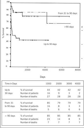
Regarding the age range, the actuarial curve showed a survival at 5 years of 42% in infants up to 30 days of age. In the ranges of 31 to 90 days and those under 90 days, it was 79% and 85%, respectively (fig. 2).
In 36 patients (24.3%) there was a recoarctation, from which 18 (50%) were under 30 days of age at the moment of the surgery. A new correction was performed in 10 patients (6.8%), with a time median from the first surgery of 53 days; dilation with balloon was carried out in 12 patients (8.1%), with a time median from the surgery of 482 days.
The average age of recoarcted patients was 63.4±12.1 days and 106±27.3 days in patients who were recoarcted, p=0.01. The average weight of the recoarcted patients was 3,908 g and 4,520 g of those who were not recoarcted (p=0.06).
Recoarctations were more frequent in males, 66.7%. Among the females they were 33.3%.
The worst results took place in patients with aortic arch hy-poplasia, in which termino-terminal anastomosis was applied, with an incidence of recoarctation of 42%.
Infants up to 90 days, in which the classic termino-terminal
Days
Time in Days 1000 2000 3000 4000
Waldhausen % of survival 69 69 69 69
Number of patients 14 9 7 6
Number of events 10 10 10 10
Termino- % of survival 70 63 63 63
Terminal Number of patients 24 11 4 1
Number of events 20 22 22 22
Isthmusplasty % of survival 56 56 56
-Number of patients 7 4 1
-Number of events 7 7 7
-2000 4000 6000 8000
% Survival
Waldhausen
Termino-Terminal
Isthmusplasty
Fig. 1 - Actuarial probability of survival in the whole population.
technique was applied, showed an increase of relative risk of recoarctation of 3.5 times (CI 0.8-13.8) in relation to those within the age range over 90 days, p=0.07. Infants under 30 days showed an increase of 6.3 times of the risk of recoarctation (p=0.01), and the normal aortic arch is shown with reduction of risk for recoarctation. Isthmusplasty increases in 8 times the risk of re-coarctation (p=0.0009).
Urgent surgery increased the risk of recoarctation in 1.8 times (CI 95% - 0.8-4.2), 33.3% of the patients who recoarctation had been submitted to urgent surgery.
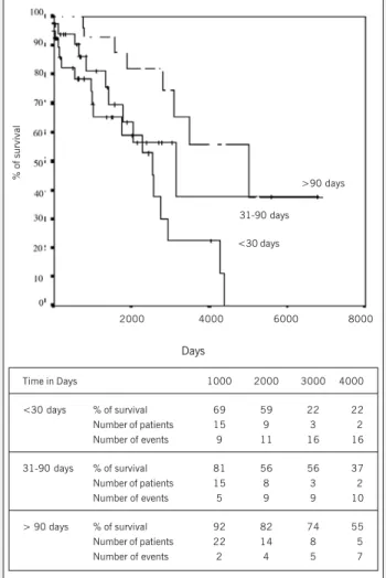
The actuarial probability free of recoarctation of the whole po-pulation at 5 and 10 years was 69% and 40%, respectively. When analyzed regarding the age range, in infants under 30 days it was 60% at 5 years; between 31 and 90 days, it was 64%, at 5 years, and for infants over 91 days, it was 83%, at 5 years (fig. 3).
In the actuarial survival probability free of recoarctation, accor-ding to the surgical technique, Waldhausen’s technique showed the best results, with 90% and 80% at 5 and 10 years, respecti-vely. (fig. 4).
Analyzing per group of associated malformations, the best re-sults from probability free of recoarctation were shown in Group I (isolated CoAo), 80% at 5 years, 48% at 10 years and 25% at 15 years.
Teles de Mendonça’s technique was employed in 13 patients (8.8%), from which 92.3% showed isthmic hypoplasia. The incidence of recoarctation was 38.5% (5 patients).
Days
Time in Days 1000 2000 3000 4000
Up to % of survival 44 42 42 42
30 days Number of patients 16 9 3 2
Number of deaths 27 28 28 28
From 31 % of survival 85 79 79 79
to 90 days Number of patients 16 8 3 3
Number of deaths 5 6 6 6
> 90 days % of survival 85 85 85 85
Number of patients 23 14 8 5
Number of deaths 7 7 7 7
2000 4000 6000 8000
% Survival
From 31 to 90 days
>90 days
Up to 30 days
4
Coarctation of the Aorta in Infants under One Year of Age. An Analysis of 20 Years of Experience
Discussion
Our retrospective cohort showed that Waldhausen’s technique – which started in our institution in June 197814 – showed the
best early and late clinical results.
Some publications have discussed the disadvantages of Wald-hausen’s technique, such as, for example, the gangrene of the limb, described in 198013, with such complication not being further
reported in the literature, or even claudication and hypogrowth8,9,
Horner’s syndrome8, late formation of aneurysms, published 5 cases
in the literature15, being the histopathologic base for its formation
the existence of a ductal origin flat muscle that further becomes fibrous16. Those complications were not found in our series.
In this series, the mortality in infants under 1 year of age with Waldhausen’s technique was 25%. In American Consortium of Pediatric Heart Care it was 7.9%, being a multicentric result in the late 1980s17. When the patients showed hypoplastic aortic arch and
isthmic hypoplasia – and the technique of Waldhausen was employed -, the incidence of total mortality was 33.3% and 21.4%, respectively, being such results very high. In infants over 30 days, the results are satisfactory, compared with results from other centers8,17-27.
In our series, the incidence of recoarctation in the first month of life was 25%, regarded as high when compared with the international average25-27. The proliferation and constriction of residual
canal tissue, still active during the first three months of life18, can
show growth after surgical and be the cause of recoarctation19.
The incidence of recoarctation, in 8 different papers with a total of 406 patients with up to a month of life, showed the average of recoarctation of 13,3%. However, in the age range of up to 3 months, the incidence lowered to 15%, and, under than 1 year of age, it was 10%, similar results to those found in the literature8,19. We believe,
as a cause factor, in the insufficient resection of the ring of the anomalous tissue of the canal, associated to other series of factors. Our actuarial survival free of recoarctation, at 5 years, was 89%. Van Heurn8 reports 57%, with Waldhausen’s technique (fig. 4).
When we analyze our results with classic termino-terminal techni-que and compare them with Waldhausen’s, we find lower results in all age ranges, concerning the mortality and recoarctation in classic termino-terminal technique, and with those results being similar to those from other publications6,27. The total mortality with classic
termino-terminal technique was 29.2%. In the literature, the results varied from 10% to 43%8,18 and are considered as unsatisfactory,
which recommends for the performance of more aggressive variations of termino-terminal technique, such as those originally described by Amato, in 1991. In the aortic arch hypoplasia, the mortality was 63.2%, showing to be imperative a more suitable reconstruction of aortic arch with extended and extended radical termino-terminal techniques to improve such results8. In patients with aortic arch
hypoplasia and isthmic hypoplasia, our incidence of recoarctation was 42.1% and 22%, respectively, conflicting with current concepts that termino-terminal technique should not be employed in such anatomic variants18. In the age range of up to 30 days, 34.6% of
Days
Time in Days 1000 2000 3000 4000
<30 days % of survival 69 59 22 22
Number of patients 15 9 3 2
Number of events 9 11 16 16
31-90 days % of survival 81 56 56 37
Number of patients 15 8 3 2
Number of events 5 9 9 10
> 90 days % of survival 92 82 74 55
Number of patients 22 14 8 5
Number of events 2 4 5 7
2000 4000 6000 8000
% of survival
>90 days
31-90 days
Fig. 3 - Actuarial probability of survival free of repair of coarctation per age range. <30 days
Days
Time in Days 1000 2000 3000 4000
Waldhausen % of survival 89 89 79 79
Number of patients 14 10 7 6
Number of events 2 2 3 3
Termino- % of survival 76 69 47 23
Terminal Number of patients 23 12 4 2
Number of events 11 13 16 18
Isthmusplastic % of survival 81 59 44
-Operation Number of patients 8 5 1
-Number of events 2 4 5
-2000 4000 6000 8000
% of survival
Fig. 4 - Actuarial probability of survival free of repair of coarctation according to surgical technique.
Waldhausen
Isthmusplastic Operation
5
recoarctation took place. In the same way, in the age range between31 and 90 days, the incidence of recoarctation of 32%, although high, is similar to those found in other publications8,19,23.
Currently it has been emphasized that the most efficient sur-gical technique to relieve the obstruction is the extended termi-no-terminal7-9, 28-33. Such procedure does not have the disadvantages
of other repair techniques, such as classic termino-terminal32,
Waldhausen20 or isthmusplasty operation with graft33. Either the
classic termino-terminal or Waldhausen’s or isthmusplasty opera-tion with graft has shown high incidence recoarctaopera-tion. Such assertions were confirmed in our series. Some authors25,34 use
the extended termino-terminal technique when the transverse aortic arch shows an index lower than 0.25 in relation to the descending aorta, which means smaller than 3 mm.
The results published in the literature with the extended ter-mino-terminal technique, in a total of 648 patients less than 3 months, in 6 different papers8,9,28,30,35,36, show an incidence of
9.1% of recoarctation. From those, 332 were under a month of age, with an incidence of recoarctation of 9.3%. In neonatal period, always some level of aortic arch hypoplasia is found in patients with aortic coarctation18. In the paper by Conte9 it was resent in
81% of the patients. Van Heurn8 reported 25% and 33% of isthmus
and transverse arch hypoplasia, respectively. Van Son and Zanini reported 44% and 100%3,29, respectively, of aortic arch hypoplasia
in children up to three months of age. Therefore, aortic arch reconstruction in neonatal period seems to be primordial, which reduces the mortality and recoarctation34.
Isthmosubclaviplasty or Teles de Mendonça’s technique was
described in 198512, Méier, in 198637, reported 28 cases with
good results, in only two patients less than 3 months. Amato24,
using the technique in 3 neonates (9, 10 and 14 days), showed 100% of recoarctation, Messmer, in 199123, showed 43% of
recoarctation (3 patients) with an average age of 2.3 months. Other authors showed high levels of recoarctation38. In our series
it was employed in 13 patients: 12 with isthmic hypoplasia, sho-wing 16.6% of early mortality (2 patients) and 38.5% (5 pa-tients) of recoarctation, in addition to 1 patient with aortic arch hypoplasia that evolved to early death. Currently, such technique is not routinely employed in our service.
Based on those results, it is possible to infer that patients under 30 days of age and those who have aortic arch hypoplasia show increased risk of mortality and recoarctation, regardless of the technique used.
The classic termino-terminal technique showed, in general terms, not being a good option to relieve the obstruction in all age ranges and in all anatomic variations of aortic arch, which makes im-perative to perform other more radical technical variations, such as the extended or extended radical termino-terminal technique.
The classic termino-terminal technique and the isthmusplasty showed twice as much death risk than Waldhausen’s technique. Teles de Mendonça’s technique and isthmusplasty operation showed poor results, especially in aortic arch hypoplasia.
Walhausen’s technique showed unsatisfactory results in the first month of life, with isthmic and aortic arch hypoplasia. In infants over one month of life, with isthmic and aortic arch hypo-plasia, it showed effectiveness.
1. Crafoord C, Nylin G. Congenital coarctation of the aorta and its surgical treatment. J
Thoracic Surg 1950; 37: 46. ApudBacker L, Mavroudis C. Glenn’s Thoracic and
Car-diovascular Surgery. 6th ed. Vol. I. Stamford: Appleton & Lange, 1996; 76: 1243.
2. Schumacker Jr HB. Use of subclavian artery in the surgical treatment of the
coarc-tation of the aorta. Surgical Gynecol Obstet 1951;93:491-5. ApudRubay JE,
Sluysmans TH, Alexandrescu V, Khelif K, Moulin D, Vliers A, Jaumin P, Chalant H. Surgical repair of coarctation of the aorta in infants under one year of age. J Cardiovasc Surg. 1992; 33: 216-22.
3. Vosschulte K. Surgical correction of coarctation of the aorta by na “isthmusplastic”
operation. Thorax 1961;16:338 apudCastañeda A, Jonas R, Mayer J, Hanley F.
Car-diac Surgery of the Neonate and Infant. Phidadelphia: WA Saunders, 1994; 22: 353.
4. Waldhausen JA, Nahrwold DL. Repair of coartaction of the aorta with a
subcla-vian flap. J Thorac Cardiovasc Surg. 1966; 51: 532.
5. Rubay JE, Sluysmans TH, Alexandrescu V, et al. Surgical repair of coarctation of
the aorta in infants under one year of age. J Cardiovasc Surg. 1992; 33: 216-22.
6. Merril WH, Hoff SJ, Stewart JR, Elkins CC, Graham TP, Bender HW. Operative risk
factors and durability of repair of coarctaition of the aorta in the neonate. Ann Thorac Surg. 1994; 58: 399-403.
7. Harlan JL, Doty DB, Brandt III B, Ehrenhaft JL. Coarctation of the aorta in
in-fants. J Thorac Cardiovasc Surg. 1984; 88: 1012-19.
8. Van Heurn LWE, Wong CM, Spiegelhalter DJ, et al. Surgical treatment of aortic
coarctation in infants younger than three months: 1985 to 1990. J Thorac Cardio-vasc Surg. 1994; 107: 74-86.
9. Conte S, Lacour-Gayet F, Serraf A, et al. Surgical management of neonatal
coarcta-tion. J Thorac Cardiovasc Surg. 1995; 109: 663-75.
10. Jahangiri M, Shinebourne EA, Zurakowski D, Rigby ML, Redington AN, Lincin C. Subclavia flap angioplasty: does the arch look after itself ? J Thorac Cardiovasc Surg. 2000; 120: 224-9.
11. Machii M, Becker AE. Hypoplastic aortic arch morphology pertinent to growth after surgical correction of aortic coarctation. Ann Thorac Surg. 1997; 64: 516-20. 12. Mendonça JT, Carvalho MR, Costa RK, Filho EF. Coarctation of the aorta. A new
surgical technique. J Thoracic Cardiovasc Surg. 1985; 90: 445-7.
13. Geiss D, Williams WG, Lindsay WK, Rowe RD. Upper extremity gangrene: a
compli-cation of subclavian artery division. Ann Thorac Surg. 1980;30:487-9. ApudConte S,
Lacour-Gayet F, Serraf A, Sousa-Uva M, Bruniaux J, Touchot A, Plaché C. Surgical management of neonatal coarctation. J Thorac Cardiovasc Surg. 1995; 109: 663-75.
References
14. Prates PR, Lucchese FA, Kalil RAK, et al. Correção da coartação da aorta pela ist-moplastia com a artéria subclávia esquerda em pacientes sintomáticos no primei-ro ano de vida. Arq Bras Cardiol. 1981; 36: 403-6.
15. Kino K, Shunji S, Sugawara E, Kohmoto T, Kamada M. Late aneurysm after subcla-vian flap aortoplasty for coarctation of the aorta. Ann Thorac Surg. 1996; 1262-4. 16. Jonas RA. Coarctation: Do we need to resect ductal tissue ? Ann Thorac Surg.
1991; 52: 604-7.
17. Norton Jr JB. Coarctation of the aorta. In: Perspectives in Pediatric Cardiology. Vol.6. Armonk: Futura Publishing, 1988; 14: 143-58.
18. Castañeda A, Jonas R, Mayer J, Hanley H. Aortic coartation. In: ___. Cardiac Surgery of the neonate and infant. Philadelphia: WB Saunders, 1994. 19. Cobanoglu A, Teply JF, Grunkmeier GL. Coarctation of the aorta in patients
youn-gher than three months. J Thorac Cardiovasc Surg. 1985; 89: 128-35. 20. Ziemer G, Jonas RA, Perry SB, Freed MD, Castañeda AR. Surgery for coartaction
of the aorta in the neonate. Circulation. 1986; 74(Suppl.I): I-25.
21. Sanchez GR, Balsara RK, Dunn JM, Mehta AV, O’Riordan AC. Recurrent obstruc-tion after subclavian flap repair of coarctaobstruc-tion of the aorta in infants. J Thorac Cardiovasc Surg. 1986; 91: 738-46.
22. Milliken JC, Braun WJ, Mee RB. Neonatal coarctation: Clinical spectrum and
im-proved results. J Am Coll Cardiol 1990;15:78A. ApudBacker L, Mavroudis C. Glenn’s
thoracic and cardiovascular surgery. 6º ed. Vol. I. Stamford: Appleton & Lange, 1996 23. Messmer BJ, Minale C, Mühler EV, Bernuth G. Surgical correction of coarctation of coarctation in early infancy; does durgical technique influence the result ? Ann
Thorac Surg 1991;52:594-603. ApudAmato JJ, Galdieri RJ, Cotroneo JV. Role of
extended aortoplasty related to the definitions of coarctation of the aorta. Ann Thorac Surg. 1991; 52: 615-20.
24. Amato JJ, Galdieri RJ, Cotroneo JV. Role of extended aortoplasty related to the definitions of coarctation of the aorta. Ann Thorac Surg. 1991; 52: 615-20. 25. Knott-Craig C, Elkins RC, Ward KE, et al. Neonatal coarctation repair – Influence
of technique on late results. Circulation. 1993; 99(part 2): 198-204. 26. Quaegebeur JM, Jonas RA, Weinberg AD, Blackstone EH, Kirklin JW, and Congenital
Heart Surgeons Society. Outcomes in seriously ill neonates with coartaction of the aorta. J Thorac Cardiovasc Surg. 1994;108: 841-54.
6
Coarctation of the Aorta in Infants under One Year of Age. An Analysis of 20 Years of Experience
28. Rubay JE, Sluysmans TH, Alexandrescu V, et al. Surgical repair of coarctation of the aorta in infants under one year of age. J Cardiovasc Surg. 1992; 33: 216-22. 29. Zanini L, Gargiulo G, Albanese SB, et al. Aortic coarctation with hypoplastic arch in neonates: a spectrum of anatomic lesions requiring different surgical options. Ann Thorac Surg. 1993; 56: 288-94.
30. Vouhé PR, Trinquet F, Lecompte Y, et al. Aortic coarctation with hypoplastic aortic arch. J Thorac Cardiovasc Surg. 1988; 96: 557-63.
31. Trinquet F, Vouhé PR, Vernant F, et al. Coarctation of the aorta in infants: Which operation ? Ann Thorac Surg. 1988; 45: 186-91.
32. Kirklin JW, Barrat-Boyes BG. Coarctation of the aorta and interrupted aortic arch. In: ___. Cardiac Surgery. 2ª ed. Londres: Churchill Livigstone, 1993; 34: 1263. 33. Backer CL, Paape K, Zales VR, Weigel TJ, Mavroudis C. Coarctation of the aorta.
Repair with polytetrafluoroethylene patch aortoplasty. Circulation. 1995; 92(suppl.II): II-132-II-6.
34. Siewers RD, Ettedgui J, Pahl E, Tallman T, del Nido PJ. Coarctation and hypoplasia of the aortic arch: will the arch grow. Ann Thorac Surg. 1991; 52: 608-14. 35. Van Son JAM, Falk V, Schneider P, Smedts F, Mohr FW. Repair of coarctation of the
aorta in neonates and young infants. J Card Surg. 1997; 12: 139-46. 36. Backer CL, Mavroudis C, Zias EA, Amin Z, Weigel TJ. Repair of coarctation with
resec-tion and extended end-to-end anastomosis. Ann Thorac Surg. 1998; 66: 1365-71. 37. Meier MA, Lucchese FA, Jazbik W, Nesralla IA, Mendonça JT. A new technique for
repair of aortic coarctation. Subclavian flap aortoplasty with preservation of arte-rial blood flow to the left arm. J Thorac Cardiovasc Surg. 1986; 92: 1005-12. 38. Nawa A, Nakayama Y, Teramoto S, Mori K, Dohi T. Coarctation restenosis after
isthmosubclavioplasty. A consideration on operative procedure and intraluminal
balloon angioplasty. Chest. 1989; 95:247-50. ApudAmato JJ, Galdieri RJ,