Comparative Study Between Intermittent
(Müller Reanimator) and Continuous Positive
Airway Pressure in the Postoperative Period of
Coronary Artery Bypass Grafting
Andréa Pires Müller, Márcia Olandoski, Rafael Macedo, Constantino Costantini, Luiz César Guarita-Souza
Hospital Costantini e Pontifícia Universidade Católica do Paraná - Curitiba, PR - Brazil
Mailing Address: Andréa Pires Müller • Universidade Católica do Paraná - Rua Imaculada Conceição, 1155 - 80215-901 - Curitiba, PR - Brazil E-mail: [email protected] Received on 04/19/05 • Accepted on 06/14/05
O
BJECTIVETo compare the effect of the use of intermittent and continuous positive airway pressure in postoperative patients undergoing coronary artery bypass grafting.
M
ETHODSThis study included forty patients divided into two groups: one undergoing continuous positive airway pressure (CPAP Group), and the other undergoing intermittent pressure (Müller Resuscitator Group). The patients were evaluated in relation to the several study variables at the following time points: preoperative, 3rd, 24th, and 48th hours.
R
ESULTSThe patient groups were homogeneous in relation to the several demographic and clinical variables. The values of pO2, pCO2 and sO2 were within normal limits and no signifi cant differences were found between the groups. Regarding respirometry, the groups showed signifi cant differences in the tidal volume and respiratory rate at the 48th postoperative hour. Dyspnea and use of accessory muscle in postoperative assessments were found with a signifi cantly higher frequency in patients undergoing CPAP. Patients undergoing Müller Resuscitator had a normal chest radiograph more frequently than did patients undergoing CPAP.
C
ONCLUSIONBoth devices were shown to be able to keep pO2, pCO2, and sO2 values within normal limits. However, when the objective was pulmonary reexpansion with less imposed workload, the Müller Resuscitator was more effective because of its prompter action and consequently lower levels of dyspnea, respiratory rate (RR) and use of accessory muscle were observed.
K
EY WORDSRespiratory complications are problems commonly found in the postoperative period of thoracic surgeries. The mechanisms leading to pulmonary injury are still unknown, but they seem to be originated during the surgical procedure, which demonstrates that the management of assisted mechanical ventilation (AMV) during and after anesthesia is a determining factor in the incidence of pulmonary complications1. The
precise incidence depends on the type of surgery and on preoperative pulmonary function conditions, most of these complications being diagnosed as pneumonia or postoperative atelectasis. These complications are frequently related to the dependence on assisted mechanic ventilation2.
Concurrently, the dependence on ventilation support is directly related to the incidence of morbidity, and prolonged stays in Intensive Care Units are related to subsequent prolonged hospitalization. The incidence of pulmonary complications and hospitalization time may be decreased by decreasing the time to start weaning and by disconnecting the mechanical ventilation support as early as possible, which usually takes more than 40% of the total AMV time3.
The deleterious effects of endotracheal intubation (nasal and orotracheal) and of assisted mechanical ventilation have been broadly disseminated in the literature. Therefore, physical therapists consider the beginning of the ventilation weaning and the discontinuation of assisted mechanical ventilation (AMV) as major factors of treatment in Intensive Care Units. These processes should therefore be started as soon as the patients present satisfactory spontaneous ventilation, that is, when they are able to sustain ventilation with an effective gas exchange4.
However, the transition between mechanical ventilation and spontaneous ventilation is the moment when many of the respiratory disorders arise and are worsened by restrictive factors such as sedation, pain, and the presence of chest and abdominal drains5.
After extubation, an important phase of the physical therapy assistance starts, with the main purpose of maintaining the patient’s spontaneous ventilation, thus avoiding reintubation. Several studies6-10 have proved
the effi ciency of non-invasive ventilation (NIV) in the weaning procedure and maintenance of spontaneous ventilation. The authors report that the utilization of this type of procedure promotes a decrease in ventilation work and in dyspnea, and an increase in residual volume, thus preventing the presence of atelectasis and favoring alveolar recruitment as well as increasing partial pressure of oxygen in the arterial blood (paO2).
In 1999, the utilization of Müller Resuscitator was proposed as an alternative to NIV. This device has a pneumatic feature that provides intermittent positive pressure. This device has some advantages similar to those of CPAP, namely, a decrease in ventilation work
and dyspnea rates and an increase in residual volume11.
Both have proven effective in post-extubation treatment, each with its own characteristics. However, there is no comparative study between these two devices reported in the literature.
Thus, the objective of this study is to compare the result of continuous positive airway pressure to that of intermittent positive pressure in postoperative patients undergoing cardiac surgery using the CPAP and Müller Resuscitator devices.
M
ETHODS
This is a quantitative prospective randomized study including forty patients undergoing coronary artery bypass grafting in a cardiology hospital from February 2004 to October 2004. Thirty three patients were males and seven were females.
The patients were divided into two groups, one undergoing continuous positive airway pressure and the other undergoing intermittent pressure. The patients were assessed in the following time points: preoperative period, 3rd, 24th and 48th hour after utilization of the devices, regarding the several variables of the study. The Goodknight 418-G with a rubber facial mask (Mallinckrodt®) was used in the group undergoing treatment with continuous positive airway pressure (CPAP group). The group undergoing treatment with intermittent positive pressure used Müller Resuscitator with a rubber facial mask (Engesp®) (Müller Resuscitator group).
Spirometry was performed in the preoperative period. Predicted and obtained values, and the percentage of the value obtained from the predicted value of the following variables were considered: FVC (forced vital capacity), FEV1 (forced expiratory volume in one second), FEV1/FVC (ratio forced expiratory volume in one second and forced vital capacity) and Peak fl ow, since to take part in the study the patients could show normal patterns, mild restrictive or obstructive ventilation disorders (79%-60% of FEV1), and moderate restrictive or obstructive ventilation disorder (59%-41% of FEV1). Likewise, the analysis of the left ventricle ejection fraction at the pre and postoperative time points was verifi ed.
the physical examination. The radiography report issued by the responsible radiologist was observed.
The protocol used for CPAP with an intermittent system at a continuous fl ow and a spring-loaded valve was applied in the postoperative time points mentioned, with the patient in fowler position (35º), and a PEEP (positive end expiratory pressure) level of 5 cm of H2O and three liters of oxygen. At the fi rst three postoperative hours, the protocol was applied for fi fteen minutes every hour, and at the 3rd hour the study variables were verifi ed. It was applied for thirteen minutes at the 24th and 48th postoperative hours, and the same variables were verifi ed in the sequence.
The use of Müller Resuscitator occurred at the 3rd, 24th, and 48th postoperative hours with the patient in a fowler position (35º). A 20 to 30 cmH2O endotracheal pressure (recommended for adult patients) was used, and only saline solution was used in the micronebulizer as a dilutant. The device was applied for fi fteen minutes per hour in the fi rst three postoperative hours, and at the 3rd hour the blood gas analysis, respirometry and radiography report variables were verifi ed. At the 24th and 48th postoperative hours it was applied for thirty minutes, in two fi fteen-minute series, and then the same variables were verifi ed at the 3rd hour.
Statistical analysis - The results obtained in the study were expressed as mean and standard deviations or as frequencies and percentages. The Student’s t test for independent samples was used in the comparison of the groups in relation to the quantitative variables, considering the homogeneity of variances, or the Mann-Whitney non-parametric test when the normality condition was not met. Fisher’s exact test was used to compare the groups in relation to the categorical variables. In all tests, a p < 0.05 value was considered statistically signifi cant.
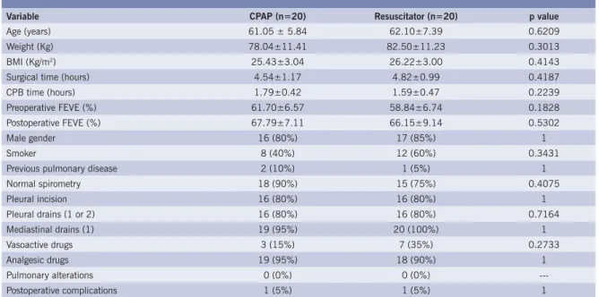
Table 1 – Patients demographic and clinical characteristics
Variable CPAP (n=20) Resuscitator (n=20) p value
Age (years) 61.05 ± 5.84 62.10±7.39 0.6209
Weight (Kg) 78.04±11.41 82.50±11.23 0.3013
BMI (Kg/m2) 25.43±3.04 26.22±3.00 0.4143
Surgical time (hours) 4.54±1.17 4.82±0.99 0.4187
CPB time (hours) 1.79±0.42 1.59±0.47 0.2239
Preoperative FEVE (%) 61.70±6.57 58.84±6.74 0.1828
Postoperative FEVE (%) 67.79±7.11 66.15±9.14 0.5302
Male gender 16 (80%) 17 (85%) 1
Smoker 8 (40%) 12 (60%) 0.3431
Previous pulmonary disease 2 (10%) 1 (5%) 1
Normal spirometry 18 (90%) 15 (75%) 0.4075
Pleural incision 16 (80%) 16 (80%) 1
Pleural drains (1 or 2) 16 (80%) 16 (80%) 0.7164
Mediastinal drains (1) 19 (95%) 20 (100%) 1
Vasoactive drugs 3 (15%) 7 (35%) 0.2733
Analgesic drugs 19 (95%) 18 (90%) 1
Pulmonary alterations 0 (0%) 0 (0%)
---Postoperative complications 1 (5%) 1 (5%) 1
R
ESULTSThe group of patients undergoing continuous pressure (CPAP) and the group of patients undergoing intermittent pressure (Müller Resuscitator) were homogeneous in relation to several demographic and clinical variables (table 1).
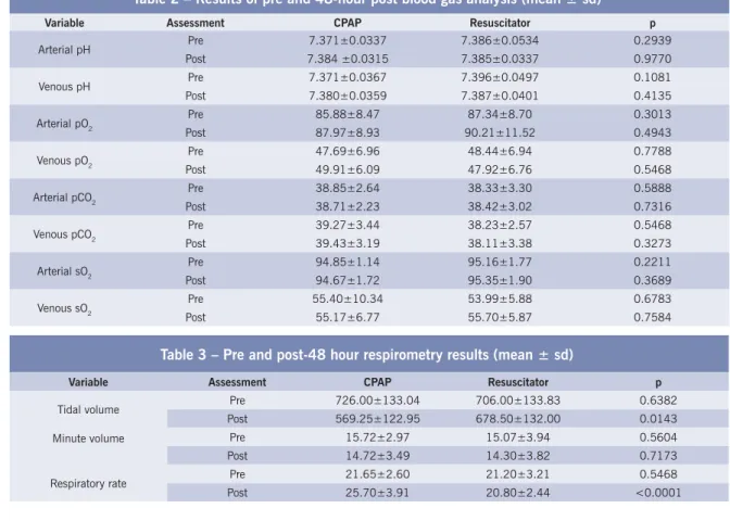
In patients’ assessment using respirometry, the preoperative tidal volume was similar in both groups, decreasing at the 3rd hour more markedly in the group undergoing CPAP, to increase again at the 24th and 48th hours. The difference is signifi cant between the groups at the 3rd hour (p = 0.0332), at the 24th hour (p = 0.0486), and at the 48th hour (p = 0.0143). The same progression was observed for the minute volume, however without a signifi cant difference between patients undergoing CPAP and patients undergoing Müller Resuscitator. In the preoperative assessment, the respiratory rate was homogeneous between the groups. However, as of the 3rd hour a signifi cant difference was found between them (p = 0.0007 at the 3rd hour, p = 0.0002 at the 24th hour, and p < 0.0001 at the 48th hour). At all these assessment time points, a higher respiratory rate in the patients undergoing CPAP was observed when compared to that of the patients undergoing Müller Resuscitator. Table 3 shows respirometry results obtained in the pre and 48 postoperative hour assessments.
Of the patients undergoing CPAP, only one presented mild dyspnea in the preoperative assessment. This number increased in the postoperative assessments (eleven patients at the 3rd hour, twelve at the 24th hour, and fi fteen at the 48th hour). Dyspnea was observed in two of the patients undergoing Müller Resuscitator in the preoperative assessment, in two patients at the 3rd hour, and in only one at the 24th and 48th hours. The
Table 2 – Results of pre and 48-hour post blood gas analysis (mean ± sd)
Variable Assessment CPAP Resuscitator p
Arterial pH Pre 7.371±0.0337 7.386±0.0534 0.2939 Post 7.384 ±0.0315 7.385±0.0337 0.9770
Venous pH Pre 7.371±0.0367 7.396±0.0497 0.1081
Post 7.380±0.0359 7.387±0.0401 0.4135
Arterial pO2 Pre 85.88±8.47 87.34±8.70 0.3013
Post 87.97±8.93 90.21±11.52 0.4943
Venous pO2 Pre 47.69±6.96 48.44±6.94 0.7788
Post 49.91±6.09 47.92±6.76 0.5468
Arterial pCO2 Pre 38.85±2.64 38.33±3.30 0.5888
Post 38.71±2.23 38.42±3.02 0.7316
Venous pCO2 Pre 39.27±3.44 38.23±2.57 0.5468
Post 39.43±3.19 38.11±3.38 0.3273
Arterial sO2 Pre 94.85±1.14 95.16±1.77 0.2211
Post 94.67±1.72 95.35±1.90 0.3689
Venous sO2 Pre 55.40±10.34 53.99±5.88 0.6783
Post 55.17±6.77 55.70±5.87 0.7584
Table 3 – Pre and post-48 hour respirometry results (mean ± sd)
Variable Assessment CPAP Resuscitator p
Tidal volume Pre 726.00±133.04 706.00±133.83 0.6382 Post 569.25±122.95 678.50±132.00 0.0143
Minute volume Pre 15.72±2.97 15.07±3.94 0.5604
Post 14.72±3.49 14.30±3.82 0.7173
Respiratory rate Pre 21.65±2.60 21.20±3.21 0.5468 Post 25.70±3.91 20.80±2.44 <0.0001
difference is statistically signifi cant at these time points (p = 0.0057 at the 3rd hour, p = 0.0004 at the 24th, and p < 0.0001 at the 48th hour). The group undergoing Müller Resuscitator had only one patient using accessory muscles at two time points – in the preoperative and at the 24th hour. The group treated with CPAP, in turn, presented a progressive increase in the participation of accessory muscles between the preoperative and the 24th hour, with a slight decrease at the 48th hour (one patient in the preoperative, nine at the 3rd hour, eleven at the 24th hour, and nine at the 48th hour).
This difference between the groups of patients is statistically signifi cant (p = 0.0012 at the 3rd, 24th, and 48th hours). In relation to chest radiograph results, in the CPAP group the number of patients with a normal report was twenty, eight, three, and nine patients, and in the Müller Resuscitator group the number was nineteen, sixteen, eighteen, and eighteen patients in the preoperative, at the 3rd, 24th, and 48th hours, respectively. A signifi cant difference was observed at the postoperative time points assessed (p = 0.0225 at the 3rd hour, p < 0.0001 at the 24th hour, and p = 0.0057 at the 48th hour).
D
ISCUSSION
48 postoperative hours, in which the patients still present much pain, thoracic instability, and CNS depression, with subsequent reduction in their chest volumes and capacities which favors the onset of atelectasis and other pulmonary conditions12.
The pulmonary repercussions resulting from the cardiac surgery may be initially correlated with the decrease in paO2 and sO2 values because of the pain resulting from the chest opening. This opening may lead to a superfi cial and low-amplitude breathing12, because the lungs remain
defl ated during cardiopulmonary bypass (CPB) allowing the formation of areas of atelectasis13, and because of
the prolonged time of this procedure14, which may be
associated with these pulmonary alterations due to the damage caused to the pulmonary capillary membrane by the infl ammatory response. The accumulation of secretions15 may also be one of the factors aggravating the
decrease in paO2 and saO2 because it impairs the passage of gases through the alveolar-capillary membranes. These alterations should be corrected early, since other complications may result from these initial manifestations, thus worsening the pulmonary conditions.
Likewise, analgesia, pain, and the presence of pleural and/or mediastinal drains are restrictive factors to breathing, and this restriction to the chest wall expansion results in a decrease in vital capacity, in functional residual capacity , and in forced expiratory volume in one second, thus favoring the onset of hypoxemia15.
In the postoperative, individuals undergoing coronary artery bypass grafting present such alterations, which may the reversed with the use of non-invasive ventilation16.
In this study, we reached similar results after using intermittent or continuous positive airway pressure, thus demonstrating that the type of pressure applied is indifferent, since these parameters remained within normal limits, and helped minimize the restriction imposed by these surgeries.
The patients’ general information demonstrated the profi le of the groups studied and their homogeneity, as well as the control of possible variables that could confound the effect of the treatment.
In relation to the quantitative variables (age, sex, weight, BMI, surgical time, CPB time, smoking, previous pulmonary disease, spirometry, pleural incision, pleural and mediastinal drains, vasoactive drugs, analgesic drugs, and postoperative complications), there were no p values indicating a statistically signifi cant difference, which corroborates the statement that the patients’ demographic and clinical characteristics in both groups studied were quite similar.
The following continuous variables were also compared between the two groups: pH, arterial and venous partial oxygen pressure, arterial and venous partial CO2 pressure, arterial and venous oxygen saturation, tidal volume, minute volume, and respiratory rate.
The results obtained in the comparison of the pH variable did not show any statistically signifi cant difference, except in the 3rd hour assessment for venous pH. This information was not relevant since the values were within normal limits.
We observed that the mean preoperative paO2 was 85.88 mmHg, remaining stable at the 3rd hour (89.33 mmHg), at the 24th hour (89.02 mmHg), and at the 48th hour (87.97 mmHg) in the group undergoing CPAP. The group undergoing Müller Resuscitator had means of 87.34 mmHg, 87.69 mmHg, 88.15 mmHg, and 90.21 mmHg at the preoperative time points, at the 3rd hour, 24th hour, and 48th hour, respectively. No statistically signifi cant difference was observed between the two groups.
Likewise, arterial and venous sO2 remained stable in the two groups at all time points, and no signifi cant differences were observed. One of the possible hemodynamic repercussions of the CPAP use may be a cardiac output alteration, and the use of continuous positive airway pressure with low pressure levels is then recommended17.
Values of venous sO2 – which may interfere with the rate of tissue perfusion – obtained in this study were within normal limits, that is, no interference in the cardiac output was observed.
In relation to arterial and venous pCO2, hypercapnia may occur in the postoperative of cardiac surgeries due to the anesthesia and the presence of pain conditions16.
With the advances in non-invasive ventilation studies, the use of NIV is necessary to prevent and correct increases in arterial pCO2 levels. These non-invasive pressure providing resources provide an increase in functional residual capacity, thus preventing the formation of areas of atelectasis, promoting an increase in alveolar dead space, and favoring gas exchanges15,18,19.
Due recommendations regarding the possibility of hypercapnia during the use of continuous or intermittent NIV were followed in this study20. Likewise, we followed
the recommendations regarding the use of Müller Resuscitator, which must follow the patient’s respiratory rhythm to keep normal pCO2 values since faster respiratory patterns may induce hypocapnia11.
The ventilation of patients undergoing this type of surgery is impaired due to the superfi cial and low-amplitude breathing they adopt in an attempt to minimize the pain. This situation was verifi ed because postoperative TV at the 3rd, 24th, and 48th hours were lower than the preoperative values, with a statistically signifi cant difference.
The indication of treatment with positive pressure in the fi rst few postoperative hours aiming at restoring pulmonary volumes and capacities is thus corroborated. Respiratory complications are frequently found in the postoperative of cardiac surgeries, and the decrease in TV in the fi rst few hours is a common fi nding that may bring serious systemic complications mainly due to cellular hypoxia21. In elderly patients (above eighty years
may precipitate digestive and neurological ischemia, in addition to important in-hospital mortality factors.
In an attempt to prevent retrogression in the weaning process and resuming ventilation support, the introduction of NIV is relevant, thus preventing the incidence of respiratory complications, prolonged ICU stays, and consequently prolonged hospitalization22. It is worth
mentioning that all subjects assessed did not fail in their weaning processes, that is, they did not resume AMV.
The treatment with CPAP provides an improvement in RV, with a subsequent maintenance of TV6,7,8,9,16.
However, the use of IPPB is recommended to increase TV directly23,11. In our study, we verifi ed an increase in TV of
patients undergoing treatment with Müller Resuscitator, with values signifi cantly higher at the 3rd hour (p = 0.0332), 24th hour (p = 0.0486), and 48th hour (p = 0.0143), when compared to the group undergoing treatment with CPAP. Based on these data, we believe that if the objective is to increase TV, the Müller Resuscitator may be more effective, since the values obtained support the statement that this device increases TV.
IPPB devices should be easy to use and simple to handle24, and these characteristics have been found in
Müller Resuscitator. The fact that the physical therapist is the agent operating the equipment enables an easier adaptation of the patient to the mask, that is, the mask can be removed at the slightest sign of discomfort, agitation or anxiety and, after these symptoms have disappeared, respiratory exercises can be resumed. Air leakage is a common situation during the use of CPAP, as well as the possibility of aerophagia. In the Müller Resuscitator this situation is prevented by the safety valve that prevents the administration of a higher pressure and enables a synchronism between the operator and the patient11,
respecting the patient’s respiratory cycle and offering a perfect mask adjustment. For these reasons it is believed that the Müller Resuscitator is more effective to obtain TV and, consequently, the pulmonary reexpansion.
In relation to the MV variable, no statistically signifi cant difference was observed between the two groups in the pre and postoperative periods. However, the mean values of respiratory rate (RR) of patients undergoing treatment with CPAP, despite remaining within normal limits, were higher at the 3rd (RR = 26.10 bpm), 24th (RR = 24,85 bpm), and 48th hours (RR = 25.70 bpm), when compared with the patients undergoing treatment with Müller Resuscitator, who had lower mean values at the 3rd hour (RR = 21.65 bpm), 24th hour (RR = 20.60 bpm), and 48th hour (RR = 20.80 bpm), respectively. In the assessment of this variable (RR), these values indicated a statistically signifi cant difference between the two groups.
When TV, MV and RR values obtained are correlated, we can verify the interrelation between them and the form of compensation used by patients undergoing treatment with CPAP in an attempt to keep an adequate MV.
These patients presented a signifi cantly lower TV, without signifi cant differences in MV, and they consequently adopted a compensatory mechanism by increasing their RR, which was signifi cantly higher.
Although some authors recommend the use of CPAP to decrease ventilation work19,10, others4,20 state that it
is possible to fi nd an increase in the respiratory work of patients treated with CPAP when they undergo high pressure levels, which can be detected by an increase in RR, resulting from a greater patient’s effort due to the marked decrease in airway pressure during inspiration, and also due to the high resistance offered by their valves during expiration.
Although we used low pressure levels in this study – only 5 cm H2O, the CPAP group presented higher rates when compared with the Müller Resuscitator group, which may suggest that there was a higher ventilation work imposed to those patients, since a decrease in TV and an increase in RR occurred. Because of these considerations, a possible hemodynamic instability resulting from the severity of the patient with coronary disease undergoing surgery may contraindicate the use of CPAP at high pressure levels. The Müller Resuscitator did not interfere with RR and MV because it acts directly in the increase of TV.
Confi rming the previous statements regarding the increase in ventilation work, we can notice that only the patients undergoing treatment with CPAP presented mild dyspnea and increase in accessory muscle use at the postoperative time points assessed, with a statistically signifi cant difference in comparison with the Müller Resuscitator group. It is worth pointing out that all parameters were assessed immediately after the use of the therapeutic device, justifying the increase in RR, a fact that did not persist in the intervals between the time points assessed, when a normal rhythm and RR were resumed.
In relation to the use of accessory muscles, the patients of the CPAP group also presented a higher prevalence of increased muscle work. This denotes an effort in the breathing activity demonstrating a signifi cant difference observed by the values obtained in the postoperative time points. In this study, when the patients presented with dyspnea and use of accessory muscles these symptoms were only mild.
remained with normal results, compared with 45% of the patients in the CPAP group. Thus, the Müller Resuscitator is suggested to have presented better results in relation to the pulmonary expansibility.
The use of CPAP improves the RV and maintains the alveoli opened, thus facilitating gas exchanges with subsequent maintenance and/or increase in paO2. Thus, a progressive reexpansion is obtained, although slower than that obtained with the devices acting directly on pulmonary reexpansion21. The present study corroborated
these statements, since an improvement in the pulmonary function and in radiograph reports of these patients can be observed, although in a slower manner when compared with those obtained with the use of Müller Resuscitator. We should also point out that Müller Resuscitator is an easy-to-handle and easy-to-keep, portable, low-cost device. These characteristics are recommended for optimal IPPB devices24.
C
ONCLUSIONS
In this study we observed that CPAP and Müller Resuscitator devices are able to keep pO2, pCO2, and sO2
values within normal limits.
With the objective of pulmonary reexpansion, aiming at reverting atelectasis or facilitating pleural effusion drainage, Müller Resuscitator was more effective because it acted faster on these alterations, which was confi rmed by the mean values with signifi cant differences presented in the radiograph report.
Likewise, we could observe that when it comes to the increase in ventilation work, the work load imposed by CPAP was higher than that imposed by Müller Resuscitator, since the patients who were treated with CPAP showed higher rates of dyspnea, RR and accessory muscle use.
In this study it was possible to verify that both CPAP and Müller Resuscitator are highly indicated for these patients. A judicious physical therapy assessment should be performed to establish the patient’s clinical conditions for the indication of one device or the other; that is, if the purpose is to keep blood gases within normal limits, both CPAP and Müller Resuscitator can be chosen. However, if the objective is the pulmonary reexpansion we indicate, based on this study, the use of Müller Resuscitator as a resource for an earlier correction.
R
EFERENCES
1. Smith M, Val B. Cardiorrespiratório para fi sioterapeutas. São Paulo: Editorial Premier, 2004.
2. Baudouin SV. Lung injury after thoracotomy. British Journal of Anaesthesia 2003; 91(1): 132-42.
3. Simeone F, Biagioli B, Dolci A, Favilli R et al. The diagnostic and prognostic value of cardiac Troponin T in bypass surgery. J Cardiovasc Surgery 1999: 40(2): 211-17.
4. Azeredo, CAC. Fisioterapia Respiratória no Hospital Geral. São Paulo: Editora Manole, 2000.
5. Pryor JA, Webber BA. Fisioterapia para Problemas Respiratórios e Cardíacos. 2ª ed. Rio de Janeiro: Guanabara Koogan, 2002; 12: 210-23.
6. Manczur T, Greenough A, Rafferty GF. Comparison of the pressure time product during synchronous intermittent mandatory ventilation and continuous positive airway pressure. Arch Dis Childhood 2000; 83(3):265-67.
7. Yan AT, Bradley D, Liu PP. The role of continuous positive airway pressure in the treatment of congestive heart failure. Chest 2001; 120(5):1675-86.
8. O’Donoghue FJ, Catcheside PG, Jordan AS, Mcevoy RD. Effect of CPAP on intrinsic PEEP, inspiratory effort, and lung volume in severe stable COPD. Thorax 2002; 57(6):533-38.
9. Reber A, Geiduschek JM, Bobbia AS, Bruppacher HR, Frei FJ. Effect of continuous positive airway pressure on the measurement of thoracoabdominal asynchrony and minute ventilation in children anesthezied with sevoflurane and nitrous oxide. Chest 2002; 122(2):473-79.
10. Sinuff T, Cook D, Randall J, Allen C. Evaluation of a practice guideline for noninvasive positive-pressure ventilation for acute respiratory failure. Chest 2003; 123(6):2062-73.
11. Müller AP. Reanimador de Müller como recurso fisioterapêutico. Revista Fisioterapia em Movimento 1999; XIII(1):9-16.
12. Hayes JP, Willians EA, Goldstraw P, Evans TW. Lung injury in patients following thoracotomy. Thorax 1995; 50:990-7.
13. Calvin SH, Song W, Yim APC, Arifi AA. Pulmonary dysfunction after cardiac surgery. Chest 2002; 121:1269-77.
14. Nozawa E, Kobayashi E, Matsumoto ME et al. Avaliação de fatores que infl uenciam no desmame de pacientes em ventilação mecânica prolongada após cirurgia cardíaca. Arq Bras Cardiol 2003; 80(3):301-5.
15. Asimakopoulos G, Smith PI, Ratnatunga CP. Lung injury and acute respiratory distress syndrome after cardiopulmonary bypass. Ann Thorac Surg 1999; 68:1107-15.
16. Simeone F, Biagioli B, Scolletta S, Manullo AC et al. Optimization of mechanical ventilation support following cardiac surgery. J Cardiovasc Surgery 2002; 43(5):633-41.
17. Umeda IIK. Quais as indicações do CPAP? Jornal do Hospital do Coração. Associação do Santoro-Sírio 1998; 4(II):10-17.
18. Keenan SP, Powers C, Cormack DG, Block G. Noninvasive positive-pressure ventilation for postextubation respiratory distress. N Engl J Med 2002; 287:3238-44.
19. Meduri GU, Conoscenci CC, Menashe P, Nair S. Noninvasive face mask ventilation in patients with acute respiratory failure. Chest 1989; 95:865-70.
20. Esteban A, Frutos-Vivar F, Ferguson NDet al. Noninvasive positive pressure ventilation for acute respiratory failure after extubation. N Engl J Med 2004; 350(24):2452-60.
21. Loeckinger A, Von Goedecke A, Brimacombe Jet al. Continuous positive airway pressure at 10cmH2O during cardiopulmonary bypass improves postoperative gas exchange. Anesth Analg 2000; 91:522-7.
23. Ayres SM, Kozam RL, Lukas DS. The effects of intermittent positive pressure breathing on intrathoracic pressure, pulmonary mechanics and the work of breathing. Am Review Respiratory Dis 1963; 87:370-9.