Neurally Mediated Syndromes
Eduardo Arrais Rocha +RVSLWDOGDV&OtQLFDVGD)DFXOGDGHGH0HGLFLQD8)&ă&(H+RVSLWDO3URQWRFDUGLR)RUWDOH]D&(%UD]LO
Mailing Address: Eduardo Arrais Rocha Ć $YHQLGD 3DGUH $QW{QLR 7RPiV ă )RUWDOH]D &( %UD]LO
Email: [email protected] 5HFHLYHGLQĆ$FFHSWHGLQ
A syncope is a transient loss of consciousness because of reduced brain blood flow with loss of postural tonus followed by fast and spontaneous recovery. It is a common medical condition that accounts for up to 6% of hospitalizations, 3% of emergency consultancy episodes and high recurrence rate (34%)1. The medical term “syncope” comes from the Greek “syncopa”, meaning “to cut short” or “faint” in English and just “faint” in Portuguese. Hippocrates, one thousand years before Christ, reported that patients who suffered frequent faints usually died, whereas Engel reports that the single difference between syncope and sudden death is the fact that the patient wakes up in the first case2.
At the inicial syncope evaluation, the physician should try to distinguish potentially fatal causes, such as hypertrophic cardiomyopathy; aortic stenosis; severe coronary insufficiency; total or advanced atrioventricular blockage; and sustained ventricular tachycardia from autonomic dysfunctions/disorders. It is known that syncopes in cardiac patients are related to a high mortality rate, whereas vasovagal syncopes point to increased morbidity1,3,4.
The study of syncopes has undergone a great deal of progress; for example, in the last decades, the understanding of the pathophysiological mechanisms of neurocardiogenic syncope and the possibility of diagnosis using the tilt table test. High resolution imaging testing, hemodynamic and eletrophysiological studies have contributed to the large variety of complementary research methods, which can make make the study highly expensive. In the past, syncopes of undetermined origin accounted for up to 50% of diagnoses. Today this rate has fallen to 20% to 30% at most5.
Clinical history and physical examination have been considered highly valuable for reaching diagnosis or for guiding adequate management. Many times these elements are considered enough, especially when combined with conventional electrocardiogram, providing diagnosis in 25% to 35% of cases and offering invaluable help in 30% to 75%
Blood pressure reduction following orthostasis over 20/10 mmHg in the first three to five minutes is called
orthostatic hypotension. Reductions below this level in the presence of clinical symptoms are called orthostatic intolerance.
Several disorders of the autonomic control have been associated with orthostatic intolerance: reflex syncopes (vasovagal or neurocardiogenic); Postural Orthostatic Tachycardia Syndrome; pure autonomic failure (acute and chronic, primary and secondary); and the multiple system atrophy (Shy-Drager Syndrome)4.
These conditions present specific characteristics and treatments very often differentiated. Orthostatic intolerance affects patients with intact automous nervous system in reflex syncopes, with the presence of transient disorders for exacerbated responses in susceptible patients. Autonomic failures have been identified in dysautonomia when the body has difficulties to compensate for the reduced venous return caused by orthostasis.
Changes in others organs and systems as well as signs and symptoms compatible with Parkinson’s disease or cerebellar disorder (Shy-Drager syndrome) can be also observed in the presence of these autonomic nervous system disorders.
In the last two decades, with the introduction and routine use of the tilt test for studying these autonomic disorders, these conditions started to receive more attention from cardiologists and neurologists and the understanding about their mechanisms has improved. Tilt test determines the likelihood that a patient is susceptible to syncope or pre-syncope episodes due to changes in the autonomic system control.
Several terms have been used in the literature4 to describe patients with orthostatic intolerance such as: irritable heart; neurocirculatory asthenia; hyperadrenergic orthostatic hypotension; vasoregulatory asthenia; idiopathic hypovolemia; orthostatic postural tachycardia syndrome; soldier’s heart; partial dysautonomia; mitral valve prolapse dysautonomia. Many of these syndromes’ manifestations and therapies are similar; however, it is very important to customize patient’s follow-up and treatment. Symptoms of some patients are mild fatigue, discomfort or dizziness, whereas some others may present severe debilitating clinical conditions with higher morbidity.
K
EY WORDSSyncope, vasovagal, dysautonomia.
both sympathetic and parasympathetic activities, followed by increased sympathetic activity before the patient faints and sudden decrease afterwards11.
Many patients with partial dysautonomia or vasovagal syncope present with decreased tonus of the vessels of the lower limbs, some denervation of the lower limbs and hypersensitivity to alpha 1 and beta 1 adrenergic receptors, in addition to reduced plasma renin activity7.
Failures of the early mechanisms described affect patients with dysautonomia, partial or not, differently from vasovagal syncopes. The failures can be aggravated by the patient’s blood volume, position during long orthostasis and use of drugs such as vasodilators, antihypertensives, anti-Parkinson drugs, tricyclic antidepressants and alpha-blockers. Several common diseases also impair the integrity of the autonomic nervous system, such as diabetes mellitus, chronic kidney diseases, neoplasia, and neurological diseases such as Parkinson’s, multiple sclerosis and Alzheimer disease –these are called secondary dysautonomias12.
Circulation is different during the passive tilt that takes place during the test and active orthostasis. The latter involves contractions of the muscles of the legs and abdomen, increasing peripheral vascular resistance (PVR) and the pressure in the right atrium, activating the heart’s low pressure receptors. This determines a subsequent reduction in the PVR, up to 40%, and more than 20mmHg decrease in the blood pressure within seconds. The compensation mechanisms that take place are the same activated during the tilt test13(Tab. 1).
C
LINICALS
YNDROMESVasovagal syncopes
Vasovagal, neurocardiogenic or reflex syncopes are responsible for the highest incidence of syncope in the non-selected global population, accounting for 50 to 80% of the episodes in some studies5. They affect patients from
several age groups, although it is more common in young individuals, with no underlying heart disease. Several recognized factors can trigger them, such as excessive heat, prolonged orthostasis, fasting, hypovolemia, alcohol intake, sight of blood, fear and strong smell. Response to these factors is individual. It can be a single, self-limiting syncope, for example, in patients with acute diarrhea or debilitating acute infections.
Some patients have reflex or situational syncope with the involvement of the autonomic nervous system in the following situations: repeated cough episodes; micturition; defecation; swallowing; and after meals. The syncopes probably occur due to sudden changes in the autonomic tonus, intravascular volume and intracranial pressure.
Prognosis is good, with no mortality increase; however, patients with a high recurrence rate usually experience poor quality of life. These patients, along with those with episodes without the presence of prodromes or with syncope with trauma, usually require pharmacological therapy in order to reduce the number and type of
The objective of this paper is to present a review of dysautonomic syndromes with their different clinical entities, pathophysiological mechanisms and treatment.
P
ATHOPHYSIOLOGYThe evolution of the species and the adoption of orthostasis has triggered several implications for the neurocirculatory system. In the orthostatic position gravity displaces approximately 500-800ml of blood to the abdomen and lower limbs, reducing venous return and ventricular filling, which can reduce the cardiac output by 40%. In normal individuals, blood pressure balance is achieved in less than one minute. This initial pressure reduction activates the high pressure receptors in the aortic and carotid sinuses as well as pulmonary and cardiac low pressure receptors. Therefore, these receptors receive less stimuli and less afferent signs are sent to the spinal chord, where information is also sent to the nucleus of the vagus nerve (solitary tract) and the sympathetic nucleus (medially and laterally in the spinal chord). As a consequence, there is an increase in the sympathetic tonus, blood pressure and heart rate.
The heart takes part in this reflex arc due to the presence of mechanoreceptors, nonmyelinated C fibers, found mainly in the atrium, in the infero-posterior region of the left ventricle and in the pulmonary artery, and via the nucleus of the vagus nerve. Following orthostasis, stimuli on these receptors are reduced, which first triggers a sympathetic response, and eventually leading to increased cardiac rate10-15, pulse, dyastolic arterial pressure (10 mmHg) and mild decrease or maintenance of the systolic arterial pressure. These reflexes are also activated by neurohumoral regulation, such as that from the renin-angiotensin system, serotonin (already recognized as a neurotransmitter that takes part in the blood pressure regulation)6, endogenous opioids and adenosine7.
In patients with predisposition to vasovagal syncope (likely to present excessive fluid retention in the lower limbs) the sustained adrenergic overestimulation activates the cardiac mechanoreceptors resulting in vigorous and fast ventricle contraction, with reduced internal volume. This eventually triggers a reflex response with parasympathetic hyper-activation and cessation of the sympathetic activity, resulting in a sudden decrease in blood pressure and cardiac rate.
It is known that the activation of the limbic system triggered by emotions or by strong stimuli, such as the sight of blood, can also trigger vasovagal responses8, suggesting that there are other central mechanisms involved in this reflex. This has already been demonstrated with the use of the transcranial Doppler ultrasound that shows reduced brain blood flow as a primary effect of the reflex, with paradoxical cerebral vasoconstriction.
episodes. Patients with vasovagal syncope present with several characteristics that distinguish them from dysautonomic patients; therefore, they should not be labeled as such. Total cardiovascular mortality in this group is not affected, but up to one third of patients may experience recurrence, especially the group with more than five episodes14.
Some signs and symptoms might suggest the diagnosis of vasovagal episodes such as clear triggering factor, malaise, visual darkening, dizziness (not vertigo), nauseas or epigastric pain followed by pale skin, intense sudoresis, “cold sweat”, feeling of “be running out of blood”, vomiting and quick recovery. Presence of tonic-clonic movements does not rule out this diagnosis, especially if it takes place after the syncope and not parallel to it15.
Carotid sinus hypersensitivity
Carotid sinus hypersensitity may affect mainly the elderly, who experience dizziness, visual darkening,
pre-syncope and pre-syncope when the carotid sinus region is compressed or manipulated, e.g. when they shave, wear shirts with tight collars or ties, for example. During the tilt test, a mild and fast (five seconds) compression is performed in the carotid region, one side at a time. This can repeated after fifteen seconds with slight increase in the exertion and result is considered positive when the symptoms mentioned are present and correlated with pauses longer than three seconds and/or associated hypotension. The procedure should be avoided in patients with carotid murmurs and significant carotid artery atherosclerotic disease. Some authors have recommended that the maneuver is performed when the table is already tilted in order to enhance the result16.
The most common responses are sinus pauses or atrioventricular block during the massage, when the symptoms are triggered, which could indicate, in some cases, the need to implant a permanent pacemaker because the cardio-inhibitory component is extremely relevant in this syndrome. Patients with pacemakers may present more than 10% recurrence because of the vasodepressor component.
Some patients may respond positively during the compression test of the carotid sinus without any history of syncope or pre-syncope. Previous cervical surgery, cancer or radiotherapy in this region should be investigated. Because they are sudden, the pauses can be mistaken as Stokes-Adams syndrome.
The vasodepressor component should be gauged continuously by pressoric measuring methods and decreases above 50 mmHg combined with symptoms should be carefully studied.
Dysautonomic syncopes
Transient failures in the mechanisms involved in the arc-reflex occur in these syndromes. Their origin might be central or peripheral, secondary to failure in the afferent or efferent loops of the baroreflex, or a minor response of the organ to neurotransmitters. Several of these patients present a dysautonomic pattern of response when submitted to tilt test, with slow and gradual decline in their blood pressure, no significant changes in their heart rate, and symptoms of orthostatic intolerance triggered. (chart 1)
Patients with dysautonomia are more likely to present recurrent events, and differently from those with vasovagal syncope, show signs of impairment in other organs, such as urinary and fecal incontinence, changes in the gastrointestinal motility, anhidrosis or hypohidrosis, impotence and changes in the pupillary reflexes. Many patients in advanced stages present permanent and persistent symptoms of orthostatic intolerance, with pre-syncope, syncope and inability to deambulate17. Prognosis
of these patients is usually reserved/poor, depending mainly on the etiology and stage of the disease18. Table 1 – Autonomic Disorders
Primary or Idiopathic Dysautonomia
1.0 Acute Pandysautonomia
2.0 Reflex Syncopes
2.1 Vasovagal Syncopes
2.2 Carotid Sinus Hypersensitivity
2.3 Situational
3.0 Pure Autonomic Failure Acute – Chronic
4.0 Multiple System Atrophy – cerebellar, parkinsonian or mixed forms (Shy-Drager Syndrome) 5.0 Postural Orthostatic Tachycardia Syndrome (POTS)
Secondary Dysautonomia
1.0 Central
1.1 Brain Cancer
1.2 Age
1.3 Brain Tumors
1.4 Multiple Sclerosis
2.0 Peripheral
2.1 Afferent
2.1.1 Tabes Dorsalis *XLOODLQ%DUUp 2.2 Efferent
2.2.1 Diabetes Mellitus
2.2.2 Enzyme Deficiencies
2.3 Medullar
2.3.1 Mielite transversa
2.3.2 Siringomielia
2.4 Renal
2.5 Paraneoplastic
2.6 Collagen Diseases
2.7 AIDS
2.8 Amyloidosis $OFRKROLVP
The multiple systemic atrophy or Shy-Drager Syndrome is a progressive, degenerative disease, with
gliosis and neuronal loss in several regions of the nervous system and clinical manifestations such as dizziness, lipothymia or syncope, postural hypotension, impairment of the cerebellar (olivopontocerebellar atrophy) or parkinsonian functions (striatonigral degeneration), with symptoms of rigidity and bradykinesia and less trembling compared to patients with Parkinson’s disease. These patients do not respond well to levodopa. It is possible that many patients diagnosed as having Parkinson actually present this syndrome.
Pure autonomic failure, a less common condition with better prognosis, also impair the sympathetic and parasympathetic systems with the involvement of peripheral post-ganglionary neurons rather than promoting central changes. Levels of basal noradrenaline are reduced in this condition, differently from the Shy-Drager; however, both have mild increase of noradrenaline during the tilt test.
Acute dysautonomias are rarer, and the clinical picture is usually more severe because it affects younger individuals and its etiology can be self-immune. Damaged digestive and urinary systems, deambulation impairment, nausea, vomiting, abdominal pain and permanent bradycardia with chronotropic incompetence are also present. We observed this clinical picture in a 16-year old boy with history of severe acute dysautonomia events which required hospitalization. This patient presented decompensation related to recent infections, and further studies revealed that it was an autonomic failure with
exclusive involvement of the circulatory system.
Therefore, in spite of presenting syncope of cardiovascular origin as their main etiology, patients with congestive heart failure may have hypotensive and dysautonomic syncopes due do the insufficient cardiac output to compensate prolonged orthostasis, in addition to the use of diuretics, vasodilators, betablockers, cardiac consumptive conditions and neurohumoral changes21.
Patients with hypertension and autonomic dysfunctions represent a therapeutic challenge because it is hard to clinically and pharmacologically manage them. Patients with no history of hypertension, but after having developed some of the mentioned clinical syndromes, usually present hypersensitivity to vasodepressors22 or
even large variations in their blood pressure when this is verified by the ABPM - Ambulatory Blood Pressure Monitoring. Thus, ABPM is a useful complementary test for following up these patients. Low plasma levels of noradrenaline found in patients with supine hypertension and orthostatic hypotension suggest the involvement of other blood pressure control mechanisms, not related to the sympathetic nervous system22.
When managing patients with recurrent clinical pictures of syncope/pre-syncope, mainly the elderly, the common causes of these autonomic dysfunctions should be investigated, such as drugs currently used or underlying diseases with dementia (Alzheimer), neurological diseases such as Parkinson’s, cerebellar disorder, multiple sclerosis, acute or chronic infections, severe diabetes mellitus with neuropathy, chronic renal failure, AIDS, adrenal gland or multiple gland insufficiency, alcoholism and cancer.
Many elderly patients can present sudden syncope
Chart 1 – Relationship between the several neurally mediated syndromes.
NÚCLEUS OF THE SOLITARY TRACT AND THE MEDULLARY
VASOMOTOR CENTER
CAROTID SINUS HYPERSENSITIVITY EXERCISE-INDUCED COUGH TILT TABLE TEST
SWALLOWING SYNCOPE AIRWAY STIMULATION
GASTROINTESTINAL STIMULATION SYNCOPE
MICTURITION SYNCOPE
EMOTIONAL
HIGHER CENTER- CORTEX
PULMONARY AND CARDIAC BARORECEPTORS IX , X CRANIAL NERVES
V, VII, VIII CRANIAL NERVES
GASTROINTESTINAL AND GENITORURINARY MECHANORECPTORES, X CRANIAL NERVE AND
THE MEDULLA
HIGHER CORTICAL CENTERS
VAGUS NERVE
X CRANIAL NERVE SPINAL CHORD
without the prodromes, contrary to what is expected in these situations, simulating Stoke-Adams Syndrome. This is usually confirmed during the tilt test, in which, in spite of the sustained hemodynamic changes patients do not show any symptoms until the syncope is suddenly triggered.
Chronic fatigue syndrome
The manifestations of the chronic fatigue syndrome are chronic fatigue and tiredness (for more than six months)23,24, changes in the cognitive functions,
myalgia, excessive discomfort, mainly following physical activity. It affects patients with no previous symptoms, when etiologies such as hypothyroidism, depression, adrenal gland insufficiency and other debilitating clinical conditions are ruled out.
Patients usually present extreme difficulty for doing their routine activities. Many individuals can tell the day symptoms started, and several patients have reported previous viral infections. It may be associated with neurally mediated hypotension23, with therapeutic
response to this condition, although most individuals chronically sustain some symptoms.
No specific treatment or precise complementary tests for diagnosis are available; however, the tilt test has been widely used in order to obtain hemodynamic and clinical findings – present in more than 80% of this type of patient – that provide diagnostic confirmation.
Postural orthostatic tachycardia syndrome
(POTS)
POTS may represent a partial, mild form of dysautonomia, non-progressive in most cases. Patients with POTS present with fatigue, discomfort, dizziness, tachycardia and palpitations, mainly postural, and symptoms are reproduced during orthostasis or during the tilt test, when relevant increases in the sinus rate are observed in the first minutes of the test, with slight blood pressure decrease25.
This condition emerges when the peripheral resistance increase fails in the attempt to compensate the blood volume loss that takes place because of orthostasis, followed by a compensatory increase of the heart rate. Many patients are misdiagnosed as having panic attacks or inappropriate sinus tachycardia and are referred for further electrophysiological studies. POTS may manifest following viral infections26.
There is evidence that shows sympathetic denervation of the lower limbs, whereas some other patients present beta-adrenergic hypersensitivity.
Treatment with exclusive use of betablockers might not be a good choice because of the already mentioned pathophysiological mechanisms involved, and mineralocorticoids may be more effective.
Tilt table test
The tilt table test is the method used to evaluate the causes of syncopes and lipothymia whose origin is related to autonomic nervous system dysfunctions. It was first described by Kenny et al.27LQDQGLW
reveals up to 50% of the undetermined origin syncopes. This test can significantly reduce the number of tests patients with syncope of possible autonomic cause have to undergo28.
The sensitivity of this method ranges from 53% to 70%, attaining up to 80 to 85% with the use of isoproterenol.
Individual susceptibility to hypotension and/or bradycardia is determined based on autonomic nervous system reflexes31 (Bezold-Jarisch reflex). Vasovagal responses for
the tilt test might be vasodepressor (predominant sudden decrease of the blood pressure, without any changes in the heart rate), cardioinhibitory (blood pressure decrease following or concurrent to significant bradycardia or prolonged pauses) or mixed (blood pressure and heart rate changes). Other types of responses described are dysautonomic pattern, such as the POTS and the psychogenic response4,5.
Slow and progressive blood pressure decline, until symptoms are triggered, is observed during the test when there is dysautonomic response. Heart rate increase in the first ten minutes above 30 heart beats or above 120 beats per minute with triggering of symptoms of orthostatic intolerance is observed in the Postural Orthostatic Tachycardia Syndrome (POTS) response. Some patients are reported as presenting cerebral or psychogenic syncope because they present multiple symptoms, including syncope, with no correlation to blood pressure changes. As it has already been mentioned, some individuals might present symptoms of orthostatic intolerance, with changes in their brain blood perfusion which is not gauged in the regular protocol of this test whereas some others have the symptoms only due to their psychosomatic condition. The standard response during the tilt test can provide significant information for the management of these patients32.
The test is performed under adequate conditions, with the presence of a doctor and a practical nurse, under appropriate temperature conditions and continuous electrocardiographic and blood pressure monitoring. A special table designed to allow 60 to 80º tilting (70º is most commonly used) is used and the test involves 20-minute rest in the supine position and 45-20-minute head up posture at the tilted table. Isoproterenol30 (1 to 2 µg/min),
sublingual nitrate (1.25 mg ) or spray (400 µg) can be used for pharmacological sensitivity enhancement in order to increase the heart rate by 30%. Higher dosages
FDQDIIHFWWKHWHVWVSHFLILFLW\UDQJLQJIURPWR
with 62% to 77% reproducibility rate in seven days at most28,31. The risks involved for undergoing the test is
extremely low. The use of the tilt table test to evaluate the therapeutic efficacy has been controversial as observed
Several other methods have been described and are being evaluated, such as the use of sensitization agents in the first phase or shorter tests (twenty minutes) followed by pharmacological sensitization28,33.
The orthostatic stress induced during the tilt test is different from that present during spontaneous manifestations. The ISSUE study compared the syncope episodes induced during the tilt test with the spontaneous episodes registered by a implantable loop recorder of the cardiac rate, and its findings revealed that more severe bradycardia is present during spontaneous events34.
(Chart 2).
S
AMPLESA study17 evaluated 152 patients with suspicion of primary or secondary dysautonomia. Fourteen patients presented with dysautonomic responses in the tilt table test, mean age of 73 years, 44% with concurrent visceral impairment, 35% with Shy-Drager Syndrome (primary pandysautonomia with neurological signs of Parkison’s or cerebellar disorders) and 23% with pure autonomic failure (component of exclusive orthostatic hypotension). Some patients had undergone chemotherapy and some presented severe conditions, such as stroke, multiple sclerosis, diabetes mellitus, alcoholism, AIDS and adrenal gland insufficiency, therefore with secondary dysautonomia. All patients presented favorable initial clinical response; however, recurrence, morbidity and mortality rates were high. We found that the severe initial clinical picture predicted therapeutic failure.
We carried out another study15, in which 117
patients were evaluated in a cardiology tertiary care hospital. They had presented 285 syncope episodes, and the cardiac causes were the most common (43.5%) – sustained ventricular tachycardia (17%), advanced DWULRYHQWULFXODU EORFNDJH VLQXV QRGH GLVHDVH XQGHWHUPLQHGHWLRORJ\DQGRQO\ related to dysautonomia. The lowest incidence of the latter, contrary to what is described in the literature3,7, was a consequence of possible pre-selection of patients because the study was conducted with patients that had sought a cardiology hospital. As expected, this group presented higher mortality rate than the groups of patients with vasovagal syncope or with dysautonomia36,37.
High mortality rate was also observed in patients with syncope of established cardiac etiologies, as the congenital long QT syndrome33 and the primary polimorphic ventricular tachycardia37.
In another study, we evaluated patients carrying the latest hi-tech cardiac pacemakers and we found that these devices can help diagnosing patients with symptoms of palpitations, pre-syncope or syncope due to the registration of endocavitary electrogram – EGM (electrocardiograms stored with information provided by implanted electrodes and recovered by telemetry by the programmers). As patients can activate the EGM with the use of a magnet, diagnoses of these symptoms due to arrhythmic causes can be clarified or ruled out.
T
HERAPEUTICA
SPECTSCustomized treatment should be provided, taking into consideration the morbidity, recurrence of episodes and
Chart 2 – Tilt Test Responses
PSYCHOGENIC RESPONSE
NORMAL RESPONSE
POTS RESPONSE POSTURAL TACHYCARDIA
DYSAUTONOMIC RESPONSE
VASODEPRESSOR RESPONSE MIXED VAGAL
RESPONSE
CARDIOINHIBITORY RESPONSE
TILT TABLE TEST
interference in life quality, in addition to increased concern of family members, mainly of those who have witnessed the syncope. Therapy approach should be based on clinical history, age, diagnostic of the syndrome involved, presence of prodromes, trauma and other concurrent diseases such as hypertension. There is little evidence suggesting drug therapy in the presence of syncope and positive tilt test, although most large treatment centers
use it when the episodes are recurrent.
Adequate hygiene and nutrition recommendations
play an important therapeutic role because they help to calm down the patient, reassuring that most of these conditions (vasovagal) are benign or that it is possible to achieve clinical improvement (dysautonomia). Liquid and salt intake and the use of compression socks have been proven to be effective treatment measures, as well as the discontinuation of drug agents or the avoidance of potentially triggering situations.
Krediet et al. have described effective maneuvers for
reducing syncope episodes, such as the handgrip and the crossing legs that can be applied at the beginning of vasovagal symptoms. These isometric contraction movements increase the systemic blood pressure.
Recently, the TILT training method40,41 has been used in order to reduce the recurrence of vasovagal episodes. It can done at home following the first five sessions at a hospital, with 10 to 50-minute duration, once a day for five days, with an increase of the test duration of 10 minutes per day, followed by twice a day, in which the scapular region of the patient should be supported and his/ her feet should be 15 cm away from the wall. The method can be also applied during the sleep. In that situation, the head rest of the bed should be at a higher position (30 to 45º) than the foot rest region42(Fig. 1).
Several drugs have been used; however, most of them show no significant scientific evidence in randomized trials that studied their efficacy or even when compared to control groups, which is extremely important in these conditions35. Atenolol, midodrine (alpha 1 adrenergic agonist of direct action) and paroxetine have shown to
be effective at least in one prospective, randomized and placebo-controlled study. Recently, at a congress of the
American Society of Arrhythmia, a study with that profile was presented, and it questioned the effect of betablockers for controlling vasovagal conditions in which a group of patients younger than 42 years presented the poorest results, contrary to what had been shown earlier43. Mineralocorticoids, commonly used by patients with dysautonomia, have been very effective in some series, ours included; however, no large controlled study has been conducted (Tab. 2).
Pacemakers are being less used for this condition, restricted only to selected and non-respondent patients. The devices used have specific functions for vasovagal syncope, such as the rate drop response function. The non-randomized VPS I, VASIS and SYDIT studies have demonstrated the pacemaker efficacy in reducing the number of syncope episodes, but two randomized, double-blind studies (VPS II e SYNPACE) did not show any significant differences44,45. Nevertheless, some studies reveal evidence of reduction of number of syncope and increase in the prodromic time. The use of pacemakers should be limited for patients with marked cardioinhibitory components or for patients with syncope of the carotid sinus hypersensitivity.
Recently, the idea of establishing syncope research units have improved the management of these patients, reduced the number of unnecessary tests that a patient has to undergo and reduced the costs of diagnostic studies46.
C
ONCLUSIONSThe clinical picture of patients with syncope is usually a consequence of changes originated in the autonomic nervous system, and it is important that cardiac etiology is ruled out as its cause due to poorer prognosis of this condition. The tilt test is very relevant for studying patients with autonomic dysfunctions, and its sensitivity, specificity and reproducibility rates are good. It can demonstrate significant hemodynamic changes and provides diagnosis
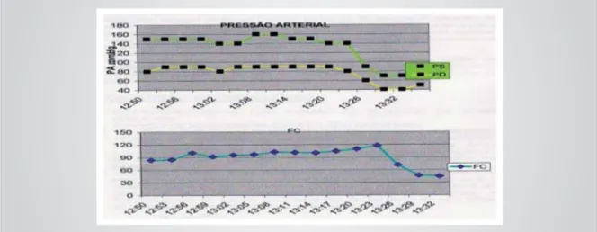
Fig. 1 – Tilt test with positive and mixed pattern response. Heart rate increase before the test is positive may suggest benefit with the use of betablockers. However, some patients with bradycardia during the TILT have their conditions worsened with these agents (pro-syncope). Pressão Arterial - Blood
of the neurally mediated syndrome involved, and it also helps to determine the therapeutic approach. Not every patient with vasovagal syncope require pharmacological treatment – only those individuals without prodromes and with history of recurrent trauma and syncope. The therapy can start with the adoption of hygiene and nutrition measures and the use of drugs (mineralocorticoids, betablockers, serotonin reuptake inhibitors, peripheral vasoconstrictors, either alone or associated with other drugs). Cardiac pacemakers are used only for treating highly refractory cases, such as patients with
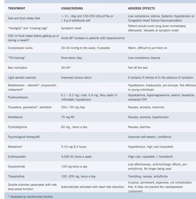
cardio-inhibitory syncope or carotid sinus hypersensitivity. The distinction between patients with vasovagal syncope and primary or secondary dysautonomia is crucial because this affects both treatment and prognosis, with increased morbidity for the second condition as well as increased mortality depending on the underlying etiology. Some patients present with Postural Orthostatic Tachycardia Syndrome (POTS) or the Chronic Fatigue Syndrome, conditions related to the neurally mediated syncope, and require specific therapeutic approach because of the major impact on their quality of life. (Figs. 2, 3 and 4). Table 2 – Treatment of the neurally mediated syndromes
TREATMENT USAGE/DOSING ADVERSE EFFECTS
Salt and fluid intake Diet !/GD\DQGP(TRI1DRU
Ĺ 3 g of additional salt
Low compliance, edema, Systemic Hypertension or Congestive Heart Failure Decompensation
“Handgrip” and “crossing legs” Symptom onset Patient should avoid lying down immediately
afterwards. Valuable at symptom onset
200 ml fluid intake before getting up or
during a meal47 Acute BP increase in patients with dysautonomia
Compression socks 30-40 mmHg to the waist, if possible Warm, difficult to put them on
“Tilt training” Once-twice /day Low compliance, trauma
Bed inclination 30-45º Fall off the bed
Light aerobic exercise Improved venous return It worsens if intense of in the absence of hydration
Betablockers – atenolol*, propranolol, metoprolol*
+\SRWHQVLRQEUDG\FDUGLDSURVtQFRSH1RWHIIHFWLYH in young individuals
Fludrocortisone ²PJPi[PJ9HU\XVHIXOLQ
orthostatic hypotension
Hypokalemia, hypomagnesemia, edema, headache, worsened CHF
Fluoxetine, paroxetine*, sertraline 20m / 50 mg /day Nausea, anorexia, insomnia
Venlafaxine 75 mg XR Nausea, anorexia, hypertension
Pyridostigmine 60 mg , twice a day Nausea, diarrhea
Psychological therapy48 Improved self-esteem, confidence
Midodrine* 5-10 mg Q 4 hours Hypertension, high cost (imported)
Erythropoietin 4,000 SC twice a week High cost, injectable, Ĺ hemotocrit
Disopiramide 150 mg twice a day Low effectiveness, anticholinergic effects,
pro-arrhythmia. No longer being used
Theophylline 100 -200 mg, twice a day Trembling, nausea, arrhythmia
Double-chamber pacemaker with rate
drop sense function Automatically activated with heart rate reduction.
Invasive, permanent, expensive, not complication-free. It does not prevent the vasodepressor component
* Assessed by randomized studies.
Fig. 2 –22-year old athlete patient, with history of recurrent syncope. The patient was admitted to a hospital with transient hemiparesis and disorientation. Neurological, hematological and immunological studies showed normal results. TILT test was clearly positive. Holter heart monitoring revealed inappropriate sinus tachycardia with mean heart rate of 103 and periods of increased sinus rhythm during orthostasis (compensatory). Major orthostatic intolerance with difficulty to ambulate. Therapeutic response to mineralocorticoid and betablocker. Control TILT test: normal. No recurrence in two-year follow-up. Pressão Arterial - Blood Pressure; Freqüencia Cardíaca - Cardiac Frequency; Tempo - Time
Fig. 3 –Elderly patient, with lipothymia and syncope with positive TILT test with dysautonomic response pattern. Supine hypertension is observed in patient previously normotense, with slow decline of blood pressure after tilting up to symptoms onset. The patient was diagnosed as having Shy-Drager Syndrome. Pressão Arterial - Blood Pressure; Freqüencia Cardíaca - Cardiac Frequency; Tempo - Time
Fig. 4 –33-year old patient, a teacher with recent history of seizures. Refractory to neurological treatment. TILT test (three times) showed severe cardio-inhibitory response with prolonged asystole during the 15-minute duration of the test, with seizures. The patient did not accept the implant of a permanent pacemaker. He is currently being treated with mineralocorticoids and serotonin reuptake inhibitor. Without any recurrence for a year.
1. Kapoor WN, Peterson J, Wieand HS, Karpft M. Diagnostic and prognostic implications of recurrences in patients with syncope. Am J 0HG
2. Engel GL. Physicologic stress, vasodepressor syncope and sudden GHDWK$QQ,QWHUQ0HG
3. Kapoor WN. Evaluation and outcome of patients with syncope. 0HGLFLQH
4. Bloomfield DM, Sheldon R, Grubb BP, Calkins H, Sutton R. A new treatment algorithm for vasovagal syncope and related disorders. Am -&DUGLRO$
5. Kapoor WN. An overview of the evaluation and management of syncope. In: Syncope: Mechanisms and management. 1ª ed. Armonk: )XWXUD3XEOLVKLQJ&RPSDQ\
6. Samoil D, Grubb BP. Neurally mediated syncope and serotonin UHXSWDNHLQKLELWRUV&OLQ$XWRQ5HV
7. Grubb BP, Olshanski B. Syncope: Mechanisms and management. $UPRQN1<)XWXUD3XEOLVKLQJ&RPSDQ\
8. Lofring V. Cardiovascular adjustments induced from the rostral cingulated gyrus: With specific reference to sympatho-inhibiting PHFKDQLVPV$FWD3DK\VLRO6FDQGVXSSO *UXEE%3*HUDUG*5RXVK.HWDO&HUHEUDOYDVRFRQVWULFWLRQGXULQJ
head upright tilt-induced vasovagal syncope: a paradoxic and XQH[SHFWHGUHVSRQVH&LUFXODWLRQ
10. Janasik D, Gomez C. Njemanze P, et al. Abnormalities in cerebral EORRGIORZDXWRUHJXODWLRQGXULQJWLOWLQGXFHGV\QFRSH3$&( 15: 542.
11. Kochiadakis GE, Papadimitriou EA, Marketou ME, et al. Is there any difference between young and older patients in vasovagal syncope? PACE 2004; 27 (10): 1371-7.
12. Consensus Committee of the American Autonomic Society and the American Academy of Neurology on the definition of orthostatic hypotension, pure autonomic failure and multiple system atrophy. 1HXURORJ\
13. Grubb, BP. Neurocardiogenic Syncope and related disorders of 2UWKRVWDWLF,QWROHUDQFH&LUFXODWLRQ
14. Savagem DD, Corwin L, Mc Gee DL, et al. Epidemiologic features of LVRODWHGV\QFRSH7KH)UDPLQJKDP6WXG\6WURNH
15. Rocha EA, Pachón JCM, Cuellar R, Medeiros P, Pachón E, Albornoz 1HWDO$YDOLDomR&OtQLFDH7HUDSrXWLFDGH3DFLHQWHVFRP6tQFRSHV 5HEODPSD
16. Hammill SC, Holmes DR, Wood DL, et al. Electrophysiological testing in the upright position: Improved evaluation of patients with rhythm GLVWXUEDQFHVXVLQJD7LOW7DEOH-$P&ROO&DUGLRO
17. Rocha EA, Farias R, Rocha A, Marques V, Paes FJN, Paes JN. 'LVDXWRQRPLDVHVtQGURPHGH6K\'UDJHU5HEODPSD 107.
18. Oldenburg O, Karliova M, Koeppen S, Weber F, Erbel R, Philipp T, et al. Shy-Drager syndrome: a rare cause of orthostatic hypotension. Dtsch 0HG:RFKHQVFKU
4XLQQ13:HQQLQJ*0DUVGHQ&'7KH6K\'UDJHUV\QGURPH:KDW GLG6K\DQG'UDJHUUHDOO\GHVFULEH"$UFK1HXURO
20. Gilman S, Quinn NP. The relationship of multiple system atrophy to sporadic olivopontocerebelar atrophy and other forms of late onset FHUHEHOODUDWURSK\1HXURORJ\
21. Keim MW, Momper R, Heck KF, Braun B, Hust MH. Neurocardiogenic syncopes in patients with implanted pacemakers. Dtsch Med :RFKHQVFKU
22. Goldestein DS, Pechnik S, Holmes C, et al. Associations between supine hypertension and orthostatic hypotension in autonomic failure. Hypertension 2003; 42 (2): 136-42.
23. Rowe PC, Bou-Holaigah I, Kan JS, et al. Is neurally mediated K\SRWHQVLRQDQXQUHFRJQL]HGFDXVHRIFKURQLFIDWLJXH"/DQFHW 345: 623-4.
24. Holmes GP, Kaplan JE, Gantz NM, et al. Chronic fatigue syndrome: A ZRUNLQJFDVHGHILQLWLRQ$QQ,QWHUQ0HG
25. Grubb BP, Klingenheben T. Postural orthostatic tachycardia syndrome 3276HWLRORJ\GLDJQRVLVDQGWKHUDS\0HG.OLQ 6.
26. Jacob G, Costa F, Shanon JR, Robertson RM, Wathen M, Stein M, et al. The neuropathic postural tachycardia syndrome. N Engl Med 2000; 343 (14): 1008-14.
27. Kenny RA, Ingram A, Bayless J, Sutton R. Head up tilt: A useful test IRULQYHVWLJDWLQJXQH[SODLQHGV\QFRSH/DQFHW
28. Brignole M, Alboni P, Benditt L, et al. Guidelines on the management, diagnosis and treatment of syncope. Eur Heart J 2004; 25: 2054-72.
0LOVWHLQ65H\HV:%HQGLWW'8SULJKWERG\WLOWIRUHYDOXDWLRQRI SDWLHQWVZLWKUHFXUUHQWV\QFRSH3$&(
30. Morillo CA, Klein G, Zandri S, Yee R. Diagnostic accuracy of a low dose
LVRSURWHUHQROKHDGXSWLOWSURWRFRO$P+HDUW-31. Benditt, DG, Fergunson DW, Grubb AP, et al. Tilt Table Testing for $VVHVVLQJ6\QFRSH-$&&
32. Hachul D, Scanavaca M, Sosa E. Does a Role Exist for Tilting-Guided Therapy in the Management of Neurocardiogenic Syncope? Arquivos Brasileiros de Cardiologia 2002; 78 (2): 167-71.
33. Raviele A, Giada F, Brignole M, Menozzi C, Marangoni E, Manzillo GF, et al. Comparison of diagnostic accuracy of sublingual niroglycerin test and low dose isoproterenol in patients with unexplained syncope. Am -&DUGLRO
34. Krahn A, Klein GJ, Yee R, Skanes AC. Randomized assessment of syncope trial: conventional diagnostic testing versus a prolonged monitoring strategy. Circulation 2001; 104: 46-51.
35. Calkins H. Pharmacologic approaches to therapy for vasovagal V\QFRSH$P-&DUGLRO4
36. Rocha EA, Farias R, Marques V, Rocha A, Pereira T, Scanavaca M, et al. 6tQGURPHGH-HUYHOO/DQJH1LHOVHQ5HEODPSD
37. Rocha EA, Marques V, Farias R, Rocha A, Rodrigues CRM, Paes )1HWDO3DSHOGRVGHVILEULODGRUHVLPSODQWiYHLVQDV7DTXLFDUGLDV Ventriculares Polimórficas. Reblampa 2001; 14 (3): 176.
38. Rocha EA, Pereira T, Farias R, Rocha A, Marques V, Ribeiro D, et DO0DUFDSDVVRVLPSODQWiYHLVFRPPRQLWRUHVGHULWPRFDUGtDFR 5HEODPSD
.UHGLHW39DQG.LMN1/LQ]HU0HWDO0DQDJHPHQWRIYDVRYDJDO syncope; controlling or aborting faints by leg crossing and muscle WHQVLQJ&LUFXODWLRQ
40. Ector H, Reybrounck T, Heidbuchel H, Gelliwig M, Van de Werf F. Tilt Training: A new treatment for recurrent neurocardiogenic syncope and VHYHUHRUWKRVWDWLFLQWROHUDQFH3$&(
41. Girolamo ED, Iorio CD, Leonzio L, Sabatini P, Barsotti A. Usefulness of a Tilt Training Program for the Prevention of Refractory Neurocardiogenic 6\QFRSHLQ$GROHVFHQWV&LUFXODWLRQ
42. Maclean AR, Allen EY. Orthostatic hypotension and orthostatic WDFK\FDUGLD7UHDWPHQWZLWK´KHDGXSµEHG-$0$ 2162.
43. Sheldon R. The Prevention of Syncope (POST) results. Paper presented at: Late breaking trials heart rhythm society meeting; May 22, 2004; San Francisco, Calif.
44. Calkins H. Think Twice Before Using Pacemaker therapy for Vasovagal Syncope. Journal Watch 2003; 2 (6).
45. Montanez A, Hennekens CH, Zebede J, Lamas GA. Pacing in vasovagal syncope: Qualitative overview and meta-analysis of the randomized trials. Heart Rhythm 2004; 1 (1S): 55.
46. Brignole M, Disertori M, Menozzi C, et al. The management of syncope referred for emergency to general hospitals with and without syncope XQLIDFLOLW\(XURSDFH
47. Jordan J, Shannon JR, Black BK, et al. The pressor resonse to water drinking in human: a sympathetic reflex? Circulation 2000; 101:
48. Giada F, Silvestri I, Rossillo A, et al. Psychiatric profile, quality of life and risk of syncopal recurrence in patients with tilt-induced vasovagal syncope. Europace; 2005 (in press)