INTRODUCTION
Keratoconus is generally a progressive, bilateral cone-like ectasia of the cornea that affects approximately 1 in 2,000 people in the gene-ral population, which is usually diagnosed in young adult patients(1). It
is characterized by localized corneal thinning and protrusion, which lead to high myopia and irregular astigmatism that reduces visual quality. The initial treatment of keratoconus is the use of glasses and/
or contact lenses(1). More advanced stages of the disease are treated
by various surgical procedures(2). However, the relief of keratoconus
symptoms using currently available treatment modalities may not be permanent, and it remains impossible to predict with certainty how long favorable results will persist.
The pathophysiology of keratoconus remains poorly defined, but its etiology is known to be multifactorial and it is an autosomal
dominant trait with variable phenotypic expression(2). It is associated
with many ophthalmic and systemic conditions, such as Down
syn-Keratoconus progression is not inhibited by reducing eyelid muscular force
with botulinum toxin A treatment: a randomized trial
A progressão do ceratocone não é inibida reduzindo a força palpebral com o uso da toxina
botulínica tipo A: ensaio clínico randomizado
AdimArAdA CAndelAriA renesto1, teissy HentonA osAki1, midori HentonA osAki1, Flávio e. HirAi1, mAuro CAmpos1
Submitted for publication: August 4, 2016 Accepted for publication: October 26, 2016
1 Department of Ophthalmology and Visual Sciences, Escola Paulista de Medicina (EPM), Universidade
Federal de São Paulo (UNIFESP), São Paulo, SP, Brazil.
Funding: No specific financial support was available for this study.
Disclosure of potential conflicts of interest: None of the authors have any potential conflict of interest to disclose.
Corresponding author: Adimara da Candelaria Renesto. Rua Botucatu, 821 - 2o andar - São Paulo,
SP - 04023-062 - Brazil - E-mail: [email protected]
Approved by the following research ethics Committee: Comitê de Ética em Pesquisa da Universi-dade Federal de São Paulo/Hospital São Paulo (#87051/2012).
Clinicaltrials.gov: NCT01691651
ABSTRACT
Purpose: To evaluate whether reducing eyelid muscular force through the administra-tion of botulinum toxin type A (BTX-A) to the orbicularis oculi muscles of patients with keratoconus affected corneal parameters indicative of disease progression. Methods: In this prospective parallel randomized clinical trial, 40eyes of 40 patients with keratoconus were randomized into equally sized control and BTX-A groups. Patients in the BTX-A group received subcutaneous BTX-A injections into the orbicularis muscle. The control group received no intervention. Palpebral fissure height, best spectacle-corrected visual acuity (BSCVA), and corneal topographic parameters were evaluated at baseline and at 3-, 6-, 12-, and 18-month follow-ups. Results: The mean ± standard deviation vertical palpebral fissure heights were 9.74 ± 1.87 mm and 9.45 ± 1.47 mm at baseline in the control and BTX-A groups, respectively, and 10.0 ± 1.49 mm and 9.62 ± 1.73 mm at 18 months, with no sig-nificant difference between the groups (p=0.337). BSCVA values were 0.63 ± 0.56 and 0.60 ± 0.27 at baseline in the control and BTX-A groups (p=0.643), and 0.52 ± 0.59 and 0.45 ± 0.26 at 18 months, again with no significant difference between the groups (p=0.452). In addition, there were no statistical differences between the groups at 18 months for the three keratometry topographic parameters: flattest (K1), steepest (K2), and mean (Km) keratometry (p=0.562).
Conclusion: BTX-A inhibition of eyelid force generation did not result in detec-table changes in corneal parameters in keratoconic patients during 18 months of follow-up.
Keywords: Corneal diseases; Keratoconus; eyelids; Botulinum toxins/therapeutic use
RESUMO
Objetivo: Avaliar se a administração da toxina botulínica tipo A (BTX-A) no músculo orbicular de pacientes com ceratocone a fim de reduzir a força muscular palpebral pode alterar os parâmetros corneanos indicativos de progressão da doença. Métodos: Ensaio clínico randomizado paralelo prospectivo. Quarenta olhos de 40 pacientes, randomizados em grupo controle ou grupo BTX-A na razão de 1:1. Pa-cientes do grupo BTX-A foram submetidos à injeção subcutânea da toxina botulínica tipo A no músculo orbicular. Os pacientes do grupo controle não sofreram nenhuma intervenção. Foram avaliados a medida da fenda palpebral, melhor acuidade visual corrigida, e topografia corneana nos momentos pré-operatório, e aos 3-,6-,12-, e 18 meses de seguimento.
Resultados: Média ± DP (desvio padrão) da fenda palpebral no pré-operatório nos grupos controle e BTX-A foram 9,74 ± 187 e 9,45 ± 1,47 mm, respectivamente; aos 18 meses, a média da altura da fenda palpebral vertical nos grupos controle e BTX-A foram 10,0 ± 1,49 mm e 9,62 ± 1,73 mm, respectivamente, sem diferença significante entre os grupos (p=0,337). A média pré-operatória da melhor acuidade visual corri-gida nos grupos controle e BTX-A foram 0,63 ± 0,56 e 0,60 ± 0,27, respectivamente (p=0,643); aos 18 meses, a média nos grupos controle e BTX-A foram 0,52 ± 0,59 e 0,45 ± 0,26, respectivamente, sem diferença significante entre os grupos (p=0,452). Não houve diferença estatística entre os grupos aos 18 meses para todos os parâ-metros topográficos ceratométricos avaliados, mais plano-(K1), mais curvo-(K2), e ceratometria média Km (p≥0,562).
Conclusão: A inibição da força muscular palpebral pela toxina botulínica tipo A não causou alterações detectáveis nos parâmetros corneanos em pacientes com ceratocone, com 18 meses de seguimento.
drome, eye rubbing, mitral valve prolapse, collagen vascular disease,
and a history of contact lens wear(2). Other conditions include eyelid
diseases such as blepharoptosis(3) and floppy eyelid syndrome(4).
Keratoconus patients present with excessive eye blinking, and usually develop a routine of chronic eye rubbing with consequent excessive manipulation of the periocular area. Augmented pressure to the eyelids may cause further topographic changes to the cor-nea(5,6), and eyelid muscular force can translate to the ocular surface
and exacerbate symptoms. It was therefore hypothesized that redu-cing eyelid muscular force may provide a novel treatment option for keratoconus that inhibited the progression of damage to the cornea.
The use of botulinum toxin type A (BTX-A) in medicine was first
reported in 1980 for treating strabismus(7). It blocks the release of
acetylcholine into the neuromuscular junction, leading to temporary
muscle weakness or paralysis(8). BTX-A is now a front-line treatment
option for focal dystonias such as essential blepharospasm and he-mifacial spasm, and it has become a major therapeutic drug with
va-luable applications in diverse medical subspecialties(8). The purpose
of this study was to evaluate, as a potential novel therapy for patients with keratoconus, whether reducing eyelid muscular force through the administration of BTX-A into the orbicularis oculi muscles could prevent the progression of corneal damage.
METHODS
D
ESIGNThis parallel randomized clinical trial was conducted between September 2012 and October 2014 in the Department of Ophthal-mology and Visual Sciences, Federal University of São Paulo, Brazil. The study was approved by the Institutional Ethics Committee and followed the tenets of the Declaration of Helsinki. All patients provi-ded written informed consent. This manuscript has been registered (NCT01691651) and is publicly available at www.clinicaltrials.gov.
P
ARTICIPANTSPatients were enrolled if they met the following inclusion criteria: documented keratoconus, best spectacle-corrected visual acuity (BSCVA) that could be measured by refraction, no major comorbi-dities such as diabetes, and aged 10-40 years. Exclusion criteria were having only one functional eye, previous ocular surgery, eyelid margin disease that did not respond to treatment (e.g., meibomian gland dysfunction and blepharitis), a known allergy to BTX-A or other medication components, a history of herpetic keratitis, concurrent corneal infection, pregnancy or nursing, and other ocular diseases that could affect visual acuity.
S
AMPLESIZEANDRANDOMIZATIONThe two main outcomes considered in this study were the dimen-sions of the palpebral fissure and keratometry measurements; the sample size was calculated on the basis of these. From a pilot study per formed in our department, we expected a mean ± standard devia-tion (SD) change in the height of the palpebral fissure of 2.0 ± 1.5 mm and an astigmatism change of 1.0 ± 0.8 diopters (D). We calculated that a sample size of 20 individuals/group would provide 90% power to de-tect these magnitudes of outcome differences in the present study.
Randomization was performed using a computer-generated randomization table (Stata v.11, Stata Corp, College Station, TX). The par -ticipants were allocated into either the control or the BTX-A injection group in a 1:1 ratio. Due to the design of this study (treatment versus no treatment), the examiner was not masked to the type of treatment during examination, although the statistician was masked during data analysis.
M
EASURESAt the baseline and post-treatment follow-up examinations at 3, 6, 12, and 18 months, all participants underwent the following
assessments: 1) uncorrected visual acuity (UCVA); 2) BSCVA; 3) mani-fest refraction in a bright environment; 4) slit-lamp biomicroscopy; 5) measurement of the palpebral fissure using ImageJ software (v.1.46s, National Institutes of Health, Bethesda, MD, USA); 6) corneal
topography and tomography (Allegro Oculyzer®, Alcon Laboratories,
Fort Worth, TX, USA) and Pentacam® optical topography assessment
(Oculus Optikgerate GmbH, Wetzlar, Germany); 7) Goldmann appla-nation tonometry (GAT; Haag-Streit, Konig, Switzerland); and 8) ocular fundus examination. All evaluations were performed by the same examiner (ACR).
M
EASUREMENTOFTHEPALPEBRALFISSUREImageJ is a public domain Java image processing program that can display, edit, analyze, process, save, and print images. It can also calculate the area and pixel value statistics of user-defined selections, measure distances and angles, and create density histograms and line profile plots.
An eyelid examination was performed with the eye image regis-tration of patients in the primary position of gaze with a portable
Sony® Cyber-shot digital camera attached to a monopod. To ensure
the standardization of all measurements, photographs were taken with the participant positioned on a chin rest (for a slit-lamp) with a ruler attached at the forehead support. The same slit-lamp was used in all evaluations, thereby maintaining the same distance from the digital camera to the chin rest. The palpebral fissure height was drawn as a vertical line from the upper to the lower eyelid margin, passing through the pupil. Multiple images were captured from the study eye, and the mean measurement from the selected images was calculated.
I
NJECTIONOFBOTULINUMTOXINTYPEA
Patients in the BTX-A injection group were treated with an
in-jection of onabotulinumtoxinA (Botox®, Allergan, Irvine, CA, USA) to
the palpebral portion of the orbicularis oculi muscle. Reconstitution of the drug and its application were performed by a single investi-gator (MHO) according to a standardized protocol. Immediately be fore appli cation, each vial was reconstituted with normal saline without preservatives to a final BTX-A concentration of 50 U/mL. The participant was seated and received a total of 12.0 U of BTX-A per application, injected using an insulin syringe in equal doses into the palpebral portion of the orbicularis oculi muscle of the upper and lower eyelids. BTX-A was applied twice: after the baseline measure-ments, and then six months after the first injection.
S
TATISTICALANALYSISThe intention-to-treat principle was used in the analysis. Pre- and post-treatment analyses were conducted using the Wilcoxon signed-rank test. Between-group analyses used the Mann-Whitney U-test for continuous variables and Fisher’s exact test for categorical variables measured at each follow-up visit. All analyses were performed using Stata v.11 software, and p-values <0.05 were considered statistically significant. Data are presented as mean ± SD unless otherwise noted.
RESULTS
A total of 40 individuals (40 eyes) were enrolled at baseline. Three participants were lost due to voluntary non-participation in some of the follow-up visits, and so only 37 eyes of 37 participants were included for some of the follow-up analyses.
All of the eyes were graded according to the Amsler-Krumeich
classification(9), based on each participant’s refraction, mean central
P
ARTICIPANTDEMOGRAPHICSParticipant demographics, study eye laterality, baseline palpebral height, corneal topography, and spherical equivalent (SE) were simi-lar between the two study groups (Table 1).
V
ISUALACUITYBoth UCVA and BSCVA, measured using a logMAR chart, were similar between the two study groups at baseline and at all follow-up time points (Figure 1).
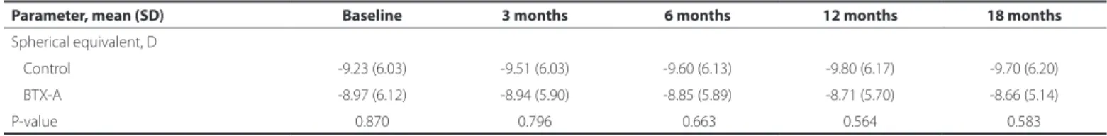
R
EFRACTIVERESULTSThe mean baseline SE was -9.23 ± 6.03 D in the control group and -8.97 ± 6.12 D in the BTX-A group (p=0.870; Tables 1 and 2). At 18 months, the mean SE was -9.70 ± 6.20 D in the control group and -8.66 ± 5.14 D in the BTX-A group, with no statistically significant difference between the groups (p=0.583; Table 2).
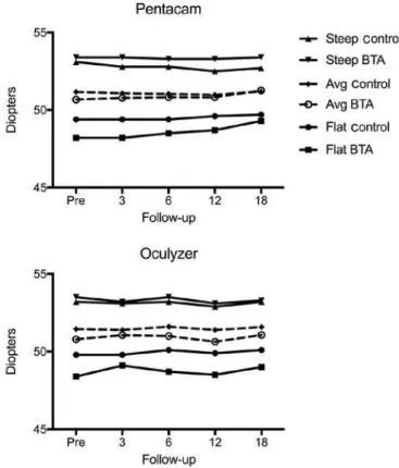
T
OPOGRAPHICRESULTSAfter adjusting for baseline levels, there was no statistically signi-ficant difference between the groups at the 18-month follow-up for
all three topography parameters assessed with the Pentacam®
(flat-test keratometry (K1), p=0.795; steepest keratometry (K2), p=0.562;
and mean keratometry (Km), p=0.903), and with the Oculyzer® (K1,
p=0.783; K2, p=0.783; and Km, p=0.842) (Figure 2).
P
ALPEBRALFISSUREThe mean height of the palpebral fissure at baseline was 9.74 ± 1.87 mm in the control group and 9.45 ± 1.47 mm in the BTX-A group (p=0.560; Tables 1 and 3). At 18 months, the respective palpebral fissure measurements were similar at 10.0 ± 1.49 mm and 9.62 ± 1.73 mm in the control and BTX-A groups, respectively (p=0.337; Table 3).
P
ACHYMETRYRESULTSTable 4 shows the pachymetry results at the central and thinnest parts of the corneas. There was no statistically significant difference between the two study groups at 18 months of follow-up in any parameter (p=0.669 and p=0.464 for the central and thinnest
cor-neal regions measured by Pentacam® pachymetry, and p=0.748 and
p=0.502 for the central and thinnest areas measured by Oculyzer®
pachymetry).
T
ONOMETRYRESULTSThe tonometry data were evaluated by GAT. Intraocular pressure (IOP) did not differ significantly between the groups at 18 months (p=0.322; Table 5).
C
OMPLICATIONSNo participant in the BTX-A group presented early or late compli-cations following the BTX-A applicompli-cations, including lagophthalmos, diplopia, or blepharoptosis.
DISCUSSION
Various factors can result in the deterioration of keratoconus, in -cluding ultraviolet-B light, atopy, mechanical eye rubbing, and
im-properly fitting contact lenses(2). Researchers have studied genetic,
histopathologic, biomechanical, and inflammatory aspects of the disease to elucidate keratoconus etiology and improve the currently
available treatments(10,11). Elevated tension within the eyelid can be
transmitted to the juxtaposed corneal surface, thereby altering the
corneal topography(5,12); thus, reducing the muscular force of the
eye-lid may result in changes to corneal measurements in keratoconus patients. With the goal of developing new treatment options for ke-ratoconus, the present study evaluated whether BTX-A injection into
Table 1. Comparison of baseline characteristics between the botuli-num toxin-A injection and control groups
Parametera Control (n=21) BTX-A (n=19) P-value Age, years -25.95 (07.33) -21.31 (07.60) 0.052
Sex, n (%) 0.087
Female -11.00 (52.40) -05.00 (26.40)
Male -10.00 (47.60) 14.00 (73.60)
Eye, n (%) 0.357
Right -11.00 (52.40) -12.00 (63.10)
Left -10.00 (47.60) -07.00 (36.90)
Palpebral fissure, mm -09.74 (01.87) -09.45 (01.47) 0.560 Steepest K, Pentacam, D -53.09 (07.29) -53.41 (05.44) 0.714
Flattest K, Pentacam, D -49.41 (05.69) -48.23 (05.19) 0.645
Mean K, Pentacam, D -51.17 (06.38) -50.67 (05.21) 0.989 Steepest K, Oculyzer, D -53.16 (07.24) -53.53 (05.04) 0.694
Flattest K, Oculyzer, D -49.86 (05.88) -48.40 (05.28) 0.606
Mean K, Oculyzer, D -51.45 (06.45) 50.80 (05.11) 0.989 Spherical equivalent, D 0-9.23 (06.03) 0-8.97 (06.12) 0.870 a= values shown are mean (standard deviation), unless noted as “n (%).” BTX-A= botuli-num toxin-A; K= keratometry measurements made with a pentacam® or Oculyzer® de vice; D= diopters.
Figure 1. Uncorrected visual acuity (UCVA) and best corrected visual acuity (BCVA) on a logMAR chart.
Table 2. Comparison of the spherical equivalent measurements between the botulinum toxin-A injection and control groups
Parameter, mean (SD) Baseline 3 months 6 months 12 months 18 months
Spherical equivalent, D
Control -9.23 (6.03) -9.51 (6.03) -9.60 (6.13) -9.80 (6.17) -9.70 (6.20)
BTX-A -8.97 (6.12) -8.94 (5.90) -8.85 (5.89) -8.71 (5.70) -8.66 (5.14)
P-value 0.870 0.796 0.663 0.564 0.583
BTX-A= botulinum toxin-A.
Figure 2. Topographic corneal indexes (steepest, mean, and lattest keratometry) by Pentacam® and Oculyzer® in the control and botulinum toxin-A injection groups.
Table 3. Comparison of the heights of the palpebral issure between the botulinum toxin-A injection and control groups
Parameter, mean (SD) Baseline 3 months 6 months 12 months 18 months
Palpebral fissure, mm
Control 9.74 (1.87) 9.14 (1.26) 9.84 (1.19) 9.66 (1.75) 10.06 (1.49)
BTX-A 9.45 (1.47) 9.79 (1.28) 9.82 (1.51) 9.84 (1.59) 0 9.62 (1.73)
SD= standard deviation; BTX-A= botulinum toxin A.
the orbicularis oculi muscles of keratoconus patients improved cor-neal topographic parameters by reducing the eyelid muscular forces that are translated to the ocular surface and exacerbate symptoms.
However, the administration of two BTX-A doses over 6 months resulted in no changes to visual acuity and refraction in our kerato-conus cohort. Previous studies have reported no success in treating keratoconus by cross-linking corneal collagen matrices to stabilize
the ocular tissue(13,14), an approach that also brought no changes in
UCVA, BSCVA, SE, or keratometry. Another keratoconus intervention involves implanting plastic intracorneal ring segments to stabilize the cornea, but this brings the risk of ocular complication, requiring ca reful selection of patients appropriate for the procedure(2,15,16).
Se-veral factors contribute to the development of corneal astigmatism, including age, sex, tear film insufficiency, IOP, and physical pressure
exerted by the eyelids(17). In a 2015 report by members of our research
team, the magnitude of astigmatism was significantly reduced and palpebral fissure height significantly increased in the affected eyes
of hemifacial spasm patients treated with BTX-A(18). In that study,
BTX-A treatment temporally alleviated eyelid tension, thereby poten-tially reducing the transmission of physical forces from the eyelids to the cornea. In the present study, no statistically significant differences were observed in the corneal topographic parameters and palpebral fissure dimensions of keratoconus eyes during the 18 months following BTX-A administration. Usually, BTX-A effects appear within 1-2 days, peak at 1-2 weeks, and gradually decline after 3-6 months, depending
on the patient’s clinical condition(8). We observed no differences in
palpebral fissure measurements 3 months after the first and second BTX-A applications, probably because the tension exerted by the eyelid on the cornea in keratoconus patients is not as high as it is in hemifacial spasm patients. Thus, BTX-A application to the orbicularis oculi muscles of keratoconus patients was not associated with significant changes in corneal astigmatism or palpebral fissure dimensions.
Methodologies for evaluating eyelid tension are not standardized and vary between studies, complicating the comparison of results
Table 4. Comparison between the botulinum toxin-A injection and control groups of corneal pachymetric measurements made using the Penta-cam® and Oculyzer® devices
Parameter, mean (SD) Baseline 3 months 6 months 12 months 18 months
Pentacam®
Central position, µm
Control 447.7 (47.6) 451.4 (42.7) 451.0 (50.3) 445.1 (46.4) 447.0 (47.9)
BTX-A 460.4 (45.7) 447.1 (48.7) 464.1(45.0) 453.4 (60.0) 454.7 (53.9)
Thinnest position, µm
Control 419.6 (53.0) 421.8 (44.2) 423.5 (55.9) 416.0 (51.1) 418.5 (52.2)
BTX-A 433.1 (46.9) 430.0 (48.9) 437.8 (45.6) 431.1 (57.3) 425.6 (52.4)
Oculyzer®
Central position, µm
Control 461.9 (46.4) 464.4 (46.8) 464.0 (50.0) 459.7 (52.5) 463.4 (47.8)
BTX-A 469.6 (49.1) 473.9 (53.8) 471.4 (59.3) 466.3 (66.1) 459.5 (63.4)
Thinnest position, µm
Control 430.7 (55.1) 430.7 (54.4) 432.4 (52.6) 430.7 (54.7) 433.6 (49.8)
BTX-A 439.8 (51.9) 443.6 (52.4) 442.4 (53.7) 432.6 (67.6) 432.5 (63.1)
from different groups. The present study indirectly evaluated the re-duction in eyelid muscular force due to BTX-A application to the orbi-cularis oculi muscles by studying the subsequent effects on the corneal topographic parameters. Other research groups have described using a lid tensiometer instrument to assess the generation of eyelid
muscu-lar force(19,20). One report indicated that, under normal circumstances,
eyelid tension was unlikely to adversely affect the shape and power of
the cornea(19). In addition, it has been noted that no difference in lid
tension exists between Asians and Caucasians(20) even though Asians
are at increased predisposition to developing keratoconus. In contrast, studies that evaluated corneal astigmatism changes after BTX-A injec-tion in patients with blepharospasm concluded that eyelid-derived
forces may play an important role in modifying corneal curvature(17,21).
The pachymetry results for the central and thinnest regions of the cornea remained similar between the groups throughout the 18 months of follow-up. This is not surprising, because we did not admi-nister BTX-A directly into the corneal tissue. Pachymetry was assessed using two different devices, but both were based on the same principle (Scheimpflug tomography). This approach has good reproducibility in differentiating normal corneas from subclinical and symptomatic keratoconus cases(22).
GAT is the gold standard for tonometry and is the most frequently used technique for IOP measurement. Eyes with corneal pathologies can present challenges for measuring IOP because of changes to the
corneal thickness, rigidity, and curvature(23). However, newer
tonome-ters such as the Pascal dynamic contour tonometer (Ziemer Ophthal-mic Systems AG, Bern, Switzerland) and the Ocular Response Analyzer and TonoPen XL (Reichert Ophthalmic Instruments, Depew, NY, USA)
have better diagnostic accuracy than GAT for keratoconic corneas(23).
We are not aware of any previous reports that have used BTX-A to reduce eyelid muscular force in patients with keratoconus. Although
several studies(17-20) have confirmed the influence of eyelid tension on
corneal shape, it appears that eyelid tension in keratoconus patients is no higher than for other conditions such as hemifacial spasm and
blepharoptosis(18,24). In the present study, BTX-A application did not
result in a significant change to corneal parameters. It remains unclear whether BTX-A application to the orbicularis oculi muscles could be a useful adjuvant approach for controlling the progression of kerato-conus. Corneal changes after the application of BTX-A in orbicularis oculi muscles, if there are any, take longer to be detected than earlier
eyelid morphometric changes(18); our follow-up period may therefore
not have been long enough to detect associated corneal changes in patients with keratoconus. Furthermore, keratoconic corneas may present structural alterations that respond differently from the corneas of patients with hemifacial spasm.
We recognize potential limitations in this study. The number of BTX-A applications was limited, and the follow-up period was rela-tively short. Future studies of keratoconus patients with a longer follow-up period and an increased number of BTX-A applications may help to elucidate whether BTX-A application in the orbicularis oculi muscle could be a valuable adjuvant treatment to help control the progression of keratoconus to some degree.
Keratoconus etiology and pathogenesis mechanisms remain unclear. The recent Global Consensus on Keratoconus and Ectatic Di
-seases(25) concluded that keratoconus was a multifactorial disease with
genetic, biochemical, biomechanical, and environmental components. The panelists found that the most important objectives of nonsurgical treatment were to halt progression and bring about visual rehabilitation.
The past two decades, in particular, have seen exciting new de-velopments that, for the first time, promise to alter the natural history
of keratoconus favorably to benefit patients. Scientific interest in keratoconus is likely to remain high in the foreseeable future, to avoid iatrogenic ectasia, unmask subclinical disease, and expand the rapeutic options(26).
In conclusion, BTX-A administration to the orbicularis oculi muscles of keratoconus patients did not cause detectable changes in corneal parameters secondary to reduced eyelid muscular force during 18 months of follow-up.
REFERENCES
1. Rabinowitz YS. Keratoconus. Surv Ophthalmol. 1998;42(4):297-319.
2. Romero-Jiménez M, Santodomingo-Rubido J, Wolffsohn JS. Keratoconus: a review. Cont Lens Anterior Eye. 2010;33(4):157-66.
3. Kim T, Khosla-Gupta B, Debacker C. Blepharoptosis-induced superior keratoconus. Am J Ophthalmol. 2000;130(2):232-4.
4. Pihlblad MS, Schaefer DP. Eyelid laxity, obesity, and obstructive sleep apnea in kera-toconus. Cornea. 2013;32(9):1232-6.
5. Lieberman DM, Grierson JW. The lids influence on corneal shape. Cornea. 2000;19(3): 336-42.
6. Collins MJ, Buehren T, Trevor T, Statham M, Hansen J, Cavanagh DA. Factors influen-cing lid pressure on the cornea. Eye Contact Lens. 2006;32(4):168-73.
7. Scott AB. Botulinum toxin injection into extraocular muscles as an alternative to strabismus surgery. Ophthalmology. 1980;87(10):1044-9.
8. Dutton JJ, Fowler AM. Botulinum toxin in ophthalmology. Surv Ophthalmol. 2007; 52(1):13-31.
9. Krumeich JH, Daniel J, Knülle A. Live-epikeratophakia for keratoconus. JCataract Re-fract Surg. 1998;24(4):456-63.
10. Kenney MC, Brown DJ. The cascade hypothesis of keratoconus. Cont LensAnterior Eye. 2003;26(3):139-46.
11. McMonnies CW. Inflammation and keratoconus. Optom Vis Sci. 2015;92(2):35-41. 12. Wilson G, Bell C, Chotai S. The effect of lifting the lids on corneal astigmatism. Am J
Optom Physiol Opt.1982;59(8):670-4.
13. Grewal DS, Brar GS, Jain R, Sood V, Singla M, Grewal SPS. Corneal collagen crosslinking using riboflavin and ultraviolet-A light for keratoconus. J CataractRefract Surg. 2009; 35(3):425-32.
14. Koller T, Mrochen M, Seiler T. Complication and failure rates after corneal crosslinking. J Cataract Refract Surg. 2009;35(8):1358-62.
15. Hofling-Lima AL, Branco BC, Romano AC, Campos MQS, Moreira H, Miranda D, et al. Corneal infections after implantation of intracorneal ring segments. Cornea. 2004;23(6):547-9. 16. Coskunseven E, Kymionis GD, Tsiklis NS, Atun S, Arslan E, Siganos CS, et al. Complica-tions of intrastromal corneal ring segment implantation using a femtosecond laser for channel creation: a survey of 850 eyes with keratoconus. Acta Ophthalmol. 2011; 89(1):54-7.
17. Moon NJ, Lee HI, Kim JC. The changes in corneal astigmatism after botulinum toxin-A injection in patients with blepharospasm. J Korean Med Sci. 2006;21(1):131-5. 18. Osaki T, Osaki MH, Osaki TH, Hirai FE, Nallasamy N, Campos M. Influence of involuntary
eyelid spasms on corneal topographic and eyelid morphometric changes in patients with hemifacial spasm. Br J Ophthalmol. 2015 Nov 5. pii: bjophthalmol-2015-307272. doi: 10.1136/bjophthalmol-2015-307272.
19. Lydon D, Tait A. Lid-pressure: its measurement and probable effects on the shape and form of the cornea-rigid contact lens system. Contact LensAnt Eyes. 1988;11(1):11-22. 20. Ehrmann K, Francis I, Stapleton F. A novel instrument to quantify the tension of upper
and lower eyelids. Cont Lens Anterior Eye. 2001;24(2):65-72.
21. Gunes A, Demirci S, Koyuncuoglu HR, Tok L, Tok O. Corneal and tear film changes after botulinum toxin-A in blepharospasm or hemifacial spasm. Cornea.2015;34(8):906-10. 22. Miháltz K, Kovács I, Takács A, Nagy ZZ. Evaluation of keratometric, pachymetric, and ele-vation parameters of keratoconic corneas with pentacam. Cornea. 2009;28(9):976-80. 23. Mollan SP, Wolffsohn JS, Nessim M, Laiquzzaman M, Sivakumar S, Hartley S, et al.
Accu-racy of Goldmann, ocular response analyser, Pascal and TonoPen XL tonometry in keratoconic and normal eyes. Br J Ophthalmol. 2008;92(12):1661-5.
24. Holck DE, Dutton JJ, Wehrly SR. Changes in astigmatism after ptosis surgery measured by corneal topography. Ophthal Plast Reconstr Surg. 1998;14(3):151-8.
25. Gomes JAP, Tan D, Rapuano CJ, Belin MW, Ambrósio Jr R, Guell JL, et al, the Group of Panelists for the Global Delphi Panel of Keratoconus and Ectatic Diseases. Global Con sensus on Keratoconus and Ectatic Diseases. Cornea. 2015;34(4):359-69. 26. Vazirani J, Basu S. Keratoconus: current perspectives. Clin Ophthalmol.2013;7:2019-30.
Table 5. Comparison of Goldmann applanation tonometry measurements between the botulinum toxin-A injection and control groups
Parameter, mean (SD) Baseline 3 months 6 months 12 months 18 months
GAT, mmHg
Control 9.1 (2.3) 8.8 (2.5) 8.9 (2.7) 9.3 (3.0) 9.1 (1.8)
BTX-A 9.3 (2.2) 8.4 (2.5) 8.6 (2.5) 7.6 (2.4) 8.3 (3.1)