Original Article
Artigo Original
Correspondence address: Angela Ruviaro Busanello-Stella Rua General Neto, 484, Santa Maria (RS), Brasil, CEP: 97050-240. E-mail: angelafonoaudiologia@yahoo. com.br
Received: 05/13/2014 Accepted: 11/03/2014
Study carried out at the Orofacial Motricity Laboratory of the Speech-Language Pathology and Audiology Department of Universidade Federal de Santa Maria – UFSM – Santa Maria (RS), Brazil.
(1) Special Coordination of Speech-Language Pathology and Audiology, Universidade Federal de Santa Catarina – UFSC – Florianópolis (SC), Brazil.
(2) Department of Physical Therapy and Graduate Program in Human Communication Disorders, Universidade Federal de Santa Maria – UFSM – Santa Maria (RS), Brazil.
(3) Undergraduate program in Speech-Language Pathology and Audiology and Graduate Program in Human Communication Disorders, Universidade Federal de Santa Maria – UFSM – Santa Maria (RS), Brazil. Financial support: Coordenação de Aperfeiçoamento de Pessoal de Nível Superior – CAPES. Conlict of interests: nothing to declare.
Angela Ruviaro Busanello-Stella1
Ana Paula Blanco-Dutra1
Eliane Castilhos Rodrigues Corrêa2
Ana Maria Toniolo da Silva3
Descritores
Fadiga Muscular Eletromiograia Criança Boca Respiração Bucal Face
Keywords
Muscular Fatigue Electromyography Child Mouth Mouth Breathing Face
Electromyographic fatigue of orbicular oris muscles
during exercises in mouth and nasal breathing children
Fadiga eletromiográica dos músculos orbiculares da boca
durante exercícios em crianças respiradoras orais e nasais
ABSTRACT
Purpose: To investigate the process of fatigue in orbicularis oris muscles by analyzing the median frequency of electromyographic signal and the referred fatigue time, according to the breathing mode and the facial pattern. Methods: The participants were 70 children, aged 6 to 12 years, who matched the established criteria. To be classiied as 36 nasal-breathing and 34 mouth-breathing children, they underwent speech-language, otorhinolaryngologic, and cephalometric evaluation. For the electromyographic assessment, the children had to sustain lip dumbbells weighing 40, 60, and 100 g and a lip exerciser, until the feeling of fatigue. Median frequency was analyzed in 5, 10, 15, and 20 seconds of activity. The referred time of the feeling of fatigue was also recorded. Data were analyzed through the analysis of variance — repeated measures (post hoc Tukey’s test), Kruskal-Wallis test, and Mann-Whitney U-test. Results: A signiicant decrease in the median frequency from 5 seconds of activity was observed, independently from the comparison between the groups. On comparison, the muscles did not show signiicant decrease. The reported time for the feeling of fatigue was shorter for mouth-breathing individuals. This feeling occurred after the signiicant decrease in the median frequency. Conclusion: There were signals that indicated myoelectric fatigue for the orbicularis oris muscles, in both groups analyzed, from the irst 5 seconds of activity. Myoelectric fatigue in the orbicularis oris muscles preceded the reported feeling of fatigue in all groups. The account for fatigue time was inluenced by only the breathing pattern, occurring more precociously in mouth-breathing children.
RESUMO
INTRODUCTION
Speech-language and audiological intervention, specii-cally in orofacial motricity, is based on action on structural and functional aspects of the orofacial and cervical regions. This intervention covers from the aspect of patient awareness to the rehabilitation of the changed aspects in structural and func-tional terms. Therefore, among the methods used are isometric and isotonic speech exercises(1), and deining the appropriate means of performing these exercises is key to achieving the treatment’s goal(2).
When it comes to rehabilitation in mouth-breathing and the various changes due to this pathology, the choice of appropri-ate exercise becomes even more important(3,4). These changes may be related to the growth of the face; the muscular tonus; posture and appearance(5); and the dificulty in performing other functions, such as swallowing, chewing, and speech(4,6), as well as basic movements, such as closing the mouth(3).
Thus, it is fundamental to understand how the mouth-breath-ing individual responds to exercises. In this context, there are not only individual strategies, as some authors recommend(2), but also standardized strategies through instruments that can be bought, such the lip dumbbells and the exerciser. These devices act on the orbicularis oris muscles of the mouth, aiming at its toning and strengthening(7).
However, in literature, little information is available on the prescription of muscle exercises(1) or on the response shown by patients to therapeutic approaches, especially with regard to muscle condition(2,8). One good example can be found in the recommended time for each exercise, which ranges between 10 seconds(1), 15-20 seconds(9), or even 30 seconds of static muscle contractions(7). The prescribed amount of exercise is too little explored. The few reports in the literature range from 10 to 20 repetitions of the exercises in each series(7,9) and between 1, 3(1), and 4 repetitions per day(7,9).
There is no consensus in the literature on what would be the best parameter to deine the choice of the prescribed exercises, but it is believed that the study of muscle fatigue can contribute to the improvement of this subject. This is because the continu-ous performance of muscle exercises, even with the presence of muscle fatigue, might interfere with the onset of muscular aches and pains(10), as well as with motor performance(11). According to the literature, muscle fatigue corresponds to the muscle’s inability to maintain high levels of strength in a given time(10-12). It can be considered as a natural mechanism of the muscle, being more easily measured in isometric exercises, due to the interruption of blood low that they cause, highlighting the metabolic changes(13).
In this context, objective methods(14), such as cephalom-etry(15) and surface electromyography (EMG)(13), and system-atized protocols, such as MBGR(5,16), may contribute to the investigation of the factors that could potentially interfere with muscle fatigue, whether muscle-related and/or functional fac-tors, such as the breathing mode, or at structural level, such as facial growth pattern. Cephalometry allows the assessment of craniofacial development of the type of occlusion and facial typology(15). Electromyography, in turn, allows the investigation
of fatigue by means of frequency spectrum, through analysis of the median frequency (MF)(17). Its decline for low values during contractions is considered as an objective measure of the muscle fatigue process, and relects mainly the peripheral fatigue(18).
Thus, this study aimed to evaluate fatigue of the orbicularis oris muscles of the mouth, through the analysis of the median frequency of the electromyographic signal and the reported time of fatigue, considering the breathing mode and the facial growth pattern.
METHODS Sample
The study sample comprised children mainly from public schools in a city in the interior of Rio Grande do Sul (RS). For inclusion in the study, the adhesion to previously established criteria was analyzed.
The inclusion criteria were the following: presenting at least three signs of mouth breathing for the mouth-breathing group (MB), such as slightly or wide open mouth, dry lips, dark circles, face with labby and saggy aspect, among others, and their absence for the nasal-breathing group (NB); being aged between 6 and 12 years; and presenting a body mass index (BMI) within the normal weight standards for their age(19).
The exclusion criteria for both groups were the following: having undergone speech-language and audiology and/or orth-odontic treatment; presence of suggestive signs of pathological bruxism, diagnosed through dental evaluation; presence of horizontal and/or vertical occlusal alterations that impaired lip closure; presence of syndromes or craniofacial malformations; and presence of neuromuscular impairment or suggestive signs. The age range was established considering that children aged under 6 years old would be potentially dificult to be condi-tioned and trained for EMG. The BMI was also limited because it is known that the potential interference layers higher fat under the skin may have on attracting the EMG signal(10,12). Only eu-trophics were included in the research, that is, those with BMI values between the 5th and 85th percentiles(19). The children had their height and weight measured, respectively, with a metric tape attached to the wall and a digital anthropometric scale (Toledo®). In both procedures, the participants were standing and were barefoot, and the approximate value of the clothes was disregarded.
To date no studies are available investigating muscle fatigue on the target population or on the muscled under study. Thus, the sample calculation from another study(8), whose variables were more similar and best described the data required for this search, was adopted as a parameter. The calculation was performed based on the highest standard deviation of the median frequency presented in the study, which was found to be 34 (left anterior temporal muscle), resulting in a minimum estimate of 29 sub-jects in each group and considering a signiicance level of 5% and sampling error of 15 Hz. The formula used to achieve these values was based on the study by Callegari-Jacques(20).
to the study criteria and completed all the stages of the re-search, making the inal sample. From then on, a new sample calculation was performed to conirm the sample size and it, if necessary. The highest standard deviation was 28.9 (upper orbicularis oris muscle), and a signiicance level of 5% and sample error of 15 Hz were maintained. In this new calculation, the value of 20.75 was reached, that is, 21 subjects in each group as minimum sample size, which had already been reached.
The children were divided into groups considering two criteria: breathing mode and facial growth pattern. In the irst stage, the MB and NB groups were formed and then each group was subdivided into brachyfacial (Br), mesofacial (Me), and dolichofacial (Do), generating six groups.
For the diagnosis of mouth breathing, the correlation between the speech and otorhinolaryngology evaluations, both held by professionals with experience in the area, was considered. Cases in which the discrepancy occurred between them should be excluded, but this did not occur in this study.
The clinical assessment was based on the MBGR Protocol(5,16), and it used the information about respiratory and occlusal aspects, besides those that point to other treatments and suggestive signs of craniofacial syndromes or neuromuscular impairments.
The otorhinolaryngologic evaluation investigated the etiology of respiratory changes and, according to the study by Berwig et al.(21), covered the oroscopy, rhinoscopy, and otoscopy examinations, followed by nasoibropharyngoscopy when needed. In those cases where the cephalometric assess-ment was suficient to determine the degree of pharyngeal tonsil hypertrophy, nasoibroscopy was not performed. From this evaluation, the classiication proposed by Berwig et al.(21) and Ritzel et al.(22) was adopted, in which the children were divided into nasal breathing (nasal breathing and no signs and symptoms of daytime and/or night mouth breathing) and mouth breathing (oronasal or oral breathing mode and symptoms of daytime and/or nighttime mouth breathing).
The standard diagnosis of facial growth was based on the Ricketts analysis cephalometric evaluation, carried out with teleradiography in lateral view, with Kodak® 18x24 cm ilm, placed on ilm chassis, coated with Kodak Lanex regular screen, in the X-Mind device with cephalostat to standardize the head position in the emission of rays, using the distance of 1.5 m. From this, the VERT index was calculated by determining the following facial types: brachyfacial, with index value higher than +0.5; mesofacial, with index value between -0.5 and +0.5; and dolichofacial, with index value lower than -0.5.
To ensure homogeneity of the groups in terms of gender and age, the χ2-test was performed, not observing any statis-tical signiicant difference in the constitution of the groups (p=0.05 for the gender and p=0.17 for age). The division of the sample’s age range into three groups aimed to group subjects with structural similarities together.
According to the standards regulated by Brazilian Resolution MS/CNS/CNEP No. 466, of December 12, 2012, this study was submitted to the Ethics and Research Committee on Health in its institution of origin, having received approval under No. 08105512.0.0000.5346. In addition, only children
who agreed to participate and whose parents or legal guardians signed the free and informed consent form participated in the sample selection process.
Electromyographic evaluation
This evaluation was conducted in upper and lower orbicu-laris oris muscles, always by the same professional, to prevent deviations and differences in the collection procedure. Children were introduced to the exam, were showed the environment and the equipment used for collection, and underwent previous training of collection procedures.
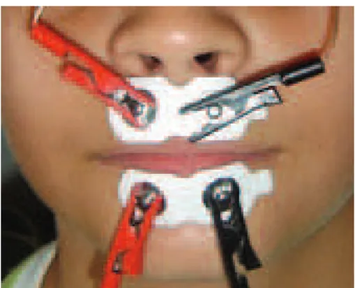
For the EMG evaluation, subjects remained seated, with lexion of 90° of hip, knees, and ankles, guided by the Frankfurt Plan. To capture the EMG signal, Miotec sensors with differ-ential input were used, connected to Ag/AgCl electrodes type DOUBLE (by Hal Indústria e Comércio). The electrodes had a disk shape, ixed distance of 20 mm between them, 10 mm in diameter, and a 2 mm contact surface, a ixed amount of conductive gel and placed by the manufacturer, 20X gain, 10 GΩ input impedance, and rate of common mode rejection >100 dB. According to international standards, the electrodes were stuck in the womb of the orbicularis oris muscles, one on the upper muscle and another on the lower muscle(12,23) (Figure 1).
Figure 1. Placement of electrodes on the upper and lower orbicularis oris muscle
Still, to avoid EMG interference, a reference electrode (connected to the earth) was placed in the region of the pa-tient’s glabella. To reduce the interference of the impedance offered by the skin(10), it was previously prepared, cleaned in the places that electrodes would be positioned with 70% ethyl alcohol and cotton, and, if necessary, the area was shaved. The location of the collections(12), of both the equipment and the assessment chair, was also treated, and the loor was covered in rubberized Pavilex. In addition, care was taken as to distance and off equipment that could interfere electro-magnetically in the exam.
Miotool equipment (Miotec) with eight input channels, 14-bit resolution A/D converter in the acquisition of EMG signals, 5,000 V electrical insulation, maximum acquisition capacity of 2,000 samples/second/channel, and 20 Hz high-pass and 500 Hz low-pass ilters. The signals were collected and ana-lyzed using the Miograph 2.0 software, used by the Orofacial Motricity Laboratory of the institution’s Speech-Language Pathology and Audiology course, and saved in an HP Pavilion dv5-204br laptop with a 500 GB HD and 4GB RAM, which was not connected to the power grid to avoid interference in the EMG signal. For the analysis of EMG signal records, the researcher had no knowledge as to the identiication of children, as they have been designated by abbreviations.
Assessment protocol of muscle fatigue
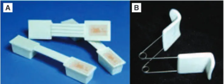
The lip dumbbells and lip exerciser were chosen as thera-peutic exercises for the orbicularis oris muscle. These instru-ments were chosen because they are standardized devices for presentation to the subjects as well as because they are used in the Speech-Language Pathologist’s practice. It is worth noting that all of these instruments were purchased with the researcher’s own funds. In the case of the lip exerciser, it was also possible to measure the force required for its closure, simu-lating a load cell for the periorbicular region, which is not found in a conventional manner in Speech-Language Pathology and Audiology equipment stores. Moreover, the literature highlights that isometric contractions show more relation with changes in MF, and therefore, with muscle fatigue(8,13).
The following collections were performed in sequence: rest for at least 10 seconds; the lifting of 40, 60, and 100 g lip dumbbells; and the lifting of the lip exerciser (Figure 2).
Program in Dental Science of the study’s institution of origin. Six apparatuses were used: three were new and three were used in approximately 100 episodes of closing. Each had three measurements, always with a 100 kgf or 1000 N load cell. Subsequently, a comparison was made between new and used equipment through the t-test, and no differences were observed in the strength used for the closing of the devices, resulting in an average of 2.5 N of strength for the closing move-ment. Therefore, for this study, each apparatus was used for a maximum of three children, always being properly cleaned with soap and water and disinfected by rubbing with gauze soaked in alcohol 70%. In this test, the subject was instructed to keep the equipment fully closed, again, until the feeling of fatigue(13), following the same guidelines and parameters of lip dumbbells.
There were three repetitions of each collection situation, with 2 minutes of rest between each of them for muscle recov-ery(12). The signal with better quality was chosen, which was ex-amined by the fast Fourier transform of the signal. After choos-ing the signal that best represented each test, the initial seconds before the onset of muscle activity were excluded. From the beginning of the requested activity, the initial 0.5 seconds were disregarded to homogenize the snippets evaluated(24).
MF was selected according to time protocol, as proposed by the researcher. In it, four moments were analyzed: T1, from 0 to 5 seconds of activity; T2, from 5.1 to 10 seconds of activity; T3, from 10.1 to 15 seconds of activity; and T4, from 15.1 to 20 seconds of activity. The analysis did not continue beyond 20 seconds, as most children did not maintain the contractions beyond that time.
Statistical analysis
Analyses were performed in the Statistica 9.0 software. Initially, the Shapiro-Wilk test was used to analyze the normal-ity of the variables. As most of the variables had normal behav-ior, to analyze FM over the collection periods, the analysis of variance test was applied for repeated measures and, as post hoc, Tukey’s test(25). For the referred time of fatigue variable, with no normal distribution, Mann-Whitney U-test was applied for the comparison between mouth and nasal breathing and between different patterns of facial growth, and the Kruskal-Wallis test was applied for comparison between groups formed from the association between the breathing mode and the facial growth pattern. The signiicance level adopted was of 5% (p<0.05).
RESULTS
The descriptive analysis of the variables gender, age, breath-ing mode, and facial growth pattern is shown in Table 1.
In the evaluation of the myoelectric fatigue of upper and lower orbicularis oris muscle, regardless of the groups, it was observed that in the four tests, there was a signiicant reduc-tion in FM over the analysis moments (T1, T2, T3, and T4), as shown in Table 2.
Means and standard deviations of the MF of the orbicu-laris oris muscle during therapy trials and interaction with the breathing pattern, facial growth pattern, and the combination
Figure 2. (A) Lip dumbbells; (B) Lip exerciser, both by Pró-Fono®
A B
For lifting the dumbbells, subjects were instructed to sus-tain them until the feeling of fatigue(13), a condition previously established as the beginning of the perception of tiredness in the evaluated muscle to keep exercising, being signaled to the researcher and recorded. After this time, the collection of EMG continued for 3 more seconds. The choice of dumbbell order was random to avoid the biasing of children.
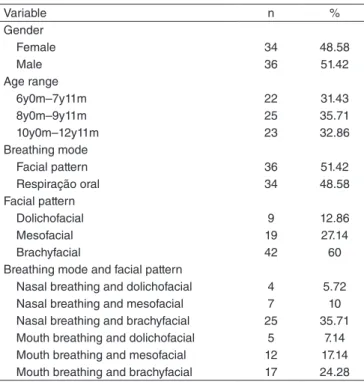
Table 1. Distribution of the descriptive analysis of the variables gender, age, breathing mode, and facial pattern of the sample
Variable n %
Gender
Female 34 48.58
Male 36 51.42
Age range
6y0m–7y11m 22 31.43
8y0m–9y11m 25 35.71
10y0m–12y11m 23 32.86
Breathing mode
Facial pattern 36 51.42
Respiração oral 34 48.58
Facial pattern
Dolichofacial 9 12.86
Mesofacial 19 27.14
Brachyfacial 42 60
Breathing mode and facial pattern
Nasal breathing and dolichofacial 4 5.72
Nasal breathing and mesofacial 7 10
Nasal breathing and brachyfacial 25 35.71
Mouth breathing and dolichofacial 5 7.14
Mouth breathing and mesofacial 12 17.14
Mouth breathing and brachyfacial 17 24.28
Caption: y = years; m = months; n= number of subjects; % =percentage
Exercise and muscles
Collection moments
p-value
T1 T2 T3 T4
Mean (standard deviation) Lip dumbbell 40 g
OU
155.6 (15.4)
150.7 (15.7)
147.1 (15.5)
147.0
(16.5) <0.01* **T1 ≠ T2. T3. T4 - T2 ≠ T3. T4
OL
139.6 (15.2)
133.6 (13.9)
130.3 (15.0)
127.6
(16.3) <0.01* **T1 ≠ T2. T3. T4 - T2 ≠ T3. T4 - T3 ≠ T4 Lip dumbbell 60 g
OU
152.9 (16.0)
149.0 (17.6)
145.4 (17.4)
143.5
(16.7) <0.01* **T1 ≠ T2. T3. T4 - T2 ≠ T3. T4
OL
135.5 (13.3)
130.7 (13.7)
126.0 (13.8)
122.3
(12.9) <0.01* **T1 ≠ T2. T3. T4 - T2 ≠ T3. T4
Lip dumbbell 100 g
OU
152.6 (15.3)
146.8 (16.3)
143.6 (16.5)
140.9
(16.1) <0.01* **T1 ≠ T2. T3. T4 - T2 ≠ T3. T4
OL
136.3 (12.8)
128.1 (12.6)
124.4 (12.4)
122.6
(12.4) <0.01* **T1 ≠ T2. T3. T4 - T2 ≠ T3. T4
Lip exerciser
OU
146.6 (13.8)
138.7 (14.2)
134.8 (13.8)
129.9
(14.0) <0.01* **T1 ≠ T2. T3. T4 - T2 ≠ T3. T4 - T3 ≠T4
OL
134.7 (11.2)
126.5 (11.0)
120.9 (10.3)
116.6
(10.8) <0.01* **T1 ≠ T2. T3. T4 - T2 ≠ T3. T4 - T3 ≠T4
Table 2. Distribution of means and standard deviations of the median frequency and statistical analysis found in the exercises for orbicular upper and lower orbicularis oris muscle, regardless of the groups, over different collection moments (T1, T2, T3, and T4)
*Statistical significance by analysis of variance (ANOVA) for repeated measures; **Post hoc analysis using the Tukey’s test;
Caption: T1 = 5 seconds of activity; T2 = 10 seconds of activity; T3 = 15 seconds of activity; T4 = 20 seconds of activity; OU = upper orbicularis oris muscle; OL = lower orbicularis oris muscle
Exercises and muscles
Nasal breathers Mouth breathers
p-value
T1 T2 T3 T4 T1 T2 T3 T4
Mean (standard deviation) Mean (standard deviation)
Lip dumbbell 40 g
OU 154.7 (12.4) 150.9 (12.7) 147.4 (14.8) 146.9 (14.0) 156.6 (18.4) 150.4 (18.6) 146.7 (16.6) 147.2 (19.3) 0.56
OL 138.7 (13.5) 133.7 (13.8) 129.5 (13.5) 125.8 (14.3) 140.5 (17.0) 133.3 (14.2) 131.3 (16.8) 129.7 (18.2) 0.19
Lip dumbbell 60 g
OU 153.5 (15.0) 150.6 (17.1) 146.1 (15.7) 144.3 (16.1) 152.3 (17.3) 147.3 (18.3) 144.6 (19.3) 142.6 (17.6) 0.78
OL 134.6 (13.1) 129.0 (12.3) 124.7 (13.8) 121.1 (13.1) 136.6 (13.6) 132.6 (15.1) 127.4 (13.9) 123.7 (12.8) 0.83
Lip dumbbell 100 g
OU 152.7 (10.5) 147.6 (15.4) 144.6 (15.7) 140.3 (14.6) 152.5 (19.8) 145.8 (17.6) 142.4 (17.6) 141.7 (18.0) 0.46
OL 135.8 (12.9) 126.2 (11.8) 123.2 (12.0) 120.4 (11.5) 137.0 (12.8) 130.5 (13.3) 125.9 (12.9) 125.3 (13.1) 0.11
Lip exerciser
OU 145.4 (13.1) 137.3 (13.7) 133.6 (13.0) 128.7 (13.8) 147.9 (14.7) 140.4 (14.9) 136.0 (14.9) 131.2 (14.4) 0.97
OL 134.1 (12.4) 125.4 (12.9) 119.1 (11.0) 115.1 (11.7) 135.5 (10.0) 127.8 (8.4) 123.0 (9.1) 118.4 (9.5) 0.37
Table 3. Distribution of means and standard deviations of the median frequency found in the exercises for upper and lower orbicularis oris muscle and statistical analysis of the interaction with the breathing mode, over different collection moments (T1, T2, T3, and T4)
Caption: T1 = 5 seconds of activity; T2 = 10 seconds of activity; T3 = 15 seconds of activity; T4 = 20 seconds of activity; OU = upper orbicularis oris muscle; OL = lower orbicularis oris muscle
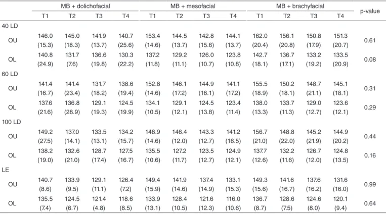
of these are given in Tables 3, 4 and 5. In the interaction with the breathing mode, although the orbicularis oris muscles of mouth and nasal breathers presented, in general, decreasing be-havior of the MF, no signiicant difference over the conduction of the isometric tests between these two groups was observed. Similarly, the interactions of these muscles with the facial growth pattern and with the association of the latter with the breathing mode, the orbicular muscles also showed a decrease in MF, but no signiicant difference.
Dolichofacial Mesofacial Brachyfacial
p-value
T1 T2 T3 T4 T1 T2 T3 T4 T1 T2 T3 T4
Mean (standard deviation) Mean (standard deviation) Mean (standard deviation)
40 LD OU 147.5 (15.1) 147.0 (15.3) 142.3 (12.7) 144.5 (19.6) 153.1 (13.4) 145.6 (12.0) 143.1 (16.1) 144.0 (13.7) 158.4 (15.8) 153.7 (16.7) 149.8 (15.6) 148.9 (17.1) 0.27 OL 140.5 (19.8) 131.5 (11.2) 135.1 (16.3) 132.1 (19.5) 138.3 (10.3) 132.1 (11.6) 128.4 (11.2) 123.8 (11.6) 139.9 (16.1) 134.7 (15.4) 130.1 (16.3) 128.3 (17.3) 0.05 60 LD OU 138.9 (16.2) 134.2 (20.7) 126.2 (14.9) 123.1 (15.2) 134.0 (9.5) 128.6 (10.2) 126.1 (12.1) 123.7 (10.8) 135.5 (14.3) 130.9 (13.4) 125.9 (14.6) 121.4 (13.5) 0.58 OL 138.9 (16.2) 134.2 (20.7) 126.2 (14.9) 123.1 (15.2) 134.0 (9.5) 128.6 (10.2) 126.1 (12.1) 123.7 (10.8) 135.5 (14.3) 130.9 (13.4) 125.9 (14.6) 121.4 (13.5) 0.13 100 LD OU 149.5 (20.6) 139.7 (13.3) 138.4 (15.6) 137.7 (16.8) 148.2 (11.8) 145.8 (13.7) 144.4 (16.6) 140.0 (15.9) 155.5 (15.0) 149.0 (17.8) 144.6 (16.8) 142.2 (16.3) 0.17 OL 137.2 (16.2) 129.4 (16.5) 127.8 (13.1) 127.1 (12.7) 135.4 (10.4) 125.7 (10.6) 123.1 (10.7) 123.4 (12.0) 136.6 (13.2) 129.0 (12.6) 124.2 (13.1) 121.1 (12.6) 0.05 LE OU 144.3 (11.4) 136.7 (11.6) 131.9 (12.8) 129.0 (9.3) 147.7 (14.6) 139.2 (13.3) 136.2 (13.6) 131.0 (15.0) 146.6 (14.3) 139.0 (15.4) 134.8 (14.4) 129.6 (14.8) 0.96 OL 138.5 (8.1) 127.0 (8.7) 122.1 (8.0) 119.4 (9.4) 132.7 (10.4) 126.8 (10.0) 120.5 (10.6) 114.7 (10.2) 134.7 (12.2) 126.3 (12.2) 120.8 (10.9) 116.8 (11.5) 0.32
Table 4. Distribution of means and standard deviations of the median frequency found in the exercises for upper and lower orbicularis oris muscle and statistical analysis of the interaction with the facial growth pattern over the different collection moments (T1, T2, T3, and T4)
Caption: T1 = 5 seconds of activity; T2 = 10 seconds of activity; T3 = 15 seconds of activity; T4 = 20 seconds of activity; OU = upper orbicularis oris muscle; OL = lower orbicularis oris muscle; 40 LD = 40 g lip dumbbell; 60 LD = 60 g lip dumbbell; 100 LD = 100 g lip dumbbell; LE = lip exerciser
Table 5. Distribution of means and standard deviations of the median frequency found in the exercises for upper and lower orbicularis oris muscle and statistical analysis of the interaction with the breathing mode associated with the facial growth pattern over the different collection times (T1, T2, T3, and T4)
NB + dolichofacial NB + mesofacial NB + brachyfacial
T1 T2 T3 T4 T1 T2 T3 T4 T1 T2 T3 T4
Mean (standard deviation) Mean (standard deviation) Mean (standard deviation)
MB + dolichofacial MB + mesofacial MB + brachyfacial
p-value
T1 T2 T3 T4 T1 T2 T3 T4 T1 T2 T3 T4
40 LD OU 146.0 (15.3) 145.0 (18.3) 141.9 (13.7) 140.7 (25.6) 153.4 (14.6) 144.5 (13.7) 142.8 (15.6) 144.1 (13.7) 162.0 (20.4) 156.1 (20.8) 150.8 (17.9) 151.3 (20.7) 0.61 OL 140.8 (24.9) 131.7 (7.6) 136.6 (19.8) 130.3 (22.2) 137.2 (11.8) 129.2 (11.1) 126.0 (10.7) 123.8 (10.8) 142.7 (18.1) 136.7 (17.1) 133.2 (19.2) 133.5 (20.9) 0.08 60 LD OU 141.4 (16.7) 141.4 (23.4) 131.7 (18.2) 138.6 (19.4) 152.8 (14.6) 146.1 (17.2) 144.9 (16.1) 141.1 (17.2) 155.5 (18.9) 150.2 (18.1) 148.7 (21.1) 145.1 (18.1) 0.31 OL 137.6 (21.6) 136.8 (28.9) 129.1 (19.3) 124.5 (19.9) 134.1 (10.5) 129.1 (12.1) 124.5 (13.8) 123.4 (11.4) 138.0 (13.3) 133.7 (11.3) 129.0 (12.7) 123.6 (12.1) 0.29 100 LD OU 149.2 (27.5) 137.0 (14.1) 133.5 (13.1) 134.2 (15.7) 148.9 (14.6) 146.4 (12.0) 143.3 (12.7) 141.2 (16.5) 156.7 (21.0) 148.8 (22.0) 145.2 (21.9) 144.9 (20.2) 0.44 OL 138.2 (19.0) 132.6 (21.0) 128.7 (17.4) 127.5 (16.7) 135.5 (10.6) 127.2 (11.7) 123.5 (12.7) 124.9 (12.1) 137.7 (12.6) 132.2 (11.6) 126.7 (12.0) 124.8 (13.5) 0.16 LE OU 140.7 (8.6) 133.9 (9.5) 129.1 (11.1) 126.4 (7.2) 149.4 (15.9) 141.9 (14.6) 137.4 (14.9) 133.1 (15.3) 149.3 (15.6) 141.6 (16.7) 137.6 (16.2) 131.6 (16.0) 0.99 OL 135.5 (7.4) 124.5 (6.7) 121.4 (4.8) 118.6 (8.5) 133.9 (13.1) 128.4 (10.5) 121.6 (12.3) 116.0 (10.6) 136.7 (8.7) 128.6 (7.5) 124.6 (8.0) 120.1 (9.4) 0.64
Caption: NB = nasal breathing; MB = mouth breathing; T1 = 5 seconds of activity; T2 = 10 seconds of activity; T3 = 15 seconds of activity; T4 = 20 seconds of activity; OU = upper orbicularis oris muscle; OL = lower orbicularis oris muscle; 40 LD = 40 g lip dumbbell; 60 LD = 60 g lip dumbbell; 100 LD = 100 g lip dumbbell; LE = lip exerciser
Table 5. Continuation
Groups
Time of fatigue 40 LD
Time of fatigue 60 LD
Time of fatigue
100 LD Time of fatigue LE
Mean (SD) p-value Mean (SD) p-value Mean (SD) p-value Mean (SD) p-value
BM
NB 78.4 (56.9)
0.03* 62.0 (36.3) 0.02* 50.1 (34.3) 0.09 35.1 (13.2) 0.27
MB 54.4 (33.6) 45.5 (26.6) 38.8 (24.8) 31.1 (11.4)
FGP
Dolichofacial 77.3 (70.9)
0.56 44.1 (17.4) 0.68 50.2 (49.8) 0.53 34.5 (14.9) 0.98
Mesofacial 54.8 (29.9) 55.9 (40.3) 37.6 (18.4) 33.2 (13.8)
Brachyfacial 69.9 (49.4) 55.2 (31.9) 46.5 (29.8) 32.8 (11.5)
BM + FGP
NBDo 98.4 (106.7)
0.38 44.8 (18.6) 0.20 72.6 (73.1) 0.47 31.0 (6.5) 0.85
NBMe 64.7 (27.1) 81.4 (49.7) 47.0 (22.9) 37.2 (17.7)
NBBr 79.1 (54.7) 59.4 (33.1) 47.4 (28.7) 35.1 (12.8)
MBDo 60.5 (26.6) 43.6 (18.6) 32.4 (7.2) 37.2 (19.8)
MBMe 49.1 (31.0) 41.1 (25.7) 32.1 (13.3) 30.8 (11.1)
MBBr 56.4 (38.0) 49.1 (29.8) 45.3 (32.3) 29.5 (8.4)
*Statistical significance by Mann-Whitney U-test
Caption: 40 LD = 40 g lip dumbbell; 60 LD = 60 g lip dumbbell; 100 LD = 100 g lip dumbbell; LE = lip exerciser; SD = standard deviation; BM = breathing mode; NB = nasal breathing; MN = mouth breathing; FGP = facial growth pattern; NBDo = nasal breathing with dolichofacial pattern; NBMe = nasal breathing with mesofacial pattern; NBBr = nasal breathing with brachyfacial pattern; MBDo = mouth breathing with dolichofacial pattern; MBMe = mouth breathing with mesofacial pattern; MBBr = mouth breathing with brachyfacial pattern
DISCUSSION
Analyzing the performance of the orbicularis oris muscles, without interaction between the groups, it was observed that there was a signiicant decrease in MF, for all therapeutic ex-ercises. This difference remained between 10 and 15 seconds of activity for all events, but, for the lip exerciser event, only between 15 and 20 seconds. According to the literature(26), the orbicular muscle of the mouth has mainly type II muscle ibers and, therefore, could be easily fatigued, as observed in this study, with the reduction of MF starting from 5 seconds of activity. The reduced time of manifestation of EMG fatigue differs from periods of isometric contractions suggested by the literature of 10(1), 15(9), 20(9), and 30(7) seconds. However, it is noteworthy that, in these studies, the signs were not based on objective data, as in this study.
When considering the interaction with the respiratory mode, only the sensation of muscular fatigue was perceived differently by the groups, with the 40 and 60 g lip dumbbells, with mouth breathers realizing it more quickly. In the mouth-breathing subject, proprioception of the whole stomatognathic system is changed(27), probably because they do not stimulate the proprioceptive receptors in the oral cavity(28), because they need to stay with their mouth open to breathe. The altered proprioception in this group may have inluenced the feeling of fatigue reported only in tests with lower load (40 to 60 g), where the variability in implementation is more likely.
The literature states that the orbicularis oris muscles are not very active in the MB, due to their need to keep the mouth open(5,27). Despite the decrease of MF in the orbicular muscles of the mouth alone, when comparing NB and MB, the ac-cumulation of substrates that the literature refers to in these situations seems to have been enough to cause a signiicant difference. Thus, it is hypothesized that characteristically less tense muscles, such as the orbicularis oris in MB, would not necessarily present more dificulty in maintaining strength levels, at least during the exercises in question. In clinical terms, this information may suggest that muscle changes the perioral region could be worked with the same demand level between NB and MB.
When analyzing the sample from the point of view of facial growth pattern, there were no differences in the behavior of the MF and the time of fatigue referred for the orbicular muscles of the mouth during the conduction of isometric tests. Each facial growth pattern has its own aesthetic, bone, muscle, and func-tional features. Although there are no studies with objective measurements of the orbicular muscles of the mouth in relation to facial type in the literature, there are studies that very well describe their clinical differentiation(15,21).
The brachyfacial type, for example, is characterized by hav-ing a wider nasal airway and a smaller lower third of the face, which favors the proper resting position of the lips and tongue. In contrast, the dolichofacial type features a narrower airway and an increased lower third of the face, making it dificult to lip-lock and to position the tongue on the hard palate during rest(15). Although it is idealized that the orbicular muscles of the mouth in these facial types could experience fatigue differently
due to having speciic muscle characteristics, this factor was not enough to promote signiicant difference in MF during the isometrics tested.
The breathing mode was also considered jointly with the facial growth pattern due to belief that the changes of these two aspects could leverage each other. However, even if the sample distribution in these new groups had detailed their sto-matognathic features better, this did not occur in EMG terms for the orbicularis oris muscle. The MF and time analysis and the feeling of fatigue for that muscle over the collection times also showed no difference.
The study of muscle fatigue has been receiving more and more contributions, but mainly for the muscles of the limbs and chest. There was no literature that has analyzed the fatigue of the orbicular muscle of the mouth in children. The orbicular muscle of mouth was the subject of other studies; however, with different populations and indings.
One study investigated(29) the fatigue of the orbicularis oris muscle in musicians during the use of wind instruments, for 90 minutes. The authors found no decrease in MF that indicated muscle fatigue after the exercise. One possibility for the activity with musicians not having caused fatigue is that muscle activ-ity proposed by wind instruments, although isometric and prolonged, was not continuous, allowing the muscles to resume blood low in the region, as well as the removal of intramuscular substrates. Another study(30) investigated the orbicular muscles of the mouth in goldsmiths and found muscle fatigue after 1 day of work. It is believed that the difference among these studies might be due to the difference in situations tested, as mentioned earlier.
Thus, in general, one can infer that the orbicularis oris muscle reaches fatigue over static contractions, regardless of the group examined. The manifestation of fatigue in the EMG signal occurred in less time than the feeling of fatigue reported by the participants, regardless of group. This fact cor-roborates the literature, which reports physiological fatigue as antecedent to the feeling of fatigue(12), but raises other issues, such as what would be the best parameter of fatigue to be considered for therapy.
Although the results of this study have contributed to the understanding of fatigue in the orbicularis oris muscle, much remains to be detailed and investigated. The study has some limitations, such as not having investigated the amplitude of the EMG signal, and, therefore, further studies are suggested, taking into account also the repetition of exercises beyond their contraction times.
CONCLUSION
*ARBS authored the research and was responsible for collecting data and drafting the manuscript; APBD assisted in data collection and elaboration of the manuscript; ECRC and AMTS were responsible for directing and drafting the manuscript.
REFERENCES
1. Coutrin GC, Guedes LU, Motta AR. Treinamento muscular na face: a prática dos fonoaudiólogos de Belo horizonte. Rev Soc Bras Fonoaudiol. 2008;13(2):127-35.
2. Ferreira TS, Mangilli LD, Sassi FC, Fortunato-Tavares T, Limongi SCO, Andrade CRF. Fisiologia do exercício fonoaudiológico: uma revisão crítica da literatura. J Soc Bras Fonoaudiol. 2011;23(3):288-96. 3. Martinelli RLC, Fornaro EF, Oliveira CJM, Ferreira LMDB, Rehder
MIBC. Correlações entre alterações de fala, respiração oral, dentição e oclusão. Rev CEFAC. 2011;13(1):17-26.
4. Hitos SF, Arakaki R, Solé D, Weckx LLM. Oral breathing and speech disorders in children. J Pediatr. 2013;89(4):361-5.
5. Silva MAA, Marchesan IQ, Ferreira LP, Schmidt R, Ramires RR. Postura, tônus e mobilidade de lábios e língua de crianças respiradoras orais. Rev CEFAC. 2012;14(5):853-60.
6. Machado Júnior AJ, Crespo AN. Avaliação cefalométrica de via aérea e do osso hioide em crianças com deglutição normal e atípica: estudo de correlações. Sao Paulo Med J. 2012;130(4):236-41.
7. Jardini RSR. Labial exerciser: preliminary study for enlarge the oral orbicular muscle. Pró-Fono R Atual Cient. 1999;11(1):8-12.
8. Lyons MF, Rouse ME, Baxendale RH. Fatigue and EMG changes in the masseter and temporalis muscles during sustained contractions. J Oral Rehabil. 1993;20:321-31.
9. Jardini RSR. Avaliação eletromiográica do músculo bucinador lácido usando o Exercitador Facial. Pró-Fono R Atual Cient. 2002;14(3):331-43. 10. Cram JR, Kasman GS, Holtz J. Introduction to surface electromyography.
Maryland: Aspen Publishers; 1998.
11. Silva BARS, Martinez FG, Pacheco AM, Pacheco I. Efeitos da fadiga muscular induzida por exercícios no tempo de reação muscular dos ibulares em indivíduos sadios. Rev Bras Med Esporte. 2006;12(2):85-9. 12. De Luca CJ. The use of surface electromyography in biomechamics. J
Applied Biomec. 1997;13(2):135-63.
13. Buzinelli RV, Bérzin F. Electromyographic analysis of fatigue in temporalis and masseter muscles during continuous chewing. J Oral Rehabil. 2001;28:1165-7.
14. Knösel M, Klein S, Bleckmann A, Engelke W. Coordination of tongue activity during swallowing in mouth-breathing children. Dysphagia. 2012;27(3):401-7.
15. Ramires RR, Ferreira LP, Marchesan IQ, Cattoni DM, Silva MAA. Tipologia facial aplicada à Fonoaudiologia: revisão de literatura. Rev Soc Bras Fonoaudiol. 2010;15(1):140-5.
16. Marchesan IQ, Berretin-Felix G, Genaro KF. MBGR Protocol of Orofacial Myofunctional evaluation with scores. Int J Orofacial Myology. 2012;38:38-77.
17. Da Silva CR, Geres BS, Kuriki HU, Negrão Filho RF, Alves N, Azevedo FM. Análise da reprodutibilidade de parâmetros no domínio da frequência do sinal EMG utilizados na caracterização da fadiga muscular localizada. Motriz Rev Educ Fís. 2012;18(3):456-64.
18. Santos MG, Dezan VH, Sarraf TA. Bases metabólicas da fadiga muscular aguda. Rev Bras Ciênc Mov. 2003;11(1):7-12.
19. Organização Mundial da Saúde, 2006 [cited 2014 Apr 30]. Available from: http://www.opas.org.br
20. Callegari-Jacques SM. Bioestatística: princípios e aplicações. Porto Alegre: Artmed; 2007.
21. Berwig LC, Silva AMT, Côrrea ECR, Moraes AB, Montenegro MM, Ritzel RA. Análise quantitativa do palato duro em diferentes tipologias faciais de respiradores nasais e orais. Rev CEFAC. 2012;14(4):616-25. 22. Ritzel RA, Berwig LC, Silva AMT, Côrrea ECR, Serpa EO. Correlação
entre a nasofibrofaringoscopia e a cefalometria no diagnóstico de hiperplasia de tonsilas faríngeas. Int Arch Otorhinolaryngol. 2012;16(2):209-16.
23. Hermens HJ, Freriks B, Disselhorst-Klug C, Rau G. Development of recommendations for SEMG sensors and sensor placement procedures. J Electr Kinesiol. 2000;10(5):361-74.
24. Silva SRD, Gonçalves M. Comparação de protocolos para veriicação da fadiga muscular pela eletromiografia de superfície. Motriz. 2003;9(1):51-8.
25. Tourinho Filho H, Puggina EF, Marini LL, Machado DRL, Barbanti VJ, Pimentel GL. Efeitos agudos do treinamento aeróbio sobre o desempenho da força muscular. Pensar a Prática. 2013;16(2):320 602.
26. Stal P, Eriksson PO, Thornell LE. Differences in capillary supply between human oro-facial, masticatory and limb muscles. J Muscle Res Cell Motil. 1996;17:183-97.
27. Marchesan IQ. Fundamentos em Fonoaudiologia: aspectos clínicos da motricidade oral. Rio de Janeiro: Guanabara-Koogan; 2005.
28. Douglas CR. Tratado de isiologia aplicada à fonoaudiologia. São Paulo: Robe Editorial; 2002.
29. Gotouda A, Yamaguchi T, Okada K, Matsuki T, Gotouda S, Inoue N. Inluence of playing wind instruments on activity of masticatory muscles. J Oral Rehabil. 2007;34(9):645-51.