C
a s eR
e p o Rt4 0 4 Arq Bras Otalmol. 2016;79(6):404-6 http://dx.doi.org/10.5935/0004-2749.20160114
INTRODUCTION
The prevalence of true polycoria is extremely low(1,2), and the
me chanism underlying its development remains unknown. A few theories have been proposed to explain the phenomenon, including segregation of a portion of the pupil margin, partial closure of a co-loboma, and differentiation of neural ectodermal cells into muscle fi-bers in abnormal situations(3-5). Only a few cases of true polycoria have
been reported in the literature, some of which have been associated with polar cataracts, glaucoma, and retinal detachment(3,6). Although
most patients with polycoria are not surgically treated because visual acuity is not significantly affected, dimmed vision can be an issue for some. In this report, we aim to present the details of a patient with true polycoria, who complained of a low vision quality and requested treatment for this condition, and to describe the surgical pupilloplasty procedure we performed for its correction.
CASE REPORT
A 44-year-old man was referred to our clinic complaining of poor vision in his left eye that had been present since childhood. He had no history of trauma or ocular surgery, no abnormal obstetric history, and no other systemic conditions. On examination, his best-correc-ted visual acuity (BCVA) was 1.0 diopters (D) (-1.00, -1.75, 105°) in the right eye and 0.5 D (-4.25, -1.75, 105°) in the left eye, with intraocular pressures of 16 mmHg and 17 mmHg in the right and left eyes, res-pectively. Specular microscopy revealed 2,817 cells/mm2 in the right
eye and 2,882 cells/mm2 in the left eye. Fundus examination was
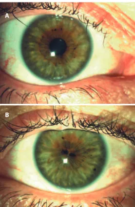
normal in both eyes, as was the right pupil, which measured 3 mm in diameter (Figure 1 A). However, there were two pupils in the left eye within a 2.5-mm central zone and measuring 1.2 and 1.1 mm in dia-meter (Figure 1 B). Both pupils in the left eye had a true iris sphincter and pigment epithelium. Direct and indirect pupillary reflexes were normal in both eyes. After the application of tropicamide 1% drops, both pupils in the left eye dilated (Figures 2 A and 2 B).
S
URGERYWe performed pupilloplasty under retrobulbar anesthesia after the pupil was dilated with 1% tropicamide. Following two 1-mm limbal incisions, the anterior chamber was filled with viscoelastic material, and a spatula was inserted through a side port. The iris tissue between the two pupils was then elevated with the spatula to avoid contact with the lens. We then cut the iris tissue using vitreoretinal scissors inserted through the other side port (Figures 3 A and 3 B). After aspirating the viscoelastic material, limbal incisions were closed with stromal hydration, and the operation was completed with the application of intracameral cefuroxime. No serious intraocular inflam-mation was reported during the early postoperative period. Postope-ratively, the patient was treated with antibiotics and steroid drops for 4 weeks. At the first follow-up 1 month after surgery, the BCVA had increased to 0.9 D (-3.75, -2.50, 95°), the intraocular pressure was 18 mmHg, and the lens was clear. The pupil was almost round and measured 3.5 mm in diameter on the first postoperative day (Figure 4 A), but had reduced to 2.7 mm one month after the surgery. The patient was satisfied with his improved vision and better quality of life.
Pupilloplasty in a patient with true polycoria: a case report
Pupiloplastia em um caso de policoria verdadeira
Handan Bardak1, nimet Yesim ercalik1, murat GunaY2, ruveYde Bolac1, Yavuz Bardak1
Submitted for publication: December 9, 2015 Accepted for publication: January 7, 2016
1 Department of Ophthalmology, HaydarpasaNumune Training and Research Hospital, Istanbul,
Turkey.
2 Department of Ophthalmology, ZeynepKamil Maternity and Children’s Diseases Training and Research
Hospital, Istanbul, Turkey.
Funding: No specific financial support was available for this study.
Disclosure of potential conflicts of interest: None of the authors have any potential conflict of interest to disclose.
Corresponding author: Handan Bardak. Haydarpasa Numune Training and Research Hospital. Tibbiye Cad. No 40, 34668 - Uskudar, Istanbul, 34716 - Turkey - E-mail: [email protected]
ABSTRACT
Here we report a case of surgical pupilloplasty in an adult with true polycoria. A 44-year old man was referred to our clinic with a best-corrected visual acuity (BCVA) of 0.5 diopters (D) in his left eye. Biomicroscopy revealed two pupils within a 2.5-mm central zone, with diameters of 1.2 and 1.1 mm. Both pupils had real iris sphincters and responded to light and chemical stimulation. Therefore, we surgically cut the bridge between the two pupils without any intraoperative or postoperative complications. One month after the surgery, BCVA had improved to 0.9 D, and the final pupil was almost round, measuring 2.7 mm in diameter.
Keywords: Iris/abnormalities; Pupil/abnormalities; Pupil/surgery
RESUMO
Relatamos um caso de pupiloplastia cirúrgica em um paciente adulto com policoria verdadeira. Um homem de 44 anos de idade foi encaminhado ao nosso serviço com acuidade visual melhor corrigida (BCVA) de 0,5 em seu olho esquerdo. Biomicroscopia revelou 2 pupilas, dentro de uma zona central de 2,5 milímetros com dimensões de 1,2 mm e 1,1 mm de diâmetro. Ambas as pupilas apresentavam esfíncteres irianos reais que respondiam à luz e a drogas. A ponte entre as 2 pupilas foi cortada cirur-gicamente. Não houve complicações transoperatórias ou pós-operatórias. A BCVA melhorou para 0,9, e a pupila ficou quase circunferencial com 2,7 mm de diâmetro, um mês após a cirurgia.
Ba r d a k H, e ta l.
4 0 5 Arq Bras Otalmol. 2016;79(6):404-6
Figure 1. A) Preoperative image of the right (normal) eye, before pupil dilatation; B) Preoperative image of the left eye with true polycoria before pupil dilatation.
A
B
Figure 2. A) Preoperative image of the right (normal) eye after pupil dilatation; B) Preoperative image of the left eye after pupil dilatation, showing true polycoria.
A
B
A
B
Figure 3. A) Intraoperative image showing cutting of the iris (upper part); B) Intraoperative image showing cutting of the iris (lower part).
A
B
Pu P i l l o P l a s t yi naPat i e n tw i t h t r u eP o ly c o r i a: ac a s er e P o rt
4 0 6 Arq Bras Otalmol. 2016;79(6):404-6 DISCUSSION
Pseudopolycoria is distinguished from true polycoria by the passive constriction of the accessory pupil when the true pupil is dilated(5) and is characteristic of essential iris atrophy that can be
asso-ciated with Seckel syndrome, posterior polymorphous dystrophy, and juvenile glaucoma(7-9). In contrast, the extra pupil in true polycoria
retains an intact sphincter muscle, is reactive to light, and synchro-nously contracts and dilates in response to medication(3). These
fin-dings were observed in our patient, supporting the diagnosis of true polycoria. Although extra pupils typically present at some distance from the principal pupil(2), the extra pupil was within a 2.5-mm central
zone in our patient.
Polycoria can decrease visual acuity. In one previous case report, a patient with true polycoria was noted to exhibit dimmed vision and decreased retinal illumination. Both these phenomena can be explained by the Airy disc effect or diffraction rings and interference fringes induced by the second pupil(5). It has been reported that, in
true polycoria, visual acuity linearly decreases with pupil diameter for pupils with diameters >1.5 mm(10). This trend may account for our
patient’s visual acuity of 0.5, considering that his pupils measured 1.2 and 1.1 mm in diameter. Although our patient was displeased with his low vision before surgery, he did not complain of either diplopia or glare symptoms. In our opinion, if his pupils had been spaced to-gether more closely, he may have experienced these conditions. It was rewarding that his visual acuity increased following surgery and that he was satisfied with the outcome.
In conclusion, we have shown that a pupilloplasty procedure for true polycoria was effective in our case. However, the efficacy of the procedure remains unknown in the wider population. We anticipate more case reports to validate our surgical approach in cases with true polycoria.
REFERENCES
1. Duke-Elder SS. System of ophthalmology. 5th ed. St Louis: Mosby; 1964. Congenital
deformities. p.592-3.
2.Loewenfeld IE. The pupil. Anatomy, physiology and clinical applications. Detroit: Wayne State University Press; 1993. Vol 1. Iris damage. p.902-6.
3.Jaffe NS, Knie P. True polycoria. Am J Ophthalmol. 1952;35(2):253-5.
4.Mann I. Developmental abnormalities of the eye. London. British Medical Association; 1957. The iris. p.252-4.
5.Islam N, Mehta JS, Plant GT. True polycoria or pseudopolycoria? Acta Ophthalmol Scand. 2007;85(7):805-6.
6.Foos RY, Kiechler RJ, Allen RA. Congenital nonattachment of the retina with hydro-phthalmia hypoplastic vitreous body and true polycoria. Am J Ophthalmol. 1968; 65(2):202-10.
7. Robbin DS. Seckel’s syndrome with pseudopolycoria. Ophthalmic Paediatr Genet. 1985;6(3):135-9.
8. Patel AK, Loh RS, Morrell AJ. Posterior polymorphous dystrophy with polycoria and corectopia. Eye (Lond). 2004;18(8):856-7.
9. Rodrigues MM, Spaeth GL, Weinreb S. Juvenile glaucoma associated with goniodys-genesis. Am J Ophthalmol. 1976;81(6):786-96.
10.Miller SD, Judisch GF. Persistent pupillary membrane: successful medical manage-ment. Arch Ophthalmol. 1979;97(10):1911-3.