330
Saraiva et al
Rheumatic Mitral valve and endomyocardial fibrosis
Arq Bras Cardiol volume 72, (nº 3), 1999
Hospital Barão de Lucena e Hospital das Clínicas da UFPE - Recife
Mailing address: Lurildo R. Saraiva - Depto de Medicina Clínica - Hospital das Clínicas - UFPE - Av. Moraes Rego, SN - 50670-420 - Recife, PE - Brazil.
Lurildo R. Saraiva, Regina W. Carneiro, Mauro B. Arruda, Djair Brindeiro F°, Vital Lira
Recife, PE - Brazil
Mitral Valve Disease with Rheumatic Appearance in the
Presence of Left Ventricular Endomyocardial Fibrosis
Case Report
This is a report of a nine-year-old boy withboth mitral stenosis and regurgitation and extensive endomyo-cardial fibrosis of the left ventricle. Focus is given to the singularity of the fibrotic process, with an emphasis on the etiopatho-genic aspects.
In Pernambuco state, in the northeastern region of Brazil, heart diseases characterized by the presence of marked ventricular endomyocardial fibrosis are practically restricted to uni- or biventricular endomyocardial fibrosis (EMF), which can also affect children and adolescents 1.
Thirty-seven years ago, in Africa, Abrahams e Brid-gen 2 reported the simultaneous occurrence of EMF of the left ventricle (LV) and rheumatic mitral regurgitation (MR) with pulmonary arterial hypertension (PAH). Necropsy of some of these patients showed active Aschoff nodules. This allowed the authors to identify a single origin for both pathological processes in the new syndrome, i. e., rheu-matic disease (RD) and EMF. According to Décourt 3, the acute rheumatic process that occurs in the myocardium can be associated with fibrosis of variable extensions during late scarring processes.
In the present case, the finding of extensive endomyo-cardial fibrosis of the LV is of note in view of its singularity. This allows considerations regarding the etiology, the pathogenesisand the clinical diagnosis of this condition.
Case report
A 9-year-old boy living in the metropolitan area of Reci-fe arrived at the Clinical Pediatrics Department of the Hospital Barão de Lucena in November, 1997, complaining of dyspnea during strenuous exertion in the previous 3 months and a re-cent episode of syncope after jogging, when he was diagno-sed with heart disease in an emergency unit. His mother reported that the patient suffered from repeated tonsillitisbut his history was not notable for fever or joint disease.
He weighed 22.7kg and his height was 121.5cm. His
blood pressure was 110/60mmHg and his heart rate was 100bpm. He was pale and showed continuous tachypnea. His face was slightly edematous, but there was no edema of the lower limbs. His peripheral pulses were regular and symmetric.
There was a precordial bulging and an increased apical impulse, as well as a slight systolic thrill in the mitral area. He had an audible ejection click in the pulmonary area (+++/4) and markedhyperphonesisof the pulmonary component of the second heart sound. There was also a pansystolic murmur in the mitral area (++/4), irradiating to the left axilla, followed by a slight diastolic rumble. Also, a pansystolic murmur that increased with inspiration was audible in the tricuspid area.
His liver was felt 3cm below the right costal margin and his respiratory sounds were rough.
Significant laboratory test results included hemoglo-bin level of 12.8g/dL, white blood cell count of 14.500/mm3, antistreptolysin O (ASO) titers of 833 Todd unit, mucopro-teins of 5.3mg/dl, a negative C reactive protein, serum albumin of 3.68g/dl, α2-globulins of 1.04 g/dl and β -globu-lins of 1.75g/dl. The levels of urea nitrogenand creatinine were within the normal range.
The electrocardiogram, which showed sinus rhythm, was consistent with hypertrophy of the four chambers. The QRS axis was deviated to + 1000 and the chest X-ray revea-led signs of severe PAH and a fourth arch in the left costal margin in the posteroanterior projection. The cardio-thoracic index was 0.62 (fig 1) .
The Doppler echocardiogram showed marked MR and moderate mitral stenosis, with thickened laciniae and doming of the anterior leaflet during valvaropening, which was consistent with a rheumatic etiology. The mitral valve area (MVA) was estimated to be 1.22cm2, the systolic pressure in the pulmonary artery was estimated to be 85mmHg and the mean pressure gradient between the left atrium (LA) and the LV during diastole was estimated to be 12mmHg. The diameter of the LA during systole was 4.7cm and the diameter of the LV during diastole was 4.6cm. The ejection fraction of the LV was 0.69.
Arq Bras Cardiol volume 72, (nº 3), 1999
Saraiva et al Rheumatic Mitral valve and endomyocardial fibrosis
331 and the tendinous chordae. According to the surgeon, the
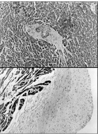
latter were very short, resembling EMFof the left ventricular chamber. The histopathological study of the resectedvalve and endomyocardial fibrous tissue showed marked fibrosis of the leaflets and tendinous chordae, with significant hyalinization, permeated by basophilic areas with a myxoma-tous aspect. In the specimen of the LV, similarly severe endo-cardial fibrosis and hyalinization were noted, with small foci of lymphocytic infiltrate. The fibrosis penetrated in the myo-cardium, adopting a fusiform aspect around the arterioles. No active Aschoff nodules were noted (fig. 2).
Three months after surgery, the child was breathing normally, and there were no heart murmurs. He was recei-ving secondary prophylaxis for RD. The echocardiogram showed normal chambers, with the exception of a slight dilation of the LA (3.1cm). MVA was estimated to be 5.5cm2.
Discussion
The physical findings and laboratory tests abnor-malities – high ASO titers, slightly increased white blood cell count, increased levels of mucoproteins - as well as the echocardiographic image of thickened mitral leaflets with a reduced opening noted in this child suggest the role of a rheumatic process in the genesis of the valvar dysfunction. The absence of both active and senescent Aschoff bodies in the valvar histopathological study does not invalidate this hypothesis.
Thirty years ago, Lira et al4, in a macroscopic and microscopic study of 52 cases of rheumatic heart disease in Pernambuco state, defined an intermediate presentation of this condition, situated between acute cases and those labeled as chronic sequelae. They were named “evolving cases”. The authors noted that these cases showed chronic inflammatory foci in the myocardial interstitium, which were also present in the endocardium and epicardium as clusters of lymphocytes. The authors described a curious finding, i.e., the presence of regressive juxtarterial lesions, resem-bling the late stages of Aschoff nodules, similar to what is seen in the image on the top of figure 2.
In the syndrome described by Abrahams and Bridgen2 the role of a rheumatic process as the sole etiology of mitral valvulitis and endomyocardial fibrosis – both leading to PAH and the latter being similar to Davies disease 5 - is clear. This finding is supported by Shaper 6. However, the fibrotic process we noted does not show the characteristics of Davies’ EMF, such as the peculiar pattern of distribution of the fibrosis in the ventricular inlet as well as the capillary neoformation close to the area of fibrosis7. In addition, the characteristic echocardiographic findings, which are almost pathognomonic of the disease, are absent 8.
This extensive endomyocardial fibrotic process of the ventricle (fig 2), may share the same etiology as mitral valve dysfunction, a phenomenon that may occur, albeit rare, and has seldomly been described 3. After all, it is stated in Occam’s classical philosophical principle: “Entia non mul-tiplicanda praeter necessitatem”- “one should not multiply the causes without need”, - thus, whenever possible, we should always search for a single explanation for a scientific question.
On the other hand, certain aspects, such as the rapidly developing PAH in the presence of a fourth arch in the chest X-ray, indicating persistent elevation of LA filling pressure (fig. 1), together with the very increased levels of β -glo-bulins (1.75g/dL), – an unusual finding in active RD 9, - may be useful in the recognition of the described association.
Fig. 1 – Chest X-ray in posteroanterior projection. An enlarged heart silhouette with clockwise rotation of the heart and clearsigns of pulmonary arterial hyper-tension are noted. Note the presence of the fourth arch.
332
Saraiva et al
Rheumatic Mitral valve and endomyocardial fibrosis
Arq Bras Cardiol volume 72, (nº 3), 1999
References
1. Saraiva LR, Tompson G, Lira V, et al. Endomiocardiofibrose na infância. Relato de três casos, um dos quais associado a CIA. Arq Bras Cardiol 1980; 34: 303–6. 2. Abrahams D, Bridgen W. Syndrome of mitral incompetence, myocarditis, and
pulmonary hypertension in Nigeria. Br Med J 1961; 2: 134-9. 3. Décourt LV. Doença Reumática. 2ª ed.São Paulo:Sarvier, 1972: 47. 4. Lira VMC, Freitas D, Maciel SM. Estudo morfológico da cardiopatia reumatismal
em Recife (Brasil). An Fac Med Univ Fed PE 1970; 30: 145–62.
5. Davies JNP. Endocardial fibrosis in Africans. A heart disease of obscure aetiology in Africans. East Afr Med J 1948; 25: 10-4.
6. Shaper AG. The aetiology of endomyocardial fibrosis. In: Valiatan MS, Somers K, Kartha CC (eds) - Endomyocardial Fibrosis. New Delhi: Oxford University Press, 1993: 111-20.
7. Lira VMC. Patologia da endomiocardiofibrose. Arq Bras Cardiol 1996; 67: 273-7. 8. Brindeiro Fº D, Cavalcanti C. O valor da ecodopplercardiografia na identificação diagnóstica e no manuseio da endomiocardiofibrose. Arq Bras Cardiol 1996; 67: 279-84.
