C
a s eR
e p o Rt4 6 Arq Bras Oftalmol. 2017;80(1):46-8 http://dx.doi.org/10.5935/0004-2749.20170012
INTRODUCTION
Chromomycosis is a fungal infection that affects the epidermis, dermis, and subcutaneous tissue and is caused by dematiaceous fun-gal species that turn black on staining. The characteristic skin le sions can be present separately or together; they are verrucous and ulcera-tive and may also exhibit dyschromia. The most commonly observed fungi are Fonsecaea pedrosoi, Phialophora verrucosa, and, more rarely,
Rhinocladiella aquaspersae. We report the case of a 50-year-old male
patient who was a rural worker and had been treated without success for three decades. Despite treatment considered appropriate by in-fectologists and dermatologists, facial lesions progressed and caused severe cicatricial retraction. As the infection evolved, the left upper eyelid developed cicatricial ectropion.
CASE REPORT
A 50-year-old male agricultural zone worker living in Anapolis, Goias (Brazil), where he was born, had a history of a trauma of the right shoulder caused by a plant 30 years before his admission to this service. The wound did not heal spontaneously and expanded slowly and continuously.
The patient did not seek treatment early in the disease evolu-tion; he simply made use of unspecified ointments and alternative treatments. Ten years after the appearance of the wound, the patient received dermatological treatment for ring worm because it was already causing anatomical deformity and functional limitations, but the treatment was only partially successful. Because of emotional
and financial aspects, the patient abandoned medical supervision and returned to seek treatment only when his condition worsened.
The patient was referred to the Tropical Diseases Hospital of the Federal University of Goias, and the dermatology team made the diag-nosis of chromomycosis caused by the fungus Fonsecaea pedrosoi(1), which was confirmed by histopathological analysis (Figures 1A and 1B). Treatment was then initiated with oral itraconazole, which is considered the drug of choice given the extensive clinical experience with this drug(2).
Treatment with different antifungals was tried on several occa-sions (Itraconazole®
, Amphotericin®
, Terbinafine®
) with only partial success because the treatment was limited by renal toxicity, which also prevented the use of higher doses of Amphotericin B®, a useful
alternative therapy in some cases. Currently the patient is being trea-ted with 500-mg terbinafine®
once a day.
When admitted to the ophthalmology service, the patient’s lesion had spread to his face and periorbital zone (Figure 2A). The severe cicatricial ectropion was treated with a skin graft combined with a tarsal strip procedure on the lower eyelids of both eyes; the cicatricial retraction in the upper eyelid of the left eye was also treated with a skin graft. The skin grafts were fixed with tie-over sutures, which were removed after 5 days. The surgical treatment performed by the oculo-plastic department of the hospital was performed to reduce corneal exposure and eye dryness, which had caused significant visual loss in the left eye as well as a risk of eye perforation (Visual acuity: right eye, 20/25; left eye, hand motion).
Chromomycosis, an unusual cause of cicatricial ectropion: a case report
Cromomicose como causa rara de ectrópio cicatricial: relato do caso
José Eduardo simarro rios1, Carolina BonfimdE Paiva1, GaBriEla mouradE Paula2, WandErlEy riBEiro BorGEs fiGuEirEdo2, Julio César dahEr arantEs1, fáBio marquEsdE almEida2, roBErto murilo limonGi1
Submitted for publication: June 25, 2015 Accepted for publication: April 14, 2016
1 Centro de Referência em Oftalmologia, Hospital de Clínica Médica, Universidade Federal de Goiás (UFG), Goiania, GO, Brazil
2 Serviço de Patologia, Hospital de Clínica Médica, Universidade Federal de Goiás (UFG), Goiania, GO, Brazil
Funding: No specific financial support was received for this study.
Disclosure of potential conflicts of interest: None of the authors have any potential conflict of interest to disclose.
Corresponding author: José Eduardo Simarro Rios. Centro de Referência em Oftalmologia. Primeira avenida, sem número - Goiânia, GO - 74605-020 - Brazil
E-mail: [email protected]
ABSTRACT
Chromomycosis is a fungal infection that affects the epidermis, dermis, and sub-cutaneous tissue and is caused by dematiaceous fungal species that turn black on staining. We report the case of a 50-year-old male patient who was a rural worker and had been treated without success for three decades. Facial lesions progressed and caused severe cicatricial retraction. As the infection evolved, the left upper eyelid developed cicatricial ectropion. The surgical treatment was performed using skin obtained from the patient’s own abdomen. Patient has developed a good postoperative appearance
Keywords: Chromoblastomycosis; Mycoses; Ectropion; Rare diseases; Neglected diseases; Case reports
RESUMO
A cromomicose é uma infecção fúngica que afeta a epiderme, derme e tecido subcutâneo. A infecção é causada por espécies de fungo dematiáceos que se coram em preto. Nós relatamos o caso de um homen de 50 anos de idade, trabalhador da zona rural, que tinha sido tratado por três décadas sem êxito conclusivo. As lesões faciais progrediram causando retração cicatricial severa. Com a evolução do quadro, houve também retração também da pálpebra superior do olho esquerdo. O tratamento cirúrgico foi realizado utilizando pele abdominal do próprio paciente. O paciente apresentou uma boa aparência pós-operatória.
Ri o s JEs, E ta l.
4 7
Arq Bras Oftalmol. 2017;80(1):46-8
A
B C
Figure 1. Anatomo-pathological biopsy performed in the face in the right temporal region of the patient. A) Skin, magniication at 4×; stained with hematoxylin and eosin (HE). Skin showing mild acanthosis, orthokeratosis, and a focus of parakeratosis, with mild perivascular lymphoplasmocytic inlammatory iniltration of the interstitial spaces, both supericial and deep, with well-formed granuloma. B) Detail of well-formed granuloma formed by epithelioid cells stained with HE and shown at 40× magniication. C) Special staining with Grocott’s silver showing the presence of structures called chromoblastomycetes.
Because the amount of skin to be grafted was large, we obtained skin from the patient’s own abdomen. There was no rejection of the transplanted tissue, and the patient has developed a good postope-rative appearance (Figure 2B).
Treatment of cicatricial ectropion secondary to chromomycosis can be performed with a skin graft from the patient. For best results, the disease must be cured or under clinical control at the time of sur-gery. In the case of the patient above, an immediate intervention was needed because of the imminent risk of perforation of the left eye.
DISCUSSION
Chromoblastomycosis is a chronic subcutaneous mycosis that typically involves the lower extremities, and most human infections
are caused by traumatic implantation. The vast majority of causative microorganisms have melanized cell walls caused by dematiaceous fungi, which belong to four genera of saprophytic fungi: Phialophora,
Fonsecaea, Rhinocladiella, and Cladophialophora. Fonsecaea pedrosoi
is the organism most commonly isolated from the chronic cutaneous mycosis and is relatively resistant to medical therapy(3).
Ch r o m o m y C o s i s, a nu n u s ua lC au s eo fC i C at r i C i a l e C t r o p i o n: aC a s er e p o rt
4 8 Arq Bras Oftalmol. 2017;80(1):46-8
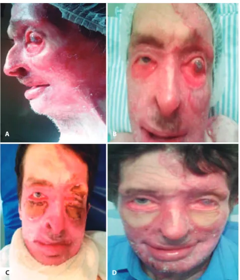
A
C
B
D
Figure 2. Preoperative and postoperative aspects showing severe ectropion leading to corneal exposure. A and B) Ectropion secondary to corneal exposition leading to corneal ulcer requiring conjunctival covering. C) Postoperative appearance after 60 days. D) Postoperative appearance with skin grafts visible after 6 months.
made by Pedroso and Gomes. Since then, the disease has been stu-died and researched, but treatment remains challenging because of the great resistance of the fungus to current therapeutic options(7).
Treatment of rare cases in which the face is affected should involve a dermatologist and an ophthalmologist because early in-tervention may be necessary to prevent loss of vision secondary to cicatricial ectropion.
REFERENCES
1. Criado PR, Valente NY, Brandt HR, Belda Junior W, Halpern I. [Pedroso and Gomes’ verrucousDermatitis (Chromoblastomycosis): 90 years on and still among us]. An Bras Dermatol. 2010;85(1):104-5.
2. Chowdhary A, Meis JF, Guarro J, de Hoog GS, Kathuria S, Arendrup MC, Arikan-Ak-da gli S, Akova M, Boekhout T, Caira M, Guinea J, Chakrabarti A, DannaouiE, van Diepeningen A, Freiberger T, Groll AH, Hope WW, Johnson E, Lackner M,Lagrou K, Lanternier F, Lass-Flörl C, Lortholary O, Meletiadis J, Muñoz P, Pagano L, Petrikkos G,
Richardson MD, Roilides E, Skiada A, Tortorano AM, Ullmann AJ, Verweij PE, Cornely OA, Cuenca-Estrella M; European Society of Clinical Microbiology and Infectious Di-seases Fungal Infection Study Group; European Confederation of Medical Mycology. ESCMID and ECMM joint clinical guidelines for the diagnosis and managementof systemic phaeohyphomycosis: diseases caused byblack fungi. Clin Microbiol Infect. 2014;20(3):47-75.
3. Bui AQ, Espana EM, Margo CE. Chromoblastomycosis of the conjunctiva mimicking melanoma of the ciliary body. Arch Ophthalmol. 2012;130(12):1615-7.
4. Fukushiro R. Chromomycosis in Japan. Int J Dermatol. 1983;22(4):221-9.
5. Minotto R, Bernardi CD, Mallmann LF, Edelweiss MI, Scroferneker ML. Chromoblas-tomycosis: a review of 100 cases in the state of Rio Grande do Sul, Brazil. J Am Acad Dermatol. 2001;44(4):585-92.
6. Kondo M, Hiruma M, Nishioka Y, Mayuzumi N, Mochida K, Ikeda S, et al. A case of chromomycosis caused by Fonsecaea pedrosoi and a review of reported cases of dematiaceous fungal infection in Japan. Mycoses. 2005;48(3):221-5.