Jemds.com
Case Report
Journal of Evolution of Medical and Dental Sciences/ eISSN- 2278-4802, pISSN- 2278-4748/ Vol. 4/ Issue 95/ Nov. 26, 2015 Page 16117
DISSEMINATED CUTANEOUS SPOROTRICHOSIS IN AN IMMUNOCOMPETENT INDIVIDUAL: A RARE
CASE REPORT
K. Venkatachalam1, J. S. N. Jyothi2, Farzana3, P. Anila Sunandini4
1Assistant Professor, Department of DVL, Andhra Medical College, Visakhapatnam. 2Post Graduate, Department of DVL, Andhra Medical College, Visakhapatnam. 3Senior Resident, Department of DVL, Andhra Medical College, Visakhapatnam. 5HOD, Department of DVL, Andhra Medical College, Visakhapatnam.
ABSTRACT
Sporotrichosis is a sub-acute or chronic fungal infection caused by the ubiquitous fungus Sporothrix schenckii. Disseminated sporotrichosis is an uncommon entity and is usually present in the immunosuppressed. Here, a case of disseminated sporotrichosis in an immune competent patient is reported. This 50-year-old woman presented with multiple painful ulcers on her upper and lower extremities of 10 months’ duration, associated with low-grade fever, night sweats, loss of appetite, and loss of weight. Histopathological examination of the skin biopsy revealed epidermal hyperplasia and granulomatous inflammation in the dermis with budding yeast. Fungal culture identified S. Schenckii. All investigations for underlying immunosuppression and internal organ involvement were negative. This case reiterates that disseminated cutaneous sporotrichosis, although common in the immunosuppressed can also be seen in immunocompetent.
KEYWORDS
Sporotrichosis, Disseminated Cutaneous, Immunocompetent, Sporothrix Schenckii.
HOW TO CITE THIS ARTICLE: K. Venkatachalam, J. S. N. Jyothi, Farzana, P. Anila Sunandini. Disseminated Cutaneous Sporotrichosis in an Immunocompetent Individual: A Rare Case Report. Journal of Evolution of Medical and Dental Sciences 2015; Vol. 4, Issue 95, November 26; Page: 16117-16119, DOI: 10.14260/jemds/2015/2358.
INTRODUCTION
Sporotrichosis is a subacute or chronic fungal infection caused by the ubiquitous fungus Sporothrix Schenckii. It is the most common yet the least severe of the deep fungal infections.1It
occurs worldwide, commonly in tropical and subtropical countries. Mexico, Central America, South America, and Africa have the highest numbers of reported cases. In India it is most common in Assam, West Bengal, Himachal Pradesh, Uttar Pradesh. Very few cases have been reported from South Indian states Tamilnadu and Karnataka. Sporotrichosis is clinically categorized into four entities, i.e., fixed cutaneous, lymphocutaneous, disseminated cutaneous, and extracutaneous.Lymphocutaneous sporotrichosis is the most common entity. Disseminated cutaneous sporotrichosis is an uncommon entity and is usually present only in the immunosuppressed. A rare case of disseminated cutaneous sporotrichosis in an immune competent female is presented here.
CASE REPORT
A 50 years old housewife presented with multiple ulcers on her upper thigh and new lesions started at lower extremities, both axillae of 10-month duration. The lesion first started 10 months ago on the left thigh and new lesions started appearing on other limbs.
Financial or Other, Competing Interest: None. Submission 02-10-2015, Peer Review 03-10-2015, Acceptance 16-11-2015, Published 26-11-2015. Corresponding Author:
Dr. J. S. N. Jyothi, Post Graduate, Dept. of DVL,
Andhra Medical College, Visakhapatnam, Andhra Pradesh.
E-mail: jsrinagajyothi@gmail .com DOI:10.14260/jemds/2015/2358.
The lesions were painful, persistent with no tendency to heal. She had history of low-grade fever, night sweats on and off, loss of appetite and weight loss since 10 months. She was a non-alcoholic, non-smoker. She was an agricultural labourer by occupation. On examination, there were multiple erythematous plaques and ulcers over upper limbs and lower limbs, axillae of which a few were crusted. The ulcers were 20 in number, size ranging from 1x1 to 8x10cm. The edges were sloping base covered with necrotic material, not indurated but tender on palpation. There was no significant lymphadenopathy.
Blood investigations revealed leukocytosis of 10100/cu mm hypochromic microcytic anemia of 8.1g/dL, and an elevated erythrocyte sedimentation rate of 30mm/h. Other blood investigations including liver function, renal function, fasting blood sugar, HIV, viral hepatitis screening and immunoglobulin level were normal. A chest radiograph, sputum for acid-fast bacilli, and sputum culture were negative. Similarly, blood and urine cultures were negative for pathogenic bacteria.
Ultrasonography of the abdomen was normal. Histo-pathological examination of the skin biopsy revealed epidermal hyperplasia and granulomatous inflammation in the dermis. Staining with Grocott’s Methenamine Silver (GMS) stain showed the presence of budding yeast. Fungal culture of the skin specimen identified Sporothrix Schenckii. The patient was diagnosed with disseminated cutaneous sporotrichosis. She was commenced on oral itraconazole 400mg daily. Repeat investigation for internal organ involvement were negative.
The patient was lost for followup.
DISCUSSION
Jemds.com
Case Report
Journal of Evolution of Medical and Dental Sciences/ eISSN- 2278-4802, pISSN- 2278-4748/ Vol. 4/ Issue 95/ Nov. 26, 2015 Page 16118 tumor necrosis factor-alpha antagonists.1,2,3 It occurs very
rarely in immunocompetent patients.
In the current case, it is postulated that the patient’s advanced age allowed for dissemination and spread of the cutaneous lesions. Disseminated cutaneous sporotrichosis can be acquired from skin inoculation or inhalation of the fungus. Cutaneous implantation usually occurs following traumatic injury, especially from vegetation and also following scratches and bites from animals, particularly cats.4 The fungus
subsequently spreads to the lymphatics and blood stream causing dissemination. Alternatively, systemic dissemination occurs via inhalation of the fungus through the respiratory tract. This route of infection is not unlike the pathogenesis of other dimorphic fungus-related deep mycoses such as disseminated histoplasmosis. It is postulated that the current case acquired the infection through skin inoculation, most likely via traumatic implantation from the thorns and splinters of vegetation as she was an agricultural labourer.
The effect of S. Schenckii on the human immune system is unknown. However, experiments in rats have shown that Th1 is activated in the initial phase of infection and Th2 in the later phase.5Th1 is activated from week 0 through week 5.
This results in the recruitment of macrophages and promotion of cell-mediated immunity. From week 5 onwards, Th2 is activated promoting the humoral immune response. This explains the higher susceptibility of patients with HIV/AIDS with deficient T helper cell function to develop disseminated sporotrichosis. Disseminated cutaneous sporotrichosis presents with multiple papules, nodules, plaques, and ulcers. Differential diagnoses of disseminated cutaneous sporotrichosis include fungal, bacterial, mycobacterial and spirochetal infections and inflammatory diseases, e.g., pyoderma gangrenosum, polyarteritis nodosa, vasculitis, sarcoidosis, Sweet’s syndrome, and prurigo nodularis.1,2
The definitive diagnosis is obtained via fungal culture for S. Schenckii. A fragment of the skin lesion is incubated at 25 8C with Sabouraud dextrose agar, producing creamy-white colonies within 5 days, which later turn into black–brown colonies.1,2The conversion of mold to yeast in vitro confirms
the diagnosis. Recent molecular evidence has shown that S. Schenckii constitutes a complex of five phylogenetic species. This complex includes Sporothrix albicans, Sporothrix brasiliensis, Sporothrix globosa, Sporothrix mexicana, and Sporothrix schenckii.6Identification of individual species is
still very academic, but will potentially explain the virulence of the infection and the different responses to antifungal agents.
Histopathological examination is usually non-specific with the presence of epithelial hyperplasia, granulomatous inflammation, histiocytic, plasma cell infiltration and occasionally asteroid bodies resulting from an antigen– antibody reaction.1,2
The fungus is not readily visualized on hematoxylin– eosin stain due to the presence of a polysaccharide coat, and staining with Periodic Acid–Schiff (PAS) and GMS offer no additional advantages in most cases.1,2 However, in the current
case it was very fortunate that the fungus was seen on GMS staining of the histopathological examination. Treatment of disseminated sporotrichosis consists of initial treatment with intravenous amphotericin B, followed by oral itraconazole. Other alternatives include oral terbinafine, Saturated Solution of Potassium Iodide (SSKI), oral fluconazole, or thermal treatment.1,2,3 This case illustrates the rarity of an uncommon
condition.
Disseminated cutaneous sporotrichosis is a rare variant of sporotrichosis and this disease occurring in an immunocompetent individual is even more uncommon. A high index of suspicion is needed for the diagnosis of this condition as the differential diagnoses are vast. Fungal culture must be added as an essential investigation in patients presenting in a similar manner.
REFERENCES
1. Felix Boon-Bin Yap. Disseminated cutaneous sporotrichosis in an immunocompetent individual. International Journal of Infectious Diseases 15 (2011) e727–e729.
2. Ramos-e-Silva M, Vasconcelos C, Carneiro S, Cestari T. Sporotrichosis. ClinDermatol 2007; 25:181–7.
3. Morris-Jones R. Sporotrichosis. Clin Exp Dermatol 2002; 27:427–31.
4. Schechtman RC. Sporotrichosis: Part 1. Skinmed 2010; 8:216–20.
5. Maia DC, Sassa´ MF, Placeres MC, Carlos IZ. Influence of Th1/Th2 cytokines and nitricoxide in murine systemic infection induced by Sporothrix schenckii. Mycopathologia 2006; 161:11–9.
6. Marimon R, Cano J, Gene´ J, Sutton DA, Kawasaki M, Guarro J. Sporothrix brasiliensis, S. globosa, and S. mexicana, three new Sporothrix species of clinical interest. J Clin Microbiol 2007; 45:3198–206.
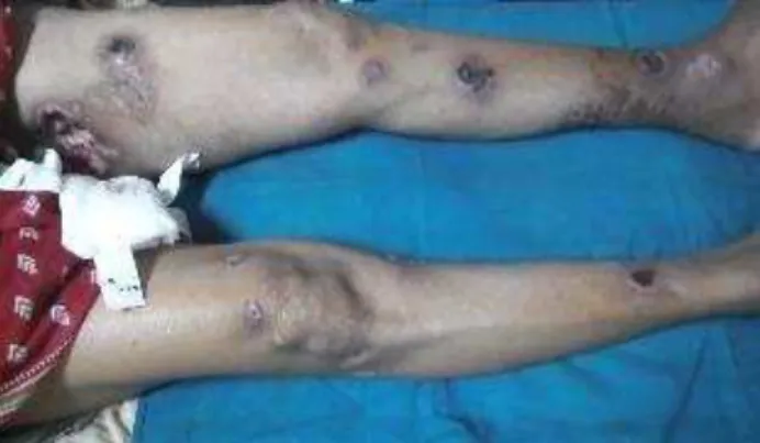
Fig. 1:Multiple Ulcers Arranged in a Linear Pattern on
Both Lower Limbs
Fig. 2: Single Large Nonhealing Ulcer of 10 months’
Jemds.com
Case Report
Journal of Evolution of Medical and Dental Sciences/ eISSN- 2278-4802, pISSN- 2278-4748/ Vol. 4/ Issue 95/ Nov. 26, 2015 Page 16119
Fig. 3: Ulcers over Right Upper Limb
Fig. 4: Ulcer over Left Cubital Fossa
Fig. 5: Large over Inner Aspect of Left Thigh
Fig. 6: Gram’s Staining of Pus from Ulcer
showing Cigarette and Ovoid Bodies
Fig. 7: Fungal Culture showing initial Creamy White Colonies which Later Turned Brown Black
Fig. 8: Lacto Phenol Cotton Blue Staining of Culture Growth showing Thin Twisted Septate Septate Hyphae with
Flower Like Arrangement of Conidia
Fig. 9: Lacto Phenol Cotton Blue Staining of Culture Growth showing Thin Twisted Septate Septate Hyphae with
Flower Like Arrangement of Conidia