In mucormycosis, the most common fungal genus is Rhizopus; however, other orga-nisms associated with human infection belong to the genus Mucor, Rhizomucor, Absidia, Apophysomyces, Saksenaea, Cunninghamella, Cokeromyces and Syncephalastrum.(7,8) The clinical presentation correlates with the predis-posing conditions of the host (Table 1). The clinical course and progression of the disease are typically fulminant due to rapid fungal growth and concomitant tissue destruction, which requires early diagnosis as well as prompt clinical and surgical treatment.(8) Most cases occur in patients with leukemia. Regarding rhinocerebral mucormycosis, the patient characteristically has
Introduction
Zygomycosis is an uncommon fungal infec-tion caused by fungi of the class Zygomycetes, order Mucorales and Entomophthorales. Entomophthoromycosis usually presents as a subcutaneous infections, occurring in tropical areas. Mucormycosis is caused by opportunistic pathogens, mainly resulting in processes that lead to neutropenia or neutrophil dysfunction. Rarely does it generate disease in immunocom-petent patients. Mucormycosis is the third most common invasive fungal infection, following aspergillosis and candidiasis, and accounts for 8.3-13.0% of all fungal infections found in autopsies of hematologic patients.(1-6)
Chapter 7 - Zygomycosis*
Capítulo 7 - Zigomicose
Cecília Bittencourt Severo, Luciana Silva Guazzelli, Luiz Carlos Severo
Abstract
Zygomycosis (mucormycosis) is a rare but highly invasive infection caused by fungi belonging to the order Mucorales, which includes the genera Rhizopus, Mucor, Rhizomucor, Absidia, Apophysomyces, Saksenaea, Cunninghamella, Cokeromyces and Syncephalastrum. This type of infection is usually associated with hematologic diseases, diabetic ketoacidosis and organ transplantation. The most common form of presentation is rhinocerebral mucormycosis, with or without pulmonary involvement. Pulmonary zygomycosis is more common in patients with profound, prolonged neutropenia and can present as segmental or lobar infiltrates, isolated nodules, cavitary lesions, hemorrhage or infarction. The clinical and radiological manifestations are often indistinguishable from those associated with invasive aspergillosis. This article describes the general characteristics of pulmonary zygomycosis, emphasizing laboratory diagnosis, and illustrates the morphology of some lesions.
Keywords: Zygomycosis; Diagnostic techniques and procedures; Mucormycosis.
Resumo
A zigomicose (mucormicose) é uma infecção rara, mas altamente invasiva, causada por fungos da ordem Mucorales (gêneros Rhizopus, Mucor, Rhizomucor, Absidia, Apophysomyces, Saksenaea, Cunninghamella, Cokeromyces e Syncephalastrum). Esse tipo de infecção é usualmente associado a doenças hematológicas, cetoacidose diabética e transplante de órgãos. A apresentação clínica mais frequente é a mucormicose rinocerebral, com ou sem envolvi-mento pulmonar. A zigomicose pulmonar ocorre mais frequentemente em pacientes com neutropenia profunda e prolongada e pode se apresentar como infiltrado lobar ou segmentar, nódulos isolados, lesões cavitárias, hemorragia ou infarto. As manifestações clínicas e radiológicas são na maioria dos casos indistinguíveis daquelas associadas com aspergilose invasiva. Este artigo descreve as características gerais da zigomicose pulmonar, com ênfase no diagnóstico laboratorial, e ilustra a morfologia de algumas lesões.
Descritores: Zigomicose; Técnicas de diagnóstico e procedimentos; Mucormicose.
* Study carried out at the Mycology Laboratory of the Santa Rita Hospital, Santa Casa Hospital Complex, and at the Universidade Federal do Rio Grande do Sul – UFRGS, Federal University of Rio Grande do Sul – Porto Alegre, Brazil.
Correspondence to: L.C. Severo. Laboratório de Micologia, Hospital Santa Rita, Santa Casa - Complexo Hospitalar, Rua Professor Annes Dias, 285, CEP 90020-090, Porto Alegre, RS, Brasil.
Tel 55 51 3214-8409. Fax 55 51 3214-8435. E-mail: [email protected] or [email protected] Financial support: None.
Clinical findings
The clinical manifestations are nonspecific: cough; fever (> 38°C); dyspnea; sputum produc-tion; weight loss; hemoptysis; and chest pain. (3) Hematologic patients can be co-infected with species of Aspergillus sp., Candida sp., cytome-galovirus and bacteria. In addition, bacteria are the reason for the occasional occurrence of an initial response to antibiotic therapy. Patients with diabetes have a tendency to develop endo-bronchial lesions. However, in the absence of this predisposing condition, the manifestation becomes less common. Among the signs, we can include hoarseness, hemoptysis, mediastinal enlargement and atelectasis.(1)
Zygomycetes have marked vascular tropism, causing thrombosis and ischemic necrosis. Consequently, hemoptysis appears as a late finding, which can be fatal if cavitation is located in the innermost area of the lungs, since it affects large diameter vessels. In such cases, surgical resection is mandatory.(1)
Pulmonary mucormycosis is uncommon in immunocompetent hosts. When infection occurs in apparently healthy individuals, its course is not so acute. Such patients can have cavitated lesions, infiltrates or slow-progressing nodules, accompanied by fever and dry cough.(1)
In hematologic patients with focal pulmo-nary infection, the mortality rate ranges from 60 to 100%, and surgical resection can mean the difference between life and death. The method of treatment most often used is the combination of surgery and therapy with amphotericin B.(1,3)
Radiological diagnosis
Conventional radiological techniques are of little use in the diagnosis of zygomycosis. (8) The use of HRCT and, more particularly, the use of magnetic resonance imaging are extre-mely useful for the diagnosis of rhinocerebral, pulmonary and disseminated zygomycosis. Chest CT scans can identify infiltrates suggestive diabetes and ketoacidosis, which, in Brazil,
cons-titutes the principal reported manifestation of mycosis. However, studies are incomplete, since they do not indicate the etiologic agent.(8)
The pulmonary manifestations occur in patients with cancer or in those submitted to bone marrow transplantation, whereas cerebral and disseminated infections predominate in intravenous drug abusers and in patients recei-ving deferoxamine.(9)
Mucormycosis is less common in patients with AIDS because T cell-mediated immunity is not considered an important factor for trigge-ring the infection.(2)
The review of clinical and radiological aspects of mucormycosis, as well as of its diagnosis and treatment, with emphasis on its pulmonary manifestation, has motivated the present study.
Pulmonary mucormycosis
The lung is the second site most affected by mucormycosis, and inhalation of spores is the main route of infection.(7) The clinical presen-tation is indistinguishable from that of invasive pulmonary aspergillosis. Patients with leukemia and lymphoma account for most of the cases (37%), followed by patients with diabetes mellitus (32%).(10) This high prevalence might be related to the large number of patients with diabetes, compared with the number of those with malignant hematologic diseases. Leukemia, lymphoma, multiple myeloma and severe neutro-penia increase the risk of developing pulmonary mucormycosis when these diseases are related to the other clinical presentations of the mycosis.(1) Patients with solid tumors rarely develop pulmo-nary mucormycosis.(1,10-13)
Mucormycosis can develop in the lungs as a result of inhalation of infected material and through hematogenous or lymphatic dissemi-nation. Frequently, if patients are not treated, there is hematogenous dissemination to other organs, particularly to the brain, resulting in death within 2-3 weeks.(8,12)
Table 1 - Clinical presentation and predisposing factors.
Clinical presentation Predisposing factors
Rhinocerebral Diabetic ketoacidosis
Pulmonary Granulocytopenia, lymphoma, leukemia, corticosteroid therapy, diabetes Gastrointestinal Severe malnutrition
Cutaneous Severe burns, cutaneous trauma
a mass-like aspect. Although HRCT is essential, in some cases, it is necessary to complete the investigation with magnetic resonance imaging in order to determine the exact location of the affected area, when surgical treatment is neces-sary, especially in cases of brain abscess.(1,20)
Laboratory diagnosis
The diagnosis is made based on the corre-lation among mycological examinations, histopathological examinations and clinical mani-festations.(8,10) Since this is the most fulminating mycosis, rapid diagnosis is extremely important for management and therapy to be successful. Unfortunately, there are few fungal elements. Therefore, this diagnosis is ultimately supported only by clinical evidence.(21) Since Zygomycetes spores are common in the environment, direct examination to identify the organism is crucial in order for culture to be given weight. However, isolated culture is highly valid in patients with diabetes or neutropenia.
The diagnosis of mucormycosis is rarely suspected in hematologic patients due to the fact that physicians make a presumptive diag-nosis of invasive aspergillosis. In hematologic patients, the diagnosis in life is made in only 23-50% of cases.(4)
Due to the clinical similarity between zygomycosis and other diseases caused by fila-mentous fungi, as well as due to the difficulty in making a specific diagnosis, many cases of of mucormycosis, and which were not detected
on chest X-rays.(14,15)
In pulmonary mucormycosis, the lesions often occur in the upper segments of the upper lobes of the lungs.(15) Using conventional radio-logical techniques, the findings of pulmonary mucormycosis are similar to those of invasive aspergillosis.(1) Both infections have a propensity to exhibit angiotropism and to cause throm-bosis.(16)
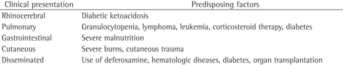
Tomographic findings demonstrate wedge-shaped infiltrates or consolidations and masses. (3) Consolidations are present in approximately 66% of cases, whereas cavitations are present in 40% (Figure 1).(1,3) The halo sign with ground-glass opacity, surrounding the pulmonary nodule, represents hemorrhage and edema, and can develop before and after the neutropenic phase.(1,17) Pleural effusion and multiple nodular pulmonary infiltrates (more than 10) are inde-pendent predictors of zygomycosis.(13,18) Chest HRCT can be sensitive in 26% of unsuspected lesions.(15,19)
Disseminated zygomycosis can begin in the lungs and then proceed to invade more than two noncontiguous organs. Disseminated lesions in the central nervous system, in the liver and in the kidneys are common, causing multiple throm-boembolism and areas of necrosis. Radiological findings vary depending on the organs invaded by Zygomycetes.(1,3,15)
In the brain, the infarcted region presents a hypodense image with areas of hemorrhage and
a b
are identified by their rhizoidal, sporangiophore-like aspect, having thermal tolerance.(23)
The development of molecular techniques, such as polymerase chain reaction, can allow earlier diagnosis in relation to conventional methods.(2)
Direct examination
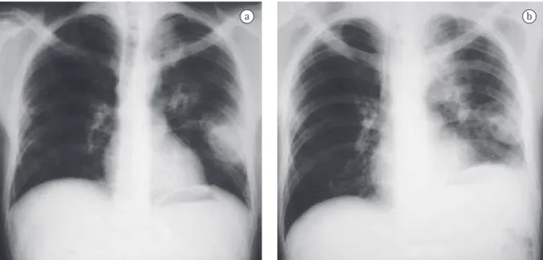
Fungal elements can be found in curettage specimens of or in aspirates from material from the nose in patients with rhinocerebral disease, whereas in patients with pulmonary disease, they can be found in sputum, in bronchial aspi-rates and in transbronchial biopsy specimens (Figure 2). Wide, sparsely septate hyphae, bran-ching at 90°, are seen when potassium hydroxide with Parker ink (Figure 3a) or calcofluor white is added to the material.(13)
Histopathology
The importance of histopathological exami-nation is indisputable, since Zygomycetes can pulmonary zygomycosis are not suspected in
their initial clinical presentation.(22) Therefore, the diagnosis of invasive mycoses requires a high degree of suspicion.(3) The clinical and radiolo-gical presentations of zygomycosis and those of aspergillosis are similar. Culture is often nega-tive in both. The radiologist should use invasive techniques to collect the clinical specimen. A CT scan reveals the extent of the lesion, indi-cates the preferential biopsy site and defines the surgical limits for lesion debridement.(13)
The differential diagnosis with other filamen-tous fungi, such as Candida sp., Aspergillus sp., Scedosporium sp., agents of hyalohyphomycosis and agents of phaeohyphomycosis, is made by histopathological examination. This diagnostic distinction can be made by direct immunofluo-rescence.(13) Fragments of tissue are preferential for diagnosis, since they confirm tissue inva-sion. In zygomycosis, culture is indispensable for an accurate etiological characterization, since microscopy only identifies the fungal class. Agents of zygomycosis show rapid growth and
a b
Figure 2 - Transbronchial biopsy. In a), tissue sample fragment ((H&E; magnification, ×10). In b), higher magnification revealing fragments of wide and irregular Zygomycetes hyphae ((H&E; magnification, ×40).
a b
agar, potato agar or Sabouraud agar medium and incubated at 25°C or 30°C.(13) Nearly all patho-gens of Mucorales are easily isolated from these materials. The culture medium should contain antibiotics for potentially contaminated speci-mens. However, media containing cycloheximide (Mycosel or Mycobiotic) are contraindicated due to the sensitivity of Zygomycetes to this subs-tance.(1,13,24)
Isolating Zygomycetes from tissue can be problematic.(25) In the literature, there are many reports of negative biopsy culture and negative autopsy culture due to maceration of the clinical specimen; since hyphae do not have septum, if there is compression, the cytoplasm will be expelled, preventing fungal growth.(7,16)
Despite the marked angiotropism of Zygomycetes, blood culture is rarely positive, especially when using liquid culture systems.(23)
Treatment
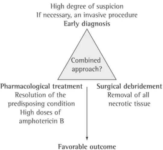
The successful treatment for zygomycosis involves a combined approach. Early diagnosis, prompt initiation of antifungal therapy, correc-tion of the metabolic disorder or resolucorrec-tion of neutropenia are fundamental for therapeutic success (Figure 5).
be found as contaminants in clinical samples. Different staining methods, such as H&E, can be used to observe Zygomycetes hyphae in tissue sections. However, the Grocott technique is the best method for demonstrating hyphae in tissue due to the high contrast with minimal background impregnation.(13) Histopathological examination reveals tissue alterations, such as neutrophilic infiltrate, necrosis, thrombosis and septic infarction, as well as invasion of blood vessels (Figure 4).(23)
Aspergillosis is the infection that most closely mimics zygomycosis (Table 2). Usually, these mycoses are distinguished by the hyphal morphology. However, this distinction is diffi-cult, particularly when Aspergillus sp. hyphae are macerated or when the lesions contain hyphal fragments.(13) There have been reports of cases of zygomycosis confirmed by culture in which the hypha was relatively uniform, with parallel borders and occasional septa. If culture had not been performed, there could have been a misdiagnosis.(22)
Culture methods
Zygomycetes grow in standard laboratory media within 12-18 h after sample inoculation, and colony maturation occurs within 4 days, forming gray to brown cotton-like colonies.(23) It is difficult to establish the diagnosis based only on culture. Pathogenic species of Zygomycetes are part of the environment, are skin and saliva contaminants and grow in nearly all organic substrates.
However, an isolated organism belonging to the order Mucorales cannot be ruled out as a contaminant. The shapes and the sources that reveal the fungus should be judged critically in order to ensure the accuracy of the diagnosis. Due to the marked saccharolytic ability that these fungi have, better fungal isolation is obtained by inoculating fragments on a slice of sterilized, humidified bread (Figure 3b). Secretion, scraping and biopsy material can be inoculated on malt
Figure 4 - Angiotropism: lung parenchyma revealing the marked angiotropism caused by Zygomycetes (Grocott; magnification, ×10).
Table 2 - Histomorphological characteristics of Aspergillus sp. and Zygomycetes.
Characteristic Aspergillus Zygomycetes
Width Narrow (3-6 µm) Wide (5-20 µm)
Caliber Uniform Varying
Branching Regular, acute angle (dichotomous) Random, right angle
Branching orientation Parallel or radial Random
tion in order to reduce the risk of exposure to spores in the air.
Most of these control measures focus on early identification in patients at risk, such as those with prolonged, deep neutropenia. Pre-transplant and chemotherapy rooms are isolated with high efficiency particulate air filters to treat the air, maintaining positive pressure and preventing the accumulation of dust.
Dust should be minimized in the environ-ment of the houses of neutropenic patients. In addition, flowers and plants should be excluded from the environments due to the fact that they can contain a variety of fungal propagules.(7)
Prevention measures for patients other than transplant recipients or patients undergoing chemotherapy include eliminating or minimizing the predisposing conditions and the risk factors for zygomycosis. Chief among these measures are the control of diabetes, the use of binders (excluding deferoxamine), the restriction of the use of aluminum contained in hemodialysis buffer solutions and the rapid detection of the agent.(7)
Final considerations
Pulmonary mucormycosis is a relatively rare disease. However, with the increase in the number of immunocompromised patients, this disease can become more common. Healthy patients with some form of trauma and a history of environmental exposure can also develop this infection.
The treatment of choice is the use of ampho-tericin B (1.0-1.5 mg/kg/day). The total dose of amphotericin B, which has ranged from 2 to 4 g, has yet to be defined. Azole antifungal agents have no proven activity in zygomycosis, although oral posaconazole therapy seems to be promising in patients with malignant hematologic diseases and submitted to stem cell transplantation as well as in those with zygomycosis that is refrac-tory to conventional treatment.(13,26,27)
The use of hematopoietic stimulation factors and hyperbaric oxygen therapy can be benefi-cial, although data supporting routine use are still limited.(13) For the patient to recover, it is important that corticosteroid therapy be reduced or discontinued.(1,28,29)
Early surgical resection has a great impact on the evolution of mucormycosis, since the mortality rate drops from 60% to 11% in these patients.(2,10) The surgical procedure depends on the extent of the disease and should be planned to remove all infected tissue. Lobectomy is the most common procedure, although pneumo-nectomy might also be necessary.(2,10)
Empirical treatment
Unlike X-ray, CT not only reveals a greater number of nodules, but also demonstrates more characteristics of the lesions, including the halo sign. When these findings are identified in immunocompromised patients, zygomycosis and aspergillosis are the main mycoses diagnosed, and empirical treatment with amphotericin B should be strongly considered.
Voriconazole is contraindicated in the empi-rical treatment of zygomycosis since Zygomycetes are not sensitive to this drug. It is suggested that the increase in the number of zygomycosis infections is a result of the increase in the use of voriconazole in patients at high risk for infection with filamentous fungi (Aspergillus sp.).(9)
Preventive measures
Measures to reduce the incidence of zygomycosis in patients at risk are extremely difficult. There is no prophylactic antifungal treatment routine available, and, due to the low prevalence of zygomycosis, there is no real indi-cation for such a routine. The most common preventive measure is environmental
modifica-Combined approach? High degree of suspicion If necessary, an invasive procedure
Early diagnosis
Surgical debridement Removal of all
necrotic tissue Pharmacological treatment
Resolution of the predisposing condition
High doses of amphotericin B
Favorable outcome
and hematopoietic stem cell transplants: an international consensus. Clin Infect Dis. 2002;34(1):7-14.
13. Chayakulkeeree M, Ghannoum MA, Perfect JR. Zygomycosis: the re-emerging fungal infection. Eur J Clin Microbiol Infect Dis. 2006;25(4):215-29.
14. McAdams HP, Rosado de Christenson M, Strollo DC, Patz EF Jr. Pulmonary mucormycosis: radiologic findings in 32 cases. AJR Am J Roentgenol. 1997;168(6):1541-8. 15. Jamadar DA, Kazerooni EA, Daly BD, White CS, Gross
BH. Pulmonary zygomycosis: CT appearance. J Comput Assist Tomogr. 1995;19(5):733-8.
16. Bouza E, Muñoz P, Guinea J. Mucormycosis: an emerging disease? Clin Microbiol Infect. 2006;12(Suppl 7):S7-S23.
17. Murphy RA, Miller WT Jr. Pulmonary mucormycosis. Semin Roentgenol. 1996;31(1):83-7.
18. Chamilos G, Marom EM, Lewis RE, Lionakis MS, Kontoyiannis DP. Predictors of pulmonary zygomycosis versus invasive pulmonary aspergillosis in patients with cancer. Clin Infect Dis. 2005;41(1):60-6.
19. Severo LC. Diagnostico radiologico. In: Palacio A, Pontón J, Guarro J, Quindós G, editors. Guia de bolsillo: Zigomicosis invasoras. Bilbao: Rev Iberoam Micol; 2008. p. 59-63.
20. Gollard R, Rabb C, Larsen R, Chandrasoma P. Isolated cerebral mucormycosis: case report and therapeutic considerations. Neurosurgery. 1994;34(1):174-7. 21. Gonzalez CE, Rinaldi MG, Sugar AM. Zygomycosis.
Infect Dis Clin North Am. 2002;16(4):895-914, vi. 22. Freifeld AG, Iwen PC. Zygomycosis. Semin Respir Crit
Care Med. 2004;25(2):221-31.
23. Chandler FW, Watts JC. Zygomycosis. In: Connor DH, Chandler FW, editors. Pathology of infectious diseases. Stamford: Appleton & Lange; 1997. p. 1113-9. 24. Chung KJ, Bennett JE. Mucormycosis. In:
Kwon-Chung KJ, Bennett JE. Medical Mycology. Philadelphia:
Lea & Febiger; 1992. p. 524-59.
25. Bigby TD, Serota ML, Tierney LM Jr, Matthay MA. Clinical spectrum of pulmonary mucormycosis. Chest. 1986;89(3):435-9.
26. Greenberg RN, Mullane K, van Burik JA, Raad I, Abzug MJ, Anstead G, et al. Posaconazole as salvage therapy for zygomycosis. Antimicrob Agents Chemother. 2006;50(1):126-33.
27. Rogers TR. Treatment of zygomycosis: current and new options. J Antimicrob Chemother. 2008;61 Suppl 1:i35-40.
28. Parfrey NA. Improved diagnosis and prognosis of mucormycosis. A clinicopathologic study of 33 cases. Medicine (Baltimore). 1986;65(2):113-23.
29. Kauffman CA. New antifungal agents. Semin Respir Crit Care Med. 2004;25(2):233-9.
Tissue invasion by hyphae can be seen by microscopy (Grocott method) and is essential for establishing the diagnosis. However, to that end, it is necessary that knowledge on tissue presen-tation of filamentous fungi be mastered.
Successful management continues to be early diagnosis, followed by systemic antifungal therapy (i.v. amphotericin B) and surgical resec-tion combined with control of the underlying disease.
References
1. Prabhu RM, Patel R. Mucormycosis and entomophthoramycosis: a review of the clinical manifestations, diagnosis and treatment. Clin Microbiol Infect. 2004;10 Suppl 1:31-47.
2. Eucker J, Sezer O, Graf B, Possinger K. Mucormycoses. Mycoses. 2001;44(7-8):253-60.
3. Lee FY, Mossad SB, Adal KA. Pulmonary mucormycosis: the last 30 years. Arch Intern Med. 1999;159(12):1301-9. 4. Nosari A, Oreste P, Montillo M, Carrafiello G, Draisci M,
Muti G, et al. Mucormycosis in hematologic malignancies: an emerging fungal infection. Haematologica. 2000;85(10):1068-71.
5. Funada H, Matsuda T. Pulmonary mucormycosis in a hematology ward. Intern Med. 1996;35(7):540-4. 6. Dromer F, Mcginnis MR. Zygomycosis. In: Anaissie EJ,
Pfaller MA, Mcginnis M, editors. Clinical mycology. New York: Churchill Linvigstone; 2003. p. 297-308. 7. Ribes JA, Vanover-Sams CL, Baker DJ. Zygomycetes in
human disease. Clin Microbiol Rev. 2000;13(2):236-301.
8. Severo LC, Oliveira FD, Dreher R, Teixeira PZ, Porto ND, Londero AT. Zygomycosis: A report of eleven cases and a review of the Brazilian literature. Rev Iberoam Micol. 2002;19(1):52-56.
9. Wingard J. Zygomycosis: epidemiology and treatment options. Johns Hopkins Adv Studies Med. 2006;6 Suppl 6:S526-30.
10. Tedder M, Spratt JA, Anstadt MP, Hegde SS, Tedder SD, Lowe JE. Pulmonary mucormycosis: results of medical and surgical therapy. Ann Thorac Surg. 1994;57(4):1044-50.
11. Rinaldi MG. Zygomycosis. Infect Dis Clin North Am. 1989;3(1):19-41.
About the authors
Cecília Bittencourt Severo
Doctoral Student. Postgraduate Program in Pulmonology, Universidade Federal do Rio Grande do Sul – UFRGS, Federal University of Rio Grande do Sul – Porto Alegre, Brazil.
Luciana Silva Guazzelli
Doctoral Student. Postgraduate Program in Pulmonology, Universidade Federal do Rio Grande do Sul – UFRGS, Federal University of Rio Grande do Sul – Porto Alegre, Brazil.
Luiz Carlos Severo