Original Article
1 9 Arq Bras Oftalmol. 2015;78(1):19-22 http://dx.doi.org/10.5935/0004-2749.20150006
INTRODUCTION
Since the 1970’s, endothelial cell loss remains a serious concern regarding a successful outcome of cataract surgery. Corneal endo-thelial damage during surgery is inevitable, and can be inluenced by several preoperative and intraoperative parameters. Preoperative parameters include older age, constricted pupil, irmness of the nu-cleus, and shorter axial length (AL)(1-2). Intraoperative factors include the incision size and design, Descemet membrane detachment, toxic intraoperative medications, phacoemulsiication technique, and the type of ophthalmic viscosurgical device (OVD) used(1-4).
Mechanisms proposed for endothelial cell damage during pha-coemulsiication include turbulence and movement of luids, direct trauma caused by instruments or lens fragments, total amount of ultrasound (US) energy used, US energy dissipated close to the cor-neal endothelium, intraoperative complications (e.g. presence of air bubbles, release of free radicals), low surgeon experience, and pos -toperative inlammation(3-6).
To reduce the energy applied, aspects related to the phacoemul-siier such as ultrasound modes, special tips, microprocessors, and high-vacuum systems must be considered. In the hands of an
expe-Mini-flared Kelman tip, reverse tip, and sidewinder tip with torsional phaco:
a prospective randomized comparative study
Faco torsional utilizando as ponteiras Kelman mini-lared, reversa e sidewinder:
um estudo prospectivo comparativo randomizado
Wilson Takashi hida1, PaTrick Frensel Tzelikis1, celso Takashi nakano2, anTonio Francisco PimenTa moTTa2, milTon ruiz alves2
Submitted for publication: May 13, 2014 Accepted for publication: November 6, 2014
1 Brasilia Ophthalmologic Hospital (HOB), Brasilia, DF, Brazil.
2 Department of Ophthalmology, University of São Paulo (USP), São Paulo, SP, Brazil.
Funding: No specific financial support was available for this study.
Disclosure of potential conflicts of interest: None of the authors have any potential conflicts of interest to disclose.
Corresponding author: Patrick Frensel de Moraes Tzelikis. SQN 203, Bloco K, Ap. 502 - Brasília, DF - 70833-110 - Brazil - E-mail: [email protected]
ClinicalTrials.gov Identifier: NCT02089698 ABSTRACT
Purpose: To compare the efficiency of surgical procedures using three phaco tip designs in torsional phacoemulsification using the bevel-down technique. Methods: In this prospective, comparative, masked study, patients were ran-domly assigned to have torsional coaxial microincision cataract surgery using the mini-flared 45-degree Kelman tip, reversed mini-flared 30-degree Kelman tip, or Sidewinder 30-degree Kelman tip. Clinical measurements included preoperative and 3-month postoperative corrected distance visual acuity (CDVA), endothelial cell counts (ECC), and preoperative and 1-day postoperative central corneal thi-ckness (CCT ). Intraoperative measurements included phaco time, torsional time, aspiration time, case time, cumulative dissipated energy (CDE), and balanced salt solution volume (BSS).
Results: The study evaluated 150 eyes of 150 patients. Intraoperatively, there was no statistically significant difference in cumulative dissipated energy, case time, torsional time, and aspiration time between the three tip configurations. However, less phaco time was used with the mini-flared 45-degree Kelman tip (p=0.02) than that with the Sidewinder 30-degree Kelman tip or reversed mini-flared 30-degree Kelman tip. The mini-flared 45-degree Kelman tip and the reversed mini-flared 30-degree Kelman tip required significantly less balanced salt solution volume than that required by the Sidewinder 30-degree Kelman tip (p=0.009). There was no statistically significant difference in corrected distance visual acuity and endothelial cell counts between tips 3 months postoperatively (p>0.05). Conclusion: All three tips were effective with no intraoperative complications. When using torsional phacoemulsification through microincisions and the prefractu re technique with the bevel-down technique, the mini-flared 45-degree Kelman tip required a lower mean phaco time than the reversed mini-flared 30-degree Kelman tip and the Sidewinder 30-degree Kelman tip.
Keywords: Phacoemulsification/methods; Cataract extraction; Drainage/instrumentation; Equipment design; Cell count; Endothelium, corneal; Visual acuity; Pros -pective studies
RESUMO
Objetivo: Comparar a eficácia cirúrgica da facoemulsificação com tecnologia torcional utilizando 3 modelos diferentes de ponteiras.
Métodos: Neste estudo prospectivo, randomizado, mascarado, os pacientes foram aleatoriamente distribuídos para serem submetidos a cirurgia de facoemulsificação coaxial torcional utilizando a ponteira Kelman mini-flared de 45 graus, ou Kelman reversed mini-flared de 30 graus ou Kelman Sidewinder de 30 graus. Os parâmetros avaliados incluíram: acuidade visual com correção (AVCC) para longe; contagem de células endoteliais (CCE) pré-operatória e pós-operatória, ao final de 3 meses; espes-sura corneana central (ECC) pré-operatória e no primeiro dia pós-operatório. Medidas intraoperatórias incluíram tempo de facoemulsificação, tempo de energia torcional, tempo da aspiração, tempo cirúrgico, energia dissipada acumulada (CDE) e volume de solução salina balanceada (BSS).
Resultados: Este estudo avaliou 150 olhos de 150 pacientes. No intraoperatório, não foram observadas diferenças significativas na energia dissipada acumulada, tempo de facoemulsificação, tempo de energia torcional, e tempo de aspiração entre os 3 modelos de ponteira. No entanto, foi utilizando menos tempo de faco com a ponteira Kelman mini-flared de 45 graus (p=0,02) quando comparado às ponteiras KelmanSidewinder de 30 graus e reversa mini-flared de 30 graus. A ponteira Kelman mini-flared de 45 graus e a reversa mini-flared de 30 graus utilizaram menos solução salina balanceada quando comparado à ponteira Sidewinder de 30 graus (p=0,009). Não foram observadas diferenças significativas na acuidade visual com correção, contagem de células endoteliais e espessura corneana central entre as diferentes ponteiras ao final do estudo (p=0,05).
Conclusão: As 3 ponteiras foram eficazes e não apresentaram complicacões intraope-ratórias. Quando foi utilizando o faco torcional através de microincisão com a técnica da pré-fratura, a ponteira Kelman mini-flared de 45 graus obteve um desempenho melhor que as ponteiras de 30 graus e Sidewinder de 30 graus, com menor tempo de faco.
Mini-flared Kelman tip, reverse tip, and sidewinder tip with torsional phaco: a prospective randomized comparative study
20 Arq Bras Oftalmol. 2015;78(1):19-22
rienced surgeon, the total amount of US energy and the hydrodyna-mic low in the anterior chamber are presumed to be the main da-maging factors to corneal endothelial cells in phacoemulsiication(1). To reduce the amount of total US energy dissipated in the eye, some surgeons use high hydrodynamic parameters (vacuum and low rate) to accelerate surgery(7). Several new phacoemulsiication strategies and new devices were developed to preserve the corneal endothe-lium by attenuating the deleterious efects of US(8). Some surgeons have attempted to alleviate the corneal load by varying the position of the phacoemulsiication tip(4,9).
The purpose of this study was to compare intraoperative and clinical parameters using the mini-lared 45-degree Kelman tip (Alcon Laboratories, Inc.), the reversed mini-lared 30-degree Kelman tip (Alcon Laboratories, Inc.), and the Sidewinder 45-degree Kelman tip (Mastel Precision Surgical, Inc.).
METHODS
P
ATIENTENROLLMENTThis was a prospective, comparative, randomized, patient-mas-ked study. A computer-generated randomization list was used to assign the phacoemulsiication cases to one of three groups with diferent tip conigurations: the mini-lared 45-degree Kelman tip, the reversed 30-degree tip, and the Sidewinder 45-degree Kelman tip, which was specially sharpened on both sides in order to increase the eiciency of torsional phaco (Figure 1).
The study complied with established ethical standards for clini-cal research from the Institutional Review Board (IRB) of the Brasilia Ophthalmologic Hospital (HOB) in Brazil, and conducted between November 2010 and February of 2011. All surgeries were performed at the HOB. Physicians conducting postoperative evaluation did not have access to patients’ medical records. Exclusion criteria were previous ocular surgery, central endothelial cell count less than 2000 cells/mm2, glaucoma or intraocular pressure greater than 21 mmHg, amblyopia, retinal abnormalities, steroid or immunosuppressive treatment, and connective tissue diseases. Enrolled patients were excluded if they had complicated cataract surgery (e.g. posterior capsule rupture, vitreous loss, or an intraocular lens not placed in the capsular bag).
S
URGICALTECHNIQUESurgeries were performed by a senior surgeon (WTH), who had used torsional mode with bevel-down technique in more than 15,000
cases with the same prefracture with preslice and prechop technique. All patients received topical anesthesia with lidocaine 2% gel before surgery. A 2.2 mm self-sealing clear corneal incision on the steepest meridian axis was created. We used a viscoelastic solution of sodium hyaluronate 3% and chondroitin sulfate 4% (Viscoat; Alcon Laborato-ries, Fort Worth, Texas, USA) to reform and stabilize the surgical planes, and to protect the endothelium. A 5.00 to 5.25 mm continuous cur-vilinear capsulorhexis was completed with Utrata forceps. We used the Akahoshi Phaco PreChopper (ASICO Products, Inc.) to fracture the nucleus in half, and removed the nucleus by phacoemulsiication using the Ininite Ozil Vision System (Alcon Surgical) device. The tip of the phaco handpiece was positioned face down during phacoe-mulsiication. The phaco settings were identical in all cases: torsional ultrasound at 20 pulse per second (minimum amplitude of 20%, maximum 80% and time on 85%), longitudinal power at zero, 100 cm irrigation bottle height, minimum 70 and maximum 350 mmHg linear vacuum, aspiration low rate 30 cc/min, dynamic rise zero, and Ozil IP settings 1.0, with 10 ms and 95% power. After cortical aspira-tion, the IOL was placed in the bag with careful centration using the Royale® (Asico, Chicago, CA, USA) or Emerald® (AMO, Santa Anna, CA, USA) delivery system.
S
TUDYPROTOCOLPreoperative Lens Opacities Classiication System III (LOCS III)(10) grading was done using a slit lamp (Topcon, SL 1E) microscope after dilating the pupil with a combination of topical tropicamide 1.0% and phenylephrine 2.5%. Nuclear opacity was graded according to nu clear opalescence (NO) and nuclear color (NC) on a scale of 0.1 to 6.9 by the same ophthalmologist. Cataract was graded using the softwa-re on the Oculus Pentacam, the Pentacam Grading System (PNS), to measure the optical density of the nucleus.
Preoperative examinations included logMAR corrected distance visual acuity (CDVA) with the early treatment of diabetic retinopathy study (ETDRS) chart, central corneal thickness (CCT) (Ocuscan Rxp, Alcon Laboratories), and endothelial cell count (ECC) (Konan Medical).
Intraoperative measurements included cumulative dissipated energy (CDE), phaco time, torsional time, aspiration time, case time, and the amount of balanced salt solution (BSS) used. The mean CDE is the mean percentage of power spent during US and is calculated in torsional mode (torsional amplitude x torsional time x 0.4). It was automatically calculated and displayed on the monitor of the phaco machine.
Hida WT, et al.
21
Arq Bras Oftalmol. 2015;78(1):19-22 The postoperative examination included CDVA, CCT, and ECC
and was performed at 1 day and approximately at 3 months after the surgery.
S
TATISTICALANALYSISWe used SPSS Statistics (version 17.0, SPSS, Inc., Chicago, Illinois, USA) for all data analyses, using analysis of variance (ANOVA). We tested group diferences in parameters using 1-way ANOVA, and used the Levene statistic to test the equality of group variances in the 1-way ANOVA. We tested multiple comparisons using the Tukey test when the group variances were equal pairwise and the Tamhane test when they were unequal. Diferences were considered statistically signiicant when the P value was less than 0.05.
RESULTS
The study included 150 eyes of 150 patients, 83 (55%) patients were women. The mean patient age was 62.54 years ± 5.81 (SD) (range 45 to 85 years). Table 1 shows the preoperative patient characteristics by phaco tip and by the LOCS III and PNS cataract
grading systems. The mean LOCS III score was 4.29 ± 0.43; the mean Scheimplug-measured lens nuclear density was 3.82 ± 0.44. There were no statistically signiicant diferences in age or sex between the three phaco-tip groups.
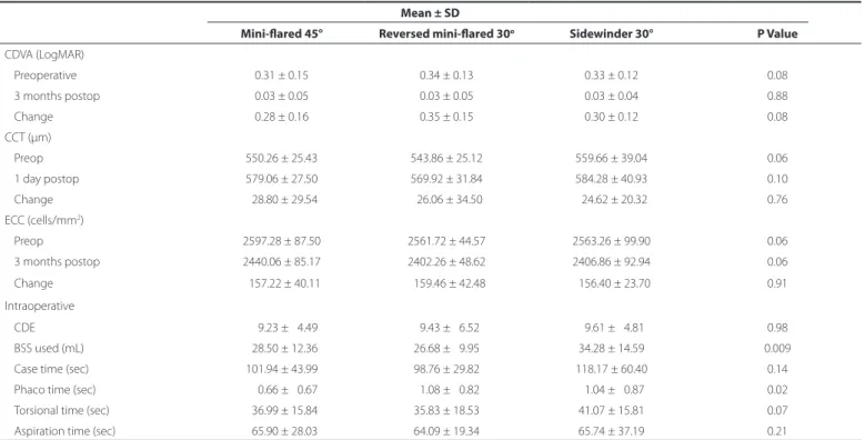
Table 2 shows the intraoperative and postoperative results. No intraoperative complications were observed. The improvement in CDVA was similar in all groups. There were no statistically signiicant diferences in the change in CCT (1-day postoperative CCT minus preoperative CCT) (p=0.76) or the change in ECC (3-month postope-rative ECC minus preopepostope-rative ECC) (p=0.91) between the three tips. When comparing CDE, case time, torsional time and aspiration time, there were no statistically signiicant diferences between any of the tips (Table 2).
The phaco time was statistically signiicantly less with the 45-de-gree Kelman tip than with the Sidewinder 30-de45-de-gree Kelman tip and the reversed mini-lared 30-degree Kelman tip (p=0.02). The mini-la-red 45-degree Kelman tip and the reversed mini-lamini-la-red 30-degree Kel-man tip required signiicantly less BSS than the Sidewinder 30-degree Kelman tip (p=0.009, Table 2).
Table 1. Preoperative patient characteristics
Phaco Tip
Mini-lared 45° Reversed mini-lared 30° Sidewinder 30° P value
Eyes (n) 50 50 50
-Mean age (y) ± SD 63.34 ± 5.21 62.78 ± 4.69 63.07 ± 7.71 0.43
Mean LOCS III score ± SD 04.22 ± 0.58 04.18 ± 0.82 04.28 ± 0.81 0.39
Mean PNS score ± SD 03.84 ± 0.81 03.66 ± 0.99 03.96 ± 0.94 0.26
LOCS III= lens opacities classiication system III; PNS= pentacam nucleus score (cataract grading system).
Table 2. Clinical and intraoperative results
Mean ± SD
Mini-lared 45° Reversed mini-lared 30o Sidewinder 30° P Value
CDVA (LogMAR)
Preoperative 0.31 ± 0.15 0.34 ± 0.13 0.33 ± 0.12 0.08
3 months postop 0.03 ± 0.05 0.03 ± 0.05 0.03 ± 0.04 0.88
Change 0.28 ± 0.16 0.35 ± 0.15 0.30 ± 0.12 0.08
CCT (μm)
Preop 550.26 ± 25.43 543.86 ± 25.12 559.66 ± 39.04 0.06
1 day postop 579.06 ± 27.50 569.92 ± 31.84 584.28 ± 40.93 0.10
Change 028.80 ± 29.54 026.06 ± 34.50 024.62 ± 20.32 0.76
ECC (cells/mm2)
Preop 2597.28 ± 87.50 2561.72 ± 44.57 2563.26 ± 99.90 0.06
3 months postop 2440.06 ± 85.17 2402.26 ± 48.62 2406.86 ± 92.94 0.06
Change 0157.22 ± 40.11 0159.46 ± 42.48 0156.40 ± 23.70 0.91
Intraoperative
CDE 009.23 ± 04.49 09.43 ± 06.52 09.61 ± 04.81 0.98
BSS used (mL) 028.50 ± 12.36 26.68 ± 09.95 34.28 ± 14.59 0.009
Case time (sec) 101.94 ± 43.99 98.76 ± 29.82 118.17 ± 60.40 0.14
Phaco time (sec) 000.66 ± 00.67 01.08 ± 00.82 01.04 ± 00.87 0.02
Torsional time (sec) 036.99 ± 15.84 35.83 ± 18.53 41.07 ± 15.81 0.07
Aspiration time (sec) 065.90 ± 28.03 64.09 ± 19.34 65.74 ± 37.19 0.21
Mini-flared Kelman tip, reverse tip, and sidewinder tip with torsional phaco: a prospective randomized comparative study
22 Arq Bras Oftalmol. 2015;78(1):19-22 DISCUSSION
Over the years, there have been signiicant advances in cataract surgery. Improvements in control of phacoemulsiication tip stroke amplitude, modulation of US “on” time via burst and pulse modes, and improved control of vacuum, postocclusion surge and aspiration low rate have allowed cataract surgical techniques to evolve to high levels of eiciency(11). Although many studies of phacoemulsiication techniques have been published, much less is published about the hardware (tips) used in the procedure. Textbooks primarily discuss various bevel angles and shapes without providing outcome data(12). Advances in phacoemulsiication surgery with diferent phaco micro-tips, sleeves, and torsional movement have resulted in a very efective procedure for patients, providing faster visual recovery and earlier return to routine daily activities(13).
Higher US power (cumulative dissipated energy) and longer phaco times have been implicated in endothelial cell loss(14,15). O’Brien et al.(14) found a signiicant association between phaco time, mean US power, and endothelial cell loss. Dick et al.(15) found a signiicant correlation between phaco time and central endothelial cell loss, but not between phaco energy and cell loss. Other studies found no correlation(16,17). In the present study, we compared clinical and intraoperative parameters of three phaco tips designs in torsional phacoemulsiication using the bevel-down technique: the Sidewinder 30-degree Kelman tip, the mini-lared 45-degree Kelman tip, and the reversed mini-lared 30-degree Kelman tip. There was a statisti-cally signiicant diference in phaco time and BSS used between the mini-lared 45-degree Kelman tip and the Sidewinder 30-degree Kel-man tip and reversed mini-lared 30-degree KelKel-man tip. However, there was no signiicant correlation between these parameters and endothelial cell loss. In addition, the cell loss was not signiicantly diferent between the tips.
The mean phaco time difered between tips. The mini-lared 45-degree Kelman tip required the least phaco time (0.66 seconds), the reversed mini-lared 30-degree Kelman tip required the most (1.08 seconds). The diference is statistically signiicant (p=0.02). The efectiveness of phacoemulsiication depends on the proportion of applied US energy and luid exchange in the anterior chamber du-ring removal of nuclear material. Dudu-ring phacoemulsiication, a hard nucleus requires more US energy than a soft nucleus. Therefore, the energy recorded as the CDE relects the eicacy of the tips. In our study, there was no statistical diference between the three groups when comparing CDE, case time, torsional time and aspiration time. Kim et al.(18) published a similar report comparing the mini-lared 30-degree Kelman tip, mini-lared 45-degree Kelman tip, and reverse mini-lared 30-degree Kelman tip in torsional phacoemulsiication cases, also using the bevel-down technique. In their study, the CDE use was statistically signiicantly less with the 45-degree Kelman tip and the reverse 30-degree Kelman tip than with the 30-degree Kelman tip (p<0.05), with no signiicant diferences in CDE use between the 45-degree Kelman tip and the reverse 30-degree Kelman tip. No case time, phaco time, torsional time and aspiration time were measured intraoperatively.
We veriied that the mini-lared 45-degree Kelman tip and the reversed mini-lared 30-degree Kelman tip required signiicantly less BSS than the Sidewinder 30-degree Kelman tip (28.50 mL and 26.68 mL versus 34.28 mL; P=0.009). The volume of luid used has been implica-ted as a risk factor for corneal endothelial damage, because irrigation low and turbulence within the anterior chamber may compromise the glycoprotein coat of the endothelium and induce stress(19). Whether the increased risk for endothelial damage is related to luidic princi-ples or simply to the fact that volume infused is a surrogate measure of needle time is unclear(20).
Regarding study weakness, the irst limitation of this study is surgeon bias. Although there is a possibility that the surgeon knew which tip he was using during each surgery, he tried to use his normal phacoemulsiication technique without variation for each surgery, regardless of tip. A second limitation is the small number of cases
analyzed. Although higher case numbers might be preferred, analysis of the diferences between sample means for tips was shown to be statistically signiicant. A third limitation is that we did not divide the cases into groups based on the nuclear opacity grade. We might observe diferent results with moderate to severe nuclear density.
CONCLUSION
Our data shows that to minimize luid use and ultrasound energy with the prefracture technique, the mini-lared 45 degrees Kelman tip was the most eicient, requiring less phaco time and BSS. The CDVA results were similar between tips, showing that all tips conigurations are safe and homogeneous, leading to fast rehabilitation and good visual quality. Additional research of phaco tips should contribute to improving the eiciency of various surgical approaches. Further studies are needed to evaluate the tip cutting geometry and the efect of refurbishing.
REFERENCES
1. Hayashi K, Hayashi H, Nakao F, Hayashi F. Risk factors for corneal endothelial injury du ring phacoemulsiication. J Cataract Refract Surg. 1996;22(8):1079-84.
2. Walkow T, Anders N, Klebe S. Endothelial cell loss after phacoemulsiication: relation to preoperative and intraoperative parameters. J Cataract Refract Surg. 2000;26(5):727-32. 3. Pirazzoli G, D’ Eliseo D, Ziosi M, Acciarri R. Efects of phacoemulsiication time on the corneal endothelium using phacofracture and phaco chop techniques. J Cataract Re-fract Surg. 1996;22(7):967-9.
4. Faramarzi A, Javadi MA, Karimian F, Jafarinasab MR, Baradaran-Raii A, Jafari F, et al. Corneal endothelial cell loss during phacoemulsiication: bevel-up versus bevel-down phaco tip. J Cataract Refract Surg. 2011;37(11):1971-6. Comment in: J Cataract Refract Surg. 2012;38(6):1113-4; author reply 1114; J Cataract Refract Surg. 2012;38(6):1114-5; author reply 1115;J Cataract Refract Surg. 2012; 38(6):1113; author reply 1113. 5. Vargas LG, Holzer MP, Solomon KD, Sandoval HP, Aufarth GU, Apple DJ. Endothelial
cell integrity after phacoemulsiication with 2 diferent handpieces. J Cataract Refract Surg. 2004;30(2):478-82.
6. Fine IH, Packer M, Hofman RS. Use of power modulations in phacoemulsiication: choo-choo chop and lip phacoemulsiication. J Cataract Refract Surg. 2001;27(2): 188-97. Comment in: J Cataract Refract Surg. 2001;27(2):175.
7. Díaz-Valle D, Benítez del Castillo Sánchez JM, Castillo A, Sayagués O, Moriche M. En dothelial damage with cataract surgery techniques. J Cataract Refract Surg. 1998; 24(7):951-5.
8. Payne M, Georgescu D, Waite AN, Olson RJ. Phacoemulsiication tip vacuum pressure: comparison of 4 devices. J Cataract Refract Surg. 2006;32(8):1374-7.
9. Frohn A, Dick HB, Fritzen CP. Corneal impact of ultrasound and bevel position in phacoemulsiication. J Cataract Refract Surg. 2002;28(9):1667-70.
10. Chylack LT Jr, Wolfe JK, Singer DM, Leske MC, Bullimore MA, Bailey IL, et al. The Lens Opacities Classiication System III. The Longitudinal Study of Cataract Study Group. Arch Ophthalmol. 1993;111(6):831-6.
11. Hofman RS, Fine IH, Packer M. New phacoemulsiication technology. Curr Opin Ophthalmol. 2005;16(1):38-43.
12. Packer M, Fishkind WJ, Fine IH, Seibel BS, Hofman RS. The physics of phaco: a review. J Cataract Refract Surg. 2005;31(2):424-31.
13. Alio J, Rodriguez-Prats JL, Galal A, Ramzy M. Outcomes of microincision cataract sur-gery versus coaxial phacoemulsiication. Ophthalmology. 2005;112(11):1997-2003. Comment in:Ophthalmology. 2006; 113(9):1687; author reply 1687.
14. O’Brien PD, Fitzpatrick P, Kilmartin D, Beatty S. Risk factors for endothelial cell loss after phacoemulsiication surgery by a junior resident. J Cataract Refract Surg. 2004; 30(4):839-43.
15. Dick HB, Kohnen T, Jacobi FK, Jacobi KW. Long-term endothelial cell loss following phacoemulsiication through a temporal clear corneal incision. J Cataract Refract Surg. 1996;22(1):63-71.
16. Kosrirukvongs P, Slade SG, Berkeley RG. Corneal endothelial changes after divide and conquer versus chip and lip phacoemulsiication. J Cataract Refract Surg. 1997;23(7):1006-12. Comment in: J Cataract Refract Surg. 1997;23(7):967-8. 17. Zetterström C, Laurell CG. Comparison of endothelial cell loss and
phacoemulsiica-tion energy during endocapsular phacoemulsiicaphacoemulsiica-tion surgery. J Cataract Refract Surg. 1995;21(1):55-8.
18. Kim EK, Jo KJ, Joo CK. Comparison of tips in coaxial microincision cataract surgery with the bevel-down technique. J Cataract Refract Surg. 2011;37(11):2028-33. Comment in: J Cataract Refract Surg. 2012;38(5):925-6.