Comparison of 7.2% hypertonic saline - 6%
hydro-xyethyl starch solution and 6% hydrohydro-xyethyl starch
solution after the induction of anesthesia in patients
undergoing elective neurosurgical procedures
Liujiazi Shao, Baoguo Wang*, Shuangyan Wang, Feng Mu, Ke Gu
Department of Anesthesiology, Beijing Sanbo Brain Hospital, Capital Medical University, Beijing, China. *corresponding author.
OBJECTIVE:The ideal solution for fluid management during neurosurgical procedures remains controversial. The aim of this study was to compare the effects of a 7.2% hypertonic saline - 6% hydroxyethyl starch (HS-HES) solution and a 6% hydroxyethyl starch (HES) solution on clinical, hemodynamic and laboratory variables during elective neurosurgical procedures.
METHODS:Forty patients scheduled for elective neurosurgical procedures were randomly assigned to the HS-HES group or the HS-HES group. After the induction of anesthesia, patients in the HS-HS-HES group received 250 mL of HS-HES (500 mL/h), whereas the patients in the HES group received 1,000 mL of HES (1000 mL/h). The monitored variables included clinical, hemodynamic and laboratory parameters. Chictr.org: ChiCTR-TRC-12002357
RESULTS:The patients who received the HS-HES solution had a significant decrease in the intraoperative total fluid input (p,0.01), the volume of Ringer’s solution required (p,0.05), the fluid balance (p,0.01) and their dural tension scores (p,0.05). The total urine output, blood loss, bleeding severity scores, operation duration and hemodynamic variables were similar in both groups (p.0.05). Moreover, compared with the HES group, the HS-HES group had significantly higher plasma concentrations of sodium and chloride, increasing the osmolality (p,0.01).
CONCLUSION:Our results suggest that HS-HES reduced the volume of intraoperative fluid required to maintain the patients undergoing surgery and led to a decrease in the intraoperative fluid balance. Moreover, HS-HES improved the dural tension scores and provided satisfactory brain relaxation. Our results indicate that HS-HES may represent a new avenue for volume therapy during elective neurosurgical procedures.
KEYWORDS: Hypertonic Saline; Hydroxyethyl Starch; Neurosurgery; Fluid Management.
Shao L, Wang B, Wang S, Mu F, Gu K. Comparison of 7.2% hypertonic saline - 6% hydroxyethyl starch solution and 6% hydroxyethyl starch solution after the induction of anesthesia in patients undergoing elective neurosurgical procedures. Clinics. 2013;68(3):323-328.
Received for publication onSeptember 27, 2012;First review completed onOctober 22, 2012;Accepted for publication onNovember 9, 2012 E-mail: [email protected]
Tel.: 86 10 62856766
& INTRODUCTION
Intraoperative fluid management is a critical component of perioperative care in neurosurgical practice (1). Patients undergoing elective neurosurgical procedures sometimes require large volumes of intravenous fluid to maintain hemodynamic homeostasis during surgery. However, these volumes may leave patients with an excessive fluid load that increases risks for various complications such as the
formation of cerebral edema, which is detrimental during neurosurgical procedures. The main goal of intraoperative fluid management is to achieve hemodynamic homeostasis via the administration of small fluid volumes while avoiding fluid overload (2). Currently, increasing investiga-tion into the ideal soluinvestiga-tion for volume therapy during neurosurgical procedures is being conducted around the world.
Since small volume resuscitation with hypertonic saline (HS) in patients suffering from hemorrhage shock was first described in the 1980s (3), HS with or without a colloid such as dextran or hydroxyethyl starch (HES) has emerged as an attractive alternative in fluid management in a variety of surgical practices (4). For example, a wealth of evidence from cardiac surgeries has shown that HS with or without a colloid can exert significant beneficial effects in maintaining hemodynamic stability and a positive fluid balance (5-9).
Copyrightß2013CLINICS– This is an Open Access article distributed under the terms of the Creative Commons Attribution Non-Commercial License (http:// creativecommons.org/licenses/by-nc/3.0/) which permits unrestricted non-commercial use, distribution, and reproduction in any medium, provided the original work is properly cited.
No potential conflict of interest was reported.
Moreover, similar benefits from HS with or without HES have also been demonstrated in aortic aneurysm surgery (10-12) and abdominal hysterectomy (13,14). However, to our knowledge in the clinical setting of neurosurgical practice, HS is mainly used to control intracranial hyperten-sion (ICP) due to its hyperosmolar nature (15), and little is known about the possible role of HS-HES in intraoperative fluid management during elective neurosurgical proce-dures.
In the prospective randomized clinical study presented here, we compared the effects of a 7.2% HS - 6% HES solution (HS-HES) and a 6% HES solution (HES) in terms of the clinical, hemodynamic and laboratory measurements in patients undergoing elective neurosurgical procedures. Our hypothesis was that HS-HES could reduce the volume of intraoperative fluid required to maintain the patients undergoing surgery and lower the dural tension during the operation.
& MATERIALS AND METHODS
Study population
Approval for the study was obtained from the Ethics Committee of Beijing Sanbo Brain Hospital, Capital Medical University, and written informed consent was obtained from all of the study participants. This study enrolled 40 consecutive American Society of Anesthesiologists (ASA) ASA I-II patients scheduled for elective neurosurgical procedures. The exclusion criteria were age ,18 years or .80 years; clinical signs of significantly increased ICP such as severe headache, blurred vision and/or papilledema; history of cardiac, pulmonary and renal dysfunction; fluid or electrolyte disturbances; preoperative coagulation dis-orders; and preoperative treatment with diuretics and/or osmotic agents. The protocol was registered at Chictr.org (ChiCTR-TRC-12002357).
Anesthetic management
Patients were premedicated with intravenous midazolam (0.05 mg/kg) 10 min before the induction of anesthesia. General anesthesia was induced with intravenous fentanyl (2mg/kg), vecuronium bromide (0.1 mg/kg) and propofol
(2 mg/kg). After the induction of anesthesia, radial arterial catheters and right internal jugular venous catheters were inserted. After endotracheal intubation, the lungs were mechanically ventilated with intermittent positive pressure ventilation to maintain the end-tidal CO2 pressure at 4.0-4.67 kPa (30-35 mmHg) during the operation. Anesthesia was maintained with isoflurane (end-tidal minimum alveo-lar concentration of 1-1.2%) combined with an intravenous bolus of fentanyl or vecuronium if necessary.
Intraoperative fluid management
After stabilization at a steady hemodynamic state after the induction of anesthesia, the patients were randomly assigned to receive 250 mL of a 7.2% HS - 6% HES solution (HS-HES group, 500 mL/h) or 1,000 mL of a 6% HES solution (HES group, 1,000 mL/h) through a central venous line. Both solutions were supplied by Fresenius Company (Bad Homburg, Germany). All of the patients in both groups were routinely given 250 mL of 20% mannitol over 10 min at 1 hr after the start of volume expansion. Ringer’s solution was used as a maintenance fluid during the study period at a rate targeted for maintaining the central venous
pressure (CVP) at 8-12 mmHg (10.7-16.0 cmH2O) and the mean arterial pressure (MAP) at$65 mmHg. Packed red blood cells (PRBC) were transfused if the hemoglobin concentration fell below 10 g/L. No additional colloid was used during the operation.
Measurements
For all of the patients, each set of measurements included clinical, hemodynamic and laboratory measurements.
The clinical variables included the volumes of Ringer’s solution and PRBC infused, intraoperative total urine output, blood loss, operative duration, intraoperative bleeding sever-ity score and dural tension score. The fluid balance was calculated as the difference between the intraoperative total input (randomized solution, 20% mannitol, Ringer’s solution and PRBC) and the output (intraoperative total urine output and blood loss). The blood loss was estimated according to the difference between the volume of fluid collected in the graduated suction bottles and surgical drapes and the volume of any washout fluids used during the operation.
The intraoperative bleeding severity was assessed in a blinded manner by the neurosurgeons according to the following grading scale: I. Mild bleeding and no suctioning of blood was required; II. Mild bleeding with occasional suctioning required, but the surgical field was not obscured; III. Moderate bleeding with frequent suctioning required, and the bleeding obscured the surgical field after the suction was removed; IV. Severe bleeding with constant suctioning required, the bleeding appeared again before it was removed by the suction and the surgical field was severely obscured, making surgery impossible.
The dural tension was used to estimate the degree of brain relaxation and was determined immediately after the opening of the dura by the neurosurgeons who were blinded to the group assignments. The dural tension scores were assigned using the following scale as described by Cold et al. (16) with a minor modification: I. Normal dural tension: the neurosurgeon easily opened the dura mater; II. Increased dural tension: the dura mater could be opened without additional procedures to lower the ICP; III. Markedly increased dural tension: additional procedures were necessary to lower the ICP to open the dura mater.
The hemodynamic measurements included the heart rate (HR), MAP (via the radial artery) and CVP (via the right internal jugular vein), which were monitored continuously using an anesthesia monitor (GE-Ohmeda S/5, USA). The laboratory measurements consisted of the hemoglobin concentration (Hb), platelet count (Plt), hematocrit (Hct), coagulation parameters (prothrombin time (PT), activated partial thromboplastin time (APTT) and fibrinogen concen-tration (Fbg)), plasma electrolyte concenconcen-trations (sodium, potassium, chloride and calcium) and osmolality. All of the hemodynamic and laboratory measurements were docu-mented before the HS-HES or HES infusion (T0) and then again at 30 min (T1), 60 min (T2), 70 min (T3,following the administration of mannitol), 120 min (T4) and 180 min (T5) after the start of infusion.
Statistical analysis
variables), a Mann-Whitney U test (intraoperative bleeding severity scores and dural tension scores) and an unpaired Student’s t test (age, weight, operation duration, intrao-perative total fluid input, volume of Ringer’s solution, PRBC, total urine output, blood loss and fluid balance). For variables with multiple measurements (hemodynamic and laboratory variables), a repeated measures analysis of variance was used to evaluate the effects of time and group assignment, while two-way analysis of variance was used to compare the difference within the groups. Significance was established atp,0.05.
& RESULTS
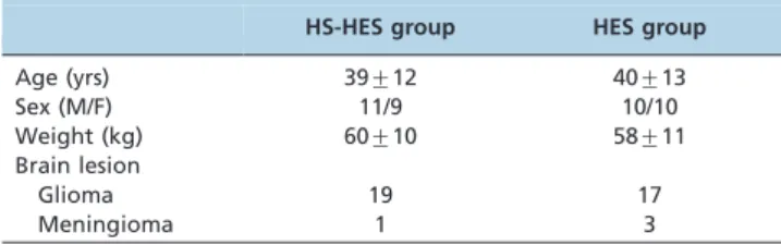
As shown in Table 1, there were no significant differences between the two groups with respect to demographic data (age, sex, weight and surgical procedures).
With regard to the clinical measurements, the two groups did not significantly differ by operation duration, volume of PRBC, intraoperative total urine output, blood loss or intraoperative bleeding severity scores (Table 2). The percentages of patients who required a PRBC transfusion in the HS-HES group and the HES group were 25% (5/20) and 20% (4/20), respectively (Table 2). Compared with HES infusion, HS-HES infusion reduced the total fluid input (p,0.01, Table 2) and the volume of Ringer’s solution (p,0.05, Table 2) required during the operation. Moreover, the fluid balance of the HS-HES group was significantly lower than the HES group (p,0.01, Table 2), and HS-HES
treatment also led to a significant decrease in the dural tension scores compared with HES infusion (p,0.05, Table 2).
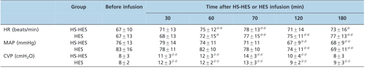
The changes in HR, MAP and CVP were similar in both groups during the study period (Table 3). Specifically, the HR and CVP in both groups significantly increased after the volume infusion and then decreased over time (Table 3), whereas the MAP was lower at T4and T5than at T0in each group.
There were no significant differences between the groups in terms of the Hb, Plt, Hct, PT, APTT and Fbg values and the plasma potassium or calcium concentrations, but the plasma levels of sodium and chloride and the osmolality were markedly increased in the HS-HES group compared with the HES group (Table 4). After the volume expansion, the Hb, Plt, Hct and Fbg values and the plasma calcium concentration in both groups decreased significantly by varying degrees, while the PT and APTT values increased significantly during the study period. The plasma potas-sium concentration displayed biphasic changes with an increase at the early time points and a decrease at the later time points after HS-HES or HES infusion. Furthermore, in the HS-HES group, the concentrations of plasma sodium and chloride peaked immediately at the end of the HS-HES infusion (T1) and remained higher than that of the HES group throughout the study period. The plasma osmolality was significantly higher after HS-HES infusion (T1-5) than prior to HS-HES infusion (T0).
& DISCUSSION
In the present study, we compared the effects of HS-HES and HES in terms of the clinical, hemodynamic and laboratory measurements in patients undergoing elective neurosurgical procedures. The main findings were as follows: 1) Administration of 250 mL of HS-HES was as efficacious as 1,000 mL of HES in maintaining hemody-namic homeostasis during the operation. 2) HS-HES treatment significantly reduced the intraoperative total fluid input and the volume of Ringer’s solution required to maintain the patients undergoing surgery and led to a significant decrease in the intraoperative fluid balance. 3) The majority of the reduction in total fluid input (61%) was due to the difference in the volume of HES (1,000 mL) and HS-HES (250 mL) initially administered. A reduction in the administration of Ringer’s solution accounted for 39% of the difference. 4) HS-HES infusion markedly reduced the dural tension scores and provided more satisfactory brain relaxa-tion for the operarelaxa-tions. 5) HS-HES infusion significantly increased the plasma concentrations of sodium and chloride and increased the plasma osmolality. These findings provide substantial evidence that the HS-HES solution is an attractive choice for fluid management during neuro-surgical procedures.
The ideal solution for intraoperative volume therapy is controversial. In neurosurgical procedures, achieving hemo-dynamic homeostasis by the administration of small fluid volumes is of major interest for avoiding fluid overload. HS has been used in the clinic for several decades; its osmotic and volume-expanding properties make it effective as a reliable solution for intraoperative volume therapy. Thus, there is great interest in the use of HS during operations. Moreover, HS is usually used in association with a colloid in most research studies because a wealth of evidence has
Table 1 -Demographic data for the patients treated with HS-HES (n = 20) or HES (n = 20).
HS-HES group HES group
Age (yrs) 39¡12 40¡13
Sex (M/F) 11/9 10/10
Weight (kg) 60¡10 58¡11
Brain lesion
Glioma 19 17
Meningioma 1 3
The data are presented as the mean¡SD. HS-HES: 7.2% hypertonic saline - 6% hydroxyethyl starch solution, HES: 6% hydroxyethyl starch solution.
Table 2 -Clinical parameters of the patients treated with HS-HES (n = 20) or HES (n = 20).
HS-HES group HES group Operation duration (min) 350¡129 360¡96 Total fluid input (mL) 4261¡674** 5498¡676
HS-HES 250 0
HES 0 1000
20% mannitol 250 250
Ringer’s solution 3687¡659* 4178¡639
PRBC 74¡141 70¡152
Percentage of patients requiring a PRBC transfusion
25% (5/20) 20% (4/20) Total urine output (mL) 1430¡572 1585¡653
Blood loss (mL) 591¡296 470¡245
Fluid balance (mL) 2240¡706** 3443¡553 Dural tension scores (Grade I/II/III) 14/5/1* 6/11/3 Bleeding severity scores
(Grade I/II/III/IV)
8/9/3/0 9/8/3/0
*
p,0.05versusthe HES group,
**
shown that a colloid such as HES can prolong the hemodynamic efficiency of HS (17).
Previous studies on models of hemorrhagic shock have indicated that HS infusion in a volume equivalent to 25% of the total blood lost can restore hemodynamic stability (18,19), whereas an HES infusion can replace the blood lost for a volume expansion at close to a 1:1 ratio (20,21). In the present study, our data showed that there were no significant differences in the hemodynamic parameters between the groups after the infusion of 250 mL of HS-HES or 1,000 mL of HS-HES, which corroborate the above results. Moreover, our results indicated that HS-HES could reduce the intraoperative total fluid input and the volume of Ringer’s solution required to maintain the patients under-going surgery, which was in agreement with the evidence that HS can be used as the fluid for small-volume resuscitation in patients with hypovolemic shock (22). However, in the present study, the majority of the total fluid input reduction was due to the initial fluid load. Thus, because the magnitude of the hemodilution and CVP were
similar in the present study, we assumed that the volume of the plasma expansion was similar in both groups in spite of the different volumes of fluid administered. Multiple mechanisms are involved in the volume-expanding proper-ties of HS, including the compartment redistribution with fluid shifts to the vascular bed (8), the positive effects on cardiac output (23) and the hormonal and immunologic effects (24). Fluid balance during an operation and the postoperative period are of considerable importance to organic function, especially to cerebral function in neuro-surgical procedures. In the present study, we investigated the effect of HS-HES on the fluid balance during surgery and found that HS-HES infusion could significantly reduce the intraoperative fluid balance. Although we did not collect any data on the effect of HS-HES on the postoperative fluid balance, the results from other authors have demonstrated that a near-zero fluid balance was observed in patients who were given an HS-dextran infusion 48 hrs after cardiac surgery (8,25), which suggests that the maximal effect of HS-HES on the reduction of the fluid balance might appear
Table 3 -Hemodynamic parameters of the patients treated with HS-HES (n = 20) or HES (n = 20).
Group Before infusion Time after HS-HES or HES infusion (min)
30 60 70 120 180
HR (beats/min) HS-HES 67¡10 71¡13 75¡12## 78
¡13## 71
¡14 73¡16#
HES 67¡13 68¡13 72¡15#
77¡15##
75¡11##
77¡13##
MAP (mmHg) HS-HES 76¡13 79¡14 74¡11 71¡11 67¡9## 68
¡9##
HES 83¡16 78¡11 82¡10 78¡10 74¡11##
69¡11##
CVP (cmH2O) HS-HES 8¡3 11¡3## 12¡3## 14¡3## 10¡4## 8¡3
HES 8¡2 12¡3##
12¡2##
13¡3##
9¡2##
9¡3## #
p,0.05versusthe measurement before infusion,
##
p,0.01versusthe measurement before infusion. The data are presented as the mean¡SD. HR: heart rate, MAP: mean arterial pressure, CVP: central venous pressure.
Table 4 -Laboratory parameters of the patients treated with HS-HES (n = 20) or HES (n = 20).
Group Before infusion Time after HS-HES or HES infusion (min)
30 60 70 120 180
Hb (g/L) HS-HES 129.1¡11.6 110.4¡10.4## 109.9
¡11.4## 101.7
¡11.0## 110.4
¡11.8## 107.9 ¡14.1##
HES 138.8¡19.9 116.2¡18.7##
113.7¡29.0##
100.2¡16.7##
105.7¡17.9##
106.9¡19.5##
Plt (6107/L) HS-HES 208.2¡53.7 197.4¡43.7## 197.0¡45.1## 184.4¡43.3## 204.4¡48.0 200.2¡46.4
HES 220.4¡42.9 205.2¡37.6##
188.0¡36.0##
173.3¡37.7##
191.3¡38.9##
198.6¡49.3##
Hct (%) HS-HES 38.4¡3.2 33.0¡3.2## 32.8¡3.4## 30.6¡3.2## 33.3¡3.5## 32.5¡3.9##
HES 41.3¡5.0 34.7¡4.9## 32.5
¡4.7## 30.3
¡4.6## 31.7
¡4.8## 32.3 ¡5.5##
PT (s) HS-HES 10.4¡0.9 12.0¡0.7##
11.9¡0.8##
12.2¡0.9##
11.8¡0.7##
12.0¡0.8##
HES 10.5¡0.7 11.8¡0.7## 12.2
¡0.7## 12.7
¡0.8## 12.2
¡1.1## 12.2 ¡1.0##
APTT (s) HS-HES 26.3¡5.8 33.4¡7.1##
33.4¡8.0##
37.6¡9.4##
33.8¡7.7##
33.1¡8.5##
HES 24.3¡3.7 30.8¡4.9## 35.3
¡7.0## 40.6
¡8.7## 36.1
¡6.9## 36.7 ¡7.1##
Fbg (g/L) HS-HES 2.0¡0.4 2.0¡0.7 1.8¡0.3 1.7¡0.3##
1.8¡0.3 1.8¡0.3
HES 2.0¡0.6 1.8¡0.6 1.6¡0.5## 1.5
¡0.5## 1.7
¡0.6## 1.6 ¡0.5##
Na+(mmol/L) HS-HES 138.7¡2.7 152.5¡4.4**##
147.4¡2.7**##
137.7¡8.3** 141.9¡2.5**#
141.5¡2.5**#
HES 139.2¡2.5 139.2¡2.1 138.0¡4.8 131.4¡2.8## 134.8¡2.6## 136.3¡3.1##
Cl-(mmol/L) HS-HES 107.6¡1.4 122.4¡4.3**##
118.5¡3.2**##
113.3¡3.3**##
114.9¡3.0**##
116.0¡2.9**##
HES 106.3¡3.0 109.8¡2.2##
110.0¡3.6##
105.2¡2.4 107.3¡2.9 109.2¡3.3##
K+(mmol/L) HS-HES 3.8¡0.3 3.5¡0.4## 4.1
¡0.5## 4.3
¡0.6## 4.8
¡0.7## 4.4 ¡0.5##
HES 3.9¡0.3 3.7¡0.3##
3.8¡0.4 3.9¡0.5 4.3¡0.5##
4.3¡0.4##
Ca2+(mmol/L) HS-HES 2.1¡0.1 1.9¡0.1## 1.9
¡0.1## 1.8
¡0.1## 1.9
¡0.1## 1.9 ¡0.1##
HES 2.2¡0.1 1.9¡0.2##
1.8¡0.3##
1.7¡0.2##
1.8¡0.1##
1.9¡0.2##
Osm (mOsm/kg H2O) HS-HES 300.7¡5.8 323.5¡8.2**## 316.5¡5.4**## 328.7¡8.7**## 320.9¡5.3**## 315.8¡5.8**##
HES 301.0¡5.7 300.1¡5.8 302.9¡6.0 313.5¡7.4##
306.7¡7.5##
307.1¡8.2##
**
p,0.01versusthe HES group,
#
p,0.05versusthe measurement before infusion,
##
during the postoperative period. Further studies investigat-ing the effect of HS-HES on the postoperative fluid balance in patients following neurosurgical procedures are needed. In our study, we used dural tension scores as an alternative tool to indirectly monitor ICP. We found that HS-HES reduced the dural tension scores and provided more satisfactory brain relaxation for the operation. In clinical practice, the ICP is not measured routinely during elective neurosurgical procedures (26), which is also a limitation of our present study. Thus, dural tension scores are always used to partially reflect the ICP in neurosurgical research and to compare the various interventions because evidence has shown a strong positive correlation between the degree of cerebral swelling and ICP (16,27-29). In the present study, the improvement in the dural tension scores in the HS-HES group may have arisen from the effect of HS on controlling the ICP. HS reduces ICP in various types of intracranial diseases, particularly following head trauma with increased ICP (30). This effect exerted by HS-HES infusion can mainly be attributed to its hyperosmotic properties. HS-HES infusion produces an osmotic gradient between the intravascular and intracellular/interstitial compartments when the blood-brain barrier is intact, leading to a reduction of brain volume and therefore reducing the ICP (31). Additional effects such as decreasing the formation of CSF may also contribute to the reduction of ICP arising from HS-HES infusion (32). Moreover, we found that HS-HES may exert additional effects to control ICP even among patients who had already received mannitol, which corroborated the evidence suggesting that HS remained effective for mannitol-resistant ICP. Opening the dura mater is a critical moment during a craniotomy because the swelling brain may protrude through the craniotomy site, which can seriously jeopardize surgical access and increase the risk of cerebral ischemia with a possible worsening of the outcome (16,28). Therefore, satisfactory brain relaxation, as reflected by a reduction in the dural tension scores in the HS-HES group, facilitates the surgical process and reduces the potential risk for poor outcomes.
In the present study, we did not find a marked diuretic effect after HS-HES infusion compared with HES infusion, although some studies have shown that HS can act as a diuretic (33). We speculated that HS-HES infusion signifi-cantly increased the plasma osmolality, which stimulated the release of antidiuretic hormones and led to the absorption of free water by the kidneys (34). Further study is needed to explain the effect of HS-HES on the hormones regulating water metabolism.
Interference with blood coagulation is another important issue that deserves attention during volume therapy (35). In the present study, although the coagulation parameters in both groups showed significant changes from the baseline values, they were still within the clinically tolerated ranges, which indicated that there was no or only slight coagulation impairment after HS-HES infusion. The finding that there was no significant difference in the intraoperative bleeding severity scores between the two groups supported the above conclusions.
Electrolyte abnormalities are common complications after the infusion of hyperosmotic drugs (15). In the present study, hypernatremia and hyperchloremia occurred imme-diately after HS-HES infusion. Normal plasma sodium levels were present at the end of study period, although
they were higher in the HS-HES group than in the HS group. By contrast, the plasma chloride levels remained higher than normal throughout the study period. In the later study periods (T3-5), the plasma sodium and chloride concentrations showed transient decreases in comparison with those concentrations at T2 before they gradually increased. In the present study, the blood sample taken after the administration of mannitol (T3) was analyzed because previous studies had indicated that mannitol can provide a transient volume expansion and subsequent diuretic action (27), which may explain the changes in the plasma concentrations of sodium and chloride at T3-5. The plasma potassium concentrations in both groups displayed biphasic changes with increases at the earlier time points and decreases at the later time points. The concentrations never exceeded the normal clinical range, which might be due to the multiple additive effects such as hemodilution from volume expansion at the earlier time points and the diuretic effect from the mannitol at the later time points. Moreover, we found that the plasma calcium concentration decreased in both groups. Two reasons may account for the hypocalcaemia: intraoperative bleeding can lead to the loss of calcium; and hemodilution occurred from the volume expansion of the HS-HES or HES treatments, which did not contain calcium. However, even though no neurologic deficits were found in the present study, it is still necessary to monitor the electrolyte concentrations when an HS-HES solution is infused. Not surprisingly, a clear increase in plasma osmolality was observed after the HS-HES infusion but not after the HES infusion. This increase is one of the known mechanisms involved in the beneficial effects of HS-HES treatment.
In conclusion, a 7.2% HS - 6% HES solution infused after the induction of anesthesia is an attractive choice for fluid management during neurosurgical procedures. HS-HES infusion can reduce the volume of intraoperative fluid required to maintain the patients undergoing surgery and can lead to a decrease in the intraoperative fluid balance. Furthermore, the majority of the total fluid input reduction (61%) was due to the difference in the volume of HES (1,000 mL) and HS-HES (250 mL) initially administered. A reduction in the administration of Ringer’s solution accounted for 39% of the difference. Moreover, HS-HES decreased the dural tension scores and provided satisfac-tory brain relaxation. Our results indicate that HS-HES may represent a new avenue for volume therapy during neurosurgical procedures.
& AUTHOR CONTRIBUTIONS
Shao L wrote the manuscript and was responsible for the data collection. Wang B is the corresponding author, conceived and designed the study and was responsible for the data collection, analysis and interpretation, as well as the preparation of the manuscript. Wang S, Mu F and Gu K were responsible for the patient recruitment and the data collection and analysis.
& REFERENCES
1. Tummala RP, Sheth RN, Heros RC. Hemodilution and fluid manage-ment in neurosurgery. Clin Neurosurg. 2006;53:238-51.
2. McAlister V, Burns KE, Znajda T, Church B. Hypertonic saline for peri-operative fluid management. Cochrane Database Syst Rev. 2010:D5576. 3. Velasco IT, Pontieri V, Rocha ESMJ, Lopes OU. Hyperosmotic NaCl and
severe hemorrhagic shock. Am J Physiol. 1980;239(5):H664-73. 4. Azoubel G, Nascimento B, Ferri M, Rizoli S. Operating room use of
hypertonic solutions: a clinical review. Clinics. 2008;63(6):833-40. 5. Prien T, Thulig B, Wusten R, Schoofs J, Weyand M, Lawin P.
200.000/0.5) in patients with coronary artery stenoses. Zentralbl Chir. 1993;118(5):257-63,264-6.
6. Molter GP, Soltesz S, Larsen R, Baumann-Noss S, Biedler A, Silomon M. Haemodynamic effects following preoperative hypervolemic haemodi-lution with hypertonic hyperoncotic colloid sohaemodi-lutions in coronary artery bypass graft surgery. Anaesthesist. 2003;52(10):905-18, http://dx.doi. org/10.1007/s00101-003-0568-x.
7. Sirieix D, Hongnat JM, Delayance S, D’Attellis N, Vicaut E, Berrebi A, et al. Comparison of the acute hemodynamic effects of hypertonic or colloid infusions immediately after mitral valve repair. Crit Care Med. 1999;27(10):2159-65, http://dx.doi.org/10.1097/00003246-199910000-00014. 8. Bueno R, Resende AC, Melo R, Neto VA, Stolf NA. Effects of hypertonic saline-dextran solution in cardiac valve surgery with cardiopulmonary bypass. Ann Thorac Surg. 2004;77(2):604-11, http://dx.doi.org/10.1016/ S0003-4975(03)01486-3.
9. Tollofsrud S, Noddeland H. Hypertonic saline and dextran after coronary artery surgery mobilises fluid excess and improves cardior-espiratory functions. Acta Anaesthesiol Scand. 1998;42(2):154-61, http:// dx.doi.org/10.1111/j.1399-6576.1998.tb05101.x.
10. Ellinger K, Fahnle M, Schroth M, Albrecht DM. Optimal preoperative titrated dosage of hypertonic-hyperoncotic solutions in cardiac risk patients. Shock. 1995;3(3):167-72, http://dx.doi.org/10.1097/00024382-199503000-00002.
11. Ragaller M, Muller M, Bleyl JU, Strecker A, Segiet TW, Ellinger K, et al. Hemodynamic effects of hypertonic hydroxyethyl starch 6% solution and isotonic hydroxyethyl starch 6% solution after declamping during abdominal aortic aneurysm repair. Shock. 2000;13(5):367-73, http://dx. doi.org/10.1097/00024382-200005000-00004.
12. Christ F, Niklas M, Kreimeier U, Lauterjung L, Peter K, Messmer K. Hyperosmotic-hyperoncotic solutions during abdominal aortic aneur-ysm (AAA) resection. Acta Anaesthesiol Scand. 1997;41(1 Pt 1):62-70, http://dx.doi.org/10.1111/j.1399-6576.1997.tb04614.x.
13. Kolsen-Petersen JA, Nielsen JO, Tonnesen EM. Effect of hypertonic saline infusion on postoperative cellular immune function: a randomized controlled clinical trial. Anesthesiology. 2004;100(5):1108-18, http://dx. doi.org/10.1097/00000542-200405000-00012.
14. Kolsen-Petersen JA, Nielsen JO, Bendtzen K, Tonnesen E. Infusion of hypertonic saline (7.5% NaCl) causes minor immunological changes in normovolaemic women. Acta Anaesthesiol Scand. 2004;48(2):224-33, http://dx.doi.org/10.1111/j.0001-5172.2004.00301.x.
15. Ogden AT, Mayer SA, Connolly EJ. Hyperosmolar agents in neurosurgical practice: the evolving role of hypertonic saline. Neurosurgery. 2005;57(2):207-15, http://dx.doi.org/10.1227/01.NEU.0000166533.79031.D8.
16. Rasmussen M, Bundgaard H, Cold GE. Craniotomy for supratentorial brain tumors: risk factors for brain swelling after opening the dura mater. J Neurosurg. 2004;101(4):621-6, http://dx.doi.org/10.3171/jns. 2004.101.4.0621.
17. Tyagi R, Donaldson K, Loftus CM, Jallo J. Hypertonic saline: a clinical review. Neurosurg Rev. 2007;30(4):277-90, http://dx.doi.org/10.1007/ s10143-007-0091-7.
18. Poli DFL, Cruz RJ, Silva E, Yada-Langui MM, Rocha ESM. Sustained gastric mucosal acidosis after hemorrhage in spite of rapid hemody-namic restoration with blood or hypertonic/hyperoncotic solution. J Invest Surg. 2005;18(5):257-64.
19. Kramer GC, Perron PR, Lindsey DC, Ho HS, Gunther RA, Boyle WA, et al. Small-volume resuscitation with hypertonic saline dextran solution. Surgery. 1986;100(2):239-47.
20. Barros JM, Do NPJ, Marinello JL, Braz LG, Carvalho LR, Vane LA, et al. The effects of 6% hydroxyethyl starch-hypertonic saline in resuscitation of dogs with hemorrhagic shock. Anesth Analg. 2011;112(2):395-404, http://dx.doi.org/10.1213/ANE.0b013e3181f2e9b2.
21. James MF, Latoo MY, Mythen MG, Mutch M, Michaelis C, Roche AM, et al. Plasma volume changes associated with two hydroxyethyl starch colloids following acute hypovolaemia in volunteers. Anaesthesia. 2004;59(8):738-42, http://dx.doi.org/10.1111/j.1365-2044.2004.03811.x. 22. Sapsford W. Hypertonic saline dextran--the fluid of choice in the
resuscitation of haemorrhagic shock? J R Army Med Corps. 2003;149(2):110-20.
23. Kien ND, Kramer GC. Cardiac performance following hypertonic saline. Braz J Med Biol Res. 1989;22(2):245-8.
24. Rizoli SB, Rhind SG, Shek PN, Inaba K, Filips D, Tien H, et al. The immunomodulatory effects of hypertonic saline resuscitation in patients sustaining traumatic hemorrhagic shock: a randomized, controlled, double-blinded trial. Ann Surg. 2006;243(1):47-57, http://dx.doi.org/ 10.1097/01.sla.0000193608.93127.b1.
25. Oliveira SA, Bueno RM, Souza JM, Senra DF, Rocha-e-Silva M. Effects of hypertonic saline dextran on the postoperative evolution of Jehovah’s Witness patients submitted to cardiac surgery with cardiopulmonary bypass. Shock. 1995;3(6):391-4.
26. Dinsmore J. Anaesthesia for elective neurosurgery. Br J Anaesth. 2007;99(1):68-74.
27. Wu CT, Chen LC, Kuo CP, Ju DT, Borel CO, Cherng CH, et al. A comparison of 3% hypertonic saline and mannitol for brain relaxation during elective supratentorial brain tumor surgery. Anesth Analg. 2010;110(3):903-7, http://dx.doi.org/10.1213/ANE.0b013e3181cb3f8b. 28. Petersen KD, Landsfeldt U, Cold GE, Petersen CB, Mau S, Hauerberg J,
et al. Intracranial pressure and cerebral hemodynamic in patients with cerebral tumors: a randomized prospective study of patients subjected to craniotomy in propofol-fentanyl, isoflurane-fentanyl, or sevoflurane-fentanyl anesthesia. Anesthesiology. 2003;98(2):329-36, http://dx.doi. org/10.1097/00000542-200302000-00010.
29. Iversen BN, Rasmussen M, Cold GE. The relationship between intracranial pressure and the degree of brain swelling in patients subjected to infratentorial surgery. Acta Neurochir (Wien). 2008;150(4):337-44, http://dx.doi.org/10.1007/s00701-008-1461-1. 30. White H, Cook D, Venkatesh B. The role of hypertonic saline in
neurotrauma. Eur J Anaesthesiol Suppl. 2008;42:104-9, http://dx.doi. org/10.1017/S0265021507003420.
31. Elkins MR, Bye PT. Mechanisms and applications of hypertonic saline. J R Soc Med. 2011;104 (Suppl 1):S2-5.
32. Gemma M, Cozzi S, Tommasino C, Mungo M, Calvi MR, Cipriani A, et al. 7.5% hypertonic saline versus 20% mannitol during elective neurosurgi-cal supratentorial procedures. J Neurosurg Anesthesiol. 1997;9(4):329-34, http://dx.doi.org/10.1097/00008506-199710000-00007.
33. Ziai WC, Toung TJ, Bhardwaj A. Hypertonic saline: first-line therapy for cerebral edema? J Neurol Sci. 2007;261(1-2):157-66, http://dx.doi.org/10. 1016/j.jns.2007.04.048.
34. Peters HP, Robben JH, Deen PM, Wetzels JF. Water in health and disease: new aspects of disturbances in water metabolism. Neth J Med. 2007;65(9):325-32.