ARTICLE
Experimental encephalitis caused by
Taenia
crassiceps cysticerci in mice
Encefalite experimental causada por cisticercos de
Taenia crassiceps
em camundongos
Hidelberto Matos-Silva1,3, Bruno Pereira Reciputti1, Élbio Cândido De Paula2, André Luiz Oliveira1, Vânia Beatriz Lopes Moura1, Marina Clare Vinaud1, Milton Adriano Pelli Oliveira1, Ruy de Souza Lino-Júnior1
Neurocysticercosis (NCC) is one of the main parasitary dis-eases of the central nervous system (CNS) caused by Taenia so-lium cysticerci1, which are responsible for signiicant morbidity
associated with seizures and hydrocephaly2,3.
It is considered an emerging disease in most developed coun-tries especially due to the increase in the number of immigrants from endemic areas and to a few documented cases of local transmission3,4. According to a recent study, with patients from
a neurologic clinic in Texas, USA, 53% of the hospitalizations
occurred due to NCC, and epilepsy was the main clinical symp-tom. From these NCC patients, 93% were Hispanic immigrants4.
As it presents great clinic diversity, NCC has been the target of several clinic, serum epidemiologic and histopathologic studies5,
which aim at understanding the mechanisms involved in the im-mune and inlammatory responses6.
he use of experimental models to the comprehension of the host-parasite relationship has become an excellent tool to study the human cysticercosis in several organs, including its severe
1 Instituto de Patologia Tropical e Saúde Pública, Universidade Federal de Goiás, Goiânia GO, Brazil; 2 Faculdade de Medicina, Universidade Federal de Goiás, Goiânia GO, Brazil;
3 Faculdade de Medicina,Centro Universitário UNIRG, Gurupi TO, Brazil.
Correspondence: Marina Clare Vinaud; Instituto de Patologia Tropical e Saúde Pública da Universidade Federal de Goiás; Rua 235 s/n / Qd. 62 / Lt. ar 1 / ST. Universitário; 74605-050 Goiânia GO - Brasil; E-mail: [email protected]
Support: National Counsel of Technological and Scientific Development(CNPq) and Fundação de Amparo à Pesquisa of Universidade Federal de Goiás
(FUNAPE).
Conflict of interest: There is no conflict of interest to declare. Received 19 October 2011; Accepted 01 November 2011
ABSTRACT
Objectives:To present the experimental model of neurocysticercosis (NCC) caused by Taenia crassiceps cysticerci, to describe the inflam-matory process, susceptibility, or resistance of BALB/c and C57BL/6 miceto this infection, and to describe the host-parasite relationship.
Methods: The animals were intracranially inoculated with initial stage T. crassiceps cysticerci. They were euthanized at 7, 30, 60, and 90 days after the inoculation. Their encephala were removed for the histopathologic analysis, classification of the parasites, and inflammatory le-sions. Results: Experimental NCC was observed on both mice lineages. BALB/c mice presented inflammatory lesions with greater intensity, inducing necrosis on late stage parasites, and with an acute inflammation pattern, while C57BL/6 mice showed greater capability on pro-voking early necrosis in the cysticerci, which showed a chronic inflammation pattern. Conclusions: This experimental model induced NCC on mice with characteristic inflammation and lesions. C57BL/6 mice were able to induce precocious necrosis of the parasites presenting inflammatory lesions with lower intensity.
Key words: neurocysticercosis, Taenia crassiceps, BALB/c, C57BL/6.
RESUMO
Objetivos:Apresentar o modelo experimental de neurocisticercose (NCC) com cisticercos de Taenia crassiceps,descrever a inflamação, sus-cetibilidade e resistência em camundongos BALB/c e C57BL/6, caracterizando melhor a relação parasito-hospedeiro. Métodos: Os animais foram inoculados intracranialmente com cisticercos de T. crassiceps em estádio inicial e eutanasiados aos 7, 30, 60 e 90 dias após a infec-ção. Retiraram-se os encéfalos para análise histopatológica, classificação dos parasitos e lesões inflamatórias. Resultados:Foi possível induzir NCC nas duas linhagens de camundongos utilizados como modelo experimental. Os animais BALB/c apresentaram lesões inflama-tórias mais intensas do que os camundongos C57BL/6 e induziram nos parasitos necrose na fase tardia com padrão inflamatório agudo. Os C57BL/6 mostraram-se mais hábeis em provocar necrose precocemente nos cisticercos, mas com padrão inflamatório crônico. Conclusões: Este modelo experimental induziu NCC nos animais com inflamações e lesões. Os camundongos C57BL/6 foram hábeis em induzir precoce-mente necrose nos parasitos, apresentando lesões inflamatórias com menor intensidade.
form, the NCC6. he parasite most used in those cysticercosis
experimental models is Taenia crassiceps as it presents rapid developing cycle, easy maintenance, and antigenic similarity to
T. solium7,8. he intraperitoneal model7 is the most difused one
as it is useful on the evaluation of genetic factors involved in the resistance or susceptibility of hosts and especially of the immu-nological mechanisms9. he most described experimental
mod-el in literature to NCC studies is the one reported by Cardona et al.10, who used Mesocestoides corti10,11.
According to the literature, BALB/c mice are less resistant than C57BL/6 to the experimental intraperitoneal infection with T. crassiceps cysticerci9. While in the most severe form of
the disease, NCC, there are few reports on the inlammatory and immunological mechanisms from the host as well as on the evasion mechanisms used by the parasite as to survive in this hostile environment. It is known that, in the beginning of the in-traperitoneal infection, there is a type 1 cytokine that cause an inlammatory response, which controls the parasite’s growth. However, this response rapidly changes to type 2 or even to a mixed type 1/type 2 proile of cytokines, which is permissive to the parasite’s growth. he type 2 immune response results in a derestrict growth of the parasite that may lead to the death of the animal in experimental cases, demonstrating little or no im-munologic resistance to the parasitary growth12,13.
he objectives of this study were to develop an experimen-tal model that could induce NCC in mice by using T. crassiceps
cysticerci, to observe the diferences in the inlammatory reac-tion and consequent lesions in BALB/c and C57BL/6 mice, and to determine their susceptibility or resistance to the infection.
METHODS
Maintenance of the parasite
he biological cycle of T. crassiceps (ORF strain) has been main-tained in the animal facility of the Tropical Pathology and Public Health Institute from the Federal University of Goias (IPTSP/ UFG), since 2002. Ten initial phase cysticerci were inoculated in the intraperitoneal cavity from 8 to 12 weeks-old female BALB/c mice, where they were multiplied by budding. Approximately 90 days after inoculation, the animals were euthanized and necrop-sied, and the initial stage cysticerci14 was removed and washed
several times with sterile saline solution and inoculated in the peritoneal cavity of noninfected BALB/c female mice15.
Animals
Matrices from conventional female BALB/c and C57BL/6 mice were maintained in the animal facility of the IPTSP/UFG. For this study, we used animals from 8 to 12 weeks-old and with 20 to 30 g. hese animals were intracranially inoculated with three to ive initial stage cysticerci. he animals were divided into four groups, containing ive animals for each experimental day (7, 30, 60, and 90 days after the inoculation) named as: Group 1 – BALB/c mice infected with cysticerci; Group 2 – BALB/c
mice inoculated with sterile saline solution; Group 3 – C57BL/6 mice infected with cysticerci; and Group 4 – C57BL/6 mice in-oculated with sterile saline solution.
he ethical principles for animal experimentation professed by the Brazilian Society of Laboratory Animal Sciences (SBCAL) were followed. his study was authorized by the Ethics Research Committee of the Federal University of Goiás (CoEp/UFG), reg-istration number 034/09.
Experimental infection
he animals were weighted and anesthetized previously to the inoculation. he anesthetics consisted of a solution of Ketamine (100 mg/mL) and Xilazine (20 mg/mL) in the propor-tion of 0.1 mL/10g16,17. After the trichotomy of the head superior
portion and the antisepsis with topic iodine, a longitudinal and median incision was made on the skin of the skull with a scalpel. he trepanation oriice was performed with a drill (44.5x2 mm) moved by a micromotor (LB100-Beltec)18 in the topography of
the right parietal bone at 3 mm, from the median line (sagittal suture), and at 3 mm posterior to the coronal suture and with 4 mm of depth. he infected animals were intracranially inocu-lated with cysticerci and afterwards the trepanation oriice was closed with sterile dental alginate and the incision was sutured.
Removal of the encephala
At 7, 30, 60, and 90 days after the inoculation (DAI), the ani-mals were intraperitoneally anesthetized with 0.1 mL/10 g of the Xilazine 2% and Ketamine 10% solution. Afterwards, the an-imals were euthanized by cervical dislocation and had their en-cephala removed to posterior analysis.
Histopathological analysis
he histopathological analysis was performed with frag-ments of the encephala. hey were ixed with 3.4% bufered for-malin; dehydrated with alcohols; clariied with Xylol; and then blocked into parain and sectioned into 5 micrometer sections; stained with hematoxylin and eosin (HE) and other histochemi-cal techniques when necessary, such as picrossirius, to ibrosis identiication, periodic acid of Schif (PAS), to glycidic radicals identiication, von Kossa, to calcium salts deposits identiica-tion, and Congo red to amyloidosis identiication19.
Quantification of the cysticerci and their classification into development stages
hrough microscopic analysis, the anatomical localization of the cysticerci was described as well as its classiication, ac-cording to its development stage into initial, larval, and inal14.
General pathologic processes analysis
gliosis, and microgliosis. he pathologic processes described were classiied into a semi-quantitative way according to the fol-lowing criteria: absent; discrete with up to 25% of the compro-mised area; moderate from 26 to 50% of the comprocompro-mised area and accentuated above 50% of the compromised area19.
Statistical analysis
he statistical analysis was performed by using the Sigma Stat 3.5 program. All variables were tested as to their normal distribution and homogenous variation. As they presented normal distribution, the variables were analyzed by the non-parametric Mann-Whitney’s test. he diferences were consid-ered signiicant when p<0.05.
RESULTS
his study described the development of an experimen-tal model to NCC studies involving two mice lineages. hese
lineages were compared as to their susceptibility or resistance to infection by T. crassiceps cysticerci, and also as to the inlam-matory process that each one developed throughout 90 days of intracranial infection.
All infected animals presented cysticerci located in the brain ventricles (lateral and dorsal) provoking inlammation, ex-pansion to this cavity and, consequently, deviation of the me-dian line. hose efects were observed with greater intensity in BALB/c mice (Fig 1 and 2, Tables 1 and 2).
On both lineages, a decrease in the consistency and hypotro-phy of the encephala parenchyma was observed. At 7 DAI, the cysticerci found were classiied into the initial development stage on both lineages, while at 30 DAI larval stage cysticer-ci were found only in C57BL/6 mice (Fig 3A to D). Final stage cysticerci were found more precociously in C57BL/6 mice, at 60 DAI (Fig 3E and F) than in BALB/c mice, at 90 DAI (Fig 3G, Table 1). Also, the inlammatory iniltration observed in BALB/c mice being more intense than the one observed in C57BL/6 mice (Fig 3H, Table 1). Final stage cysticerci also presented discrete areas of ibrosis, decreased glycidic material deposit, and necrosis.
When analyzing the host-parasite interface from infected BALB/c mice, it was possible to observe an intra-parenchyma-tous inlammatory iniltration with predominance of polymor-phonuclear cells. In C57BL/6 mice, the inlammatory cells pre-dominance was of mononuclear cells. At 30 DAI the perivasculitis was signiicantly greater in BALB/c mice. Meanwhile, the hyper-emia was greater in C57BL/6 mice throughout the experimental period (Table 2). In animals from the Control Group, which were inoculated with sterile saline solution, only a discrete hyperemia and edema were observed at seven DAI, and it was self-limited and was not observed at other experimental days.
When analyzing the reactions on the tissues from the host in some regions, such as the parenchyma near the brain ventricles, it was possible to observe that BALB/c mice presented more in-tense ependymitis, edema, and meningitis than what was ob-served in C57BL/6 mice. Only the hyperemia was more intense in C57BL/6 mice (Table 3). At 60 and 90 DAI, all infected animals presented areas of ibrosis in the ependyma region close to the parasites (Table 3).
Fig 1. Mesoscopic image of an encephalum from BALB/c mouse at 60 DAI with a larval stage cysticerci inside the third dorsal ventricle, hyperemia in the adjacent blood vessels, and inflammatory infiltration (HE, scale=5 mm).
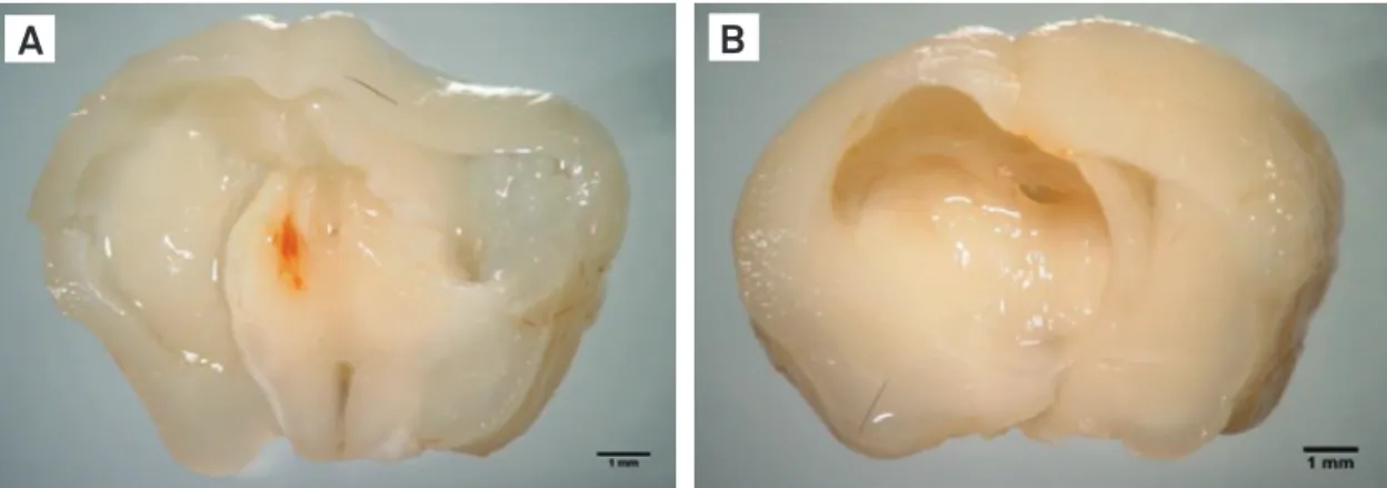
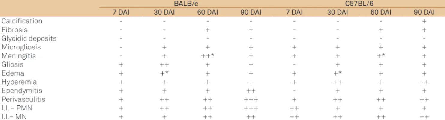
Fig 2. Mesosocopic image from encephala of BALB/c (A) and C57BL/6 (B) mice in a coronal section, presenting a cysticercus that induced ventriculomegaly and deviation of the median line (scale=1 mm).
considered the irst susceptible to T. crassiceps cysticerci infec-tion. We believe that this lower capability to induce necrosis on the parasites from BALB/c mice may have occurred due to the predominance of polymorphonuclear cells in the inlamma-tory iniltration throughout the infection. On the other hand, C57BL/6 mice already presented mononuclear cells in the in-lammatory iniltration at 60 DAI. Probably due to this difer-ence, the intensity in the ventriculomegaly, the destruction of the adjacent parenchyma, the deviation of the median line, and the gliosis have been greater in BALB/c mice.
In the initial phase of the inlammation (7 and 30 DAI), BALB/c mice presented lesions such as gliosis in the paren-chyma adjacent to the lateral ventricles. his may have oc-curred due to the triggered type of immune response (cel-lular or humoral), the control of the parasitary growth, the ventricle expansion or the presence of circulating antibod-ies. Reports from the literature describe that it is not neces-sary the presence of intraventricular cysts for the development of lesions, such as ventriculomegaly, and even the obstruction of the cerebrospinal luid low as the presence of circulating
Table 3. Pathological processes found in the host tissue from BALB/c and C57BL/6 mice experimentally infected with T. crassiceps cysticerci.
BALB/c C57BL/6
7 DAI 30 DAI 60 DAI 90 DAI 7 DAI 30 DAI 60 DAI 90 DAI
Calcification - - - +
Fibrosis - - + + - - + +
Glycidic deposits - - - -Microgliosis - + + + + + + + Meningitis - + ++* + + + +* +
Gliosis + ++ + + - + + +
Edema + +* + + + +* + +
Hyperemia + + + + + ++ + ++
Ependymitis + + + ++ - + + + Perivasculitis + ++ ++ +++ + ++ ++ ++ I.I. – PMN + ++ ++ +++ ++ + + + I.I.– MN + + ++ ++ ++ ++ ++ ++ DAI: days after the inoculation; I.I.: inflammatory infiltration; PMN: polymorphonuclear cells; MN: mononuclear cells; +: discrete; ++: moderate; +++: accentuated; n=5 per each experimental day. *p<0.05.
Table 1. Inflammatory reaction in parasites found in BALB/c and C57BL/6 mice experimentally infected with T. crassiceps cysticerci.
BALB/c C57BL/6
7 DAI 30 DAI 60 DAI 90 DAI 7 DAI 30 DAI 60 DAI 90 DAI
Development stage INI INI LAR FIN INI LAR FIN FIN Location LV LV LV/ 3rdDV LV LV LV LV LV
I.I. (PMN/MN) - - ++ +++ + + ++ ++ Calcification - - -
-Fibrosis - - - + - - - +
Glycidic deposit - - +++ + + +++ + + DAI: days after the inoculation; INI: initial; LAR: larval; FIN: final; LV: lateral ventricle; DV: dorsal ventricle; PMN: polymorphonuclear cells; MN: mononuclear cells; I.I.: inflammatory infiltration; +: discrete; ++: moderate; +++: accentuated; n=5 per each experimental day.
Table 2. Pathological processes found in the host-parasite interface in BALB/c and C57BL/6 mice experimentally infected with T. crassiceps cysticerci.
BALB/c C57BL/6
7 DAI 30 DAI 60 DAI 90 DAI 7 DAI 30 DAI 60 DAI 90 DAI
Ventriculomegaly + ++ +++ +++ + ++ ++ +++ Hyperemia + + ++ + + ++ ++ ++ Perivasculitis + ++* ++ +++ + +* ++ ++ I.I. – PMN + ++ ++ +++ + + + + I.I. – MN + + ++ ++ + ++ ++ ++ DAI: days after the inoculation; I.I.: inflammatory infiltration; PMN: polymorphonuclear cells; MN: mononuclear cells; +: discrete; ++: moderate; +++: accentuated; n=5 per each experimental day. *p<0.05.
Microgliosis was observed at 7 DAI in C57BL/6 mice, while in BALB/c mice it was observed only at 30 DAI. Also, in the irst ones a more intense gliosis in relation to the latter was observed. Discrete areas of calciication were found in the brain paren-chyma in the region of the caudal putamen in infected C57BL/6 mice at 90 DAI (Table 3).
DISCUSSION
Both lineages of mice used in this study could induce necro-sis on the parasites at 90 DAI. However, in C57BL/6 mice, this destruction occurred in a more eicient manner since 60 DAI. In spite of that, the inlammation composed mainly by poly-morphonuclear cells associated with edema, perivasculitis, and meningitis in BALB/c mice was signiicantly greater than what was observed in the C57BL/6 ones during the experimen-tal period. Fragoso et al.9, when inoculating BALB/c mice via
antigens from the parasite are enough to cause those reac-tions, even without the inlammatory reaction in the cavity20-22.
he activation and proliferation of the microglia, which is named microgliosis, were discretely higher in C57BL/6 mice through-out the experiment. hese data are in accordance to the cellu-lar proile of response, type 1, observed in these animals23. hese
cells are the main guard ones of the CNS. Once there is a lesion in the encephalic tissue, the microglia goes through an activa-tion process in which there is the modiicaactiva-tion of its morphol-ogy, surface phenotype characteristics, hypertrophy, increase in the expression of complement receptors such as CR3, increase in the expression of molecules from the main histocompatibil-ity complex (MHC), transforming the cell into one more capable of defending and stimulating the regeneration of the destroyed nervous tissue24,25.
In the late phase of the inlammation (60 and 90 DAI), in spite of BALB/c mice presented a greater intensity in the inlammato-ry iniltration than C57BL/6 ones, the latter were more capable of containing the growth of the cysticerci and of inducing their death. he inlammatory iniltration with the predominance of polymorphonuclear cells was a remarkable characteristic of the infected BALB/c mice, while in C57BL/6 mice the predominance was of mononuclear cells. Fragoso et al.23, when evaluating the
susceptibility and resistance of those both lineages against T. crassiceps cysticerci intraperitoneal infections, reported that C57BL/6 mice presented lower parasitary growth and develop-ment of lesions and a predominance of type 1 immune response. On the other hand, BALB/c mice presented a predominance of type 2 immune response which is humoral26,27. All infected
ani-mals from both lineages from this study presented discrete areas of ibrosis, which demonstrates tissue destruction due to the ac-tion of the parasite and the host inlammatory response aiming at eliminating the pathogen and the inalization of the inlam-matory process. In the animals from the Control Group, we also observed discrete edema and hyperemia in the initial days of the experiment. hese reactions were not observed in the subse-quent days of the experiment and we believe that they occurred due to the sterile saline inoculation procedure.
he dystrophic calciication areas observed in infected C57BL/6 mice at 90 DAI are in accordance to the indings from other authors19,28. According to the human NCC classiication
proposed by Cuetter et al.28, in the intraventricular active form
there may be an obstruction of the cerebrospinal luid low and hypodensic areas in the nuclear magnetic resonance examina-tion, while in the inactive form there is the late hydrocephaly without adjacent calciication areas. Probably the strong type 1 immune response, which is characteristic of the C57BL/6 mice6,9,
may be responsible for the injuries to the adjacent tissues right at the initial phase of the infection, resulting in the contention of the parasitary growth, its death and calciication.
he experimental model presented in this study, which used
T. crassiceps cysticerci, may become a reproducible method for human NCC studies because the parasite caused a dynamic in-lammatory response from the host, which evolved throughout
the experimental days and also because the Control Group pre-sented minor lesions, such as discrete hyperemia and edema, due to the inoculation procedures only in the initial days of the experiment. T. crassiceps cysticerci present other advantages such as its rapid development cycle, facilities in maintenance, and antigenic similarities to T. solium cysticerci7,8.
he previously described NCC model in the literature uses
Mesocestoides corti10,11, which are not even from the Taenia
ge-nus and do not present the cysticercus evolutive form, they only present the cysticercoid and tetrathyridium forms. Also, the M. corti experimental model presents another drawback as this parasite can proliferate and invade brain tissue11,17, which was
not observed in the T. crassiceps model. he main studies relat-ed to the immune and inlammatory responses in experimental
cysticercosis caused by T. crassiceps report their indings through the intraperitoneal model9. he model proposed in this
study may reproduce NCC and the inoculation of the parasite could occur in several areas of the encephalon such as the hip-pocampus, which may be correlated to seizures that represent the most common human clinic form of NCC.
herefore, it was possible to induce NCC in both mice lin-eages proving it to be a good experimental model. he lesions and alterations observed were ventriculomegaly, perivasculitis,
meningitis, microgliosis, and inlammation. he observation of the pathological processes in the encephala removed from the infected animals it is possible to conclude that BALB/c mice are less eicient in inducing precocious necrosis of the parasite and they present an acute inlammatory proile. While the C57BL/6 mice are more capable of provoking the parasite’s death, they have a chronic inlammatory proile, less intensity of the altera-tions and lesions and therefore are considered more resistant to
T. crassiceps cysticerci infection.
1, Agapejev S. Aspectos clínico-epidemiológicos da neurocisticercose no Brasil: Análise crítica. Arq Neuropsiquiatr 2003;61:822-828. 2. White Jr AC. Neurocysticercosis: updates on epidemiology,
pathogenesis, diagnosis, and management. Ann Rev Med 2000;51: 187-206.
3. Prasad KN, Prasad A, Gupta RK, et al. Neurocysticercosis in Patients with Active Epilepsy From a Pig Farming Community. Trans R Soc Trop Med Hyg 2009;103:144-150.
4. White Jr AC, Serpa JA, Graviss EA, Kass JS. Neurocysticercosis in Houston, Texas an update. Medicine 2011;90:81-86.
5. Del Brutto OH, Santibanez R, Idrovo L, et al. Epilepsy and neurocysticercosis in Atahualpa: a door-to-door survey in rural coastal Ecuador. Epilepsia 2005;46:583-587.
6. Terrazas IL. The Complex role of pro- and anti-inflammatory cytokines in cysticercosis: immunological lessons from experimental and natural hosts. Curr Top Med Chem 2008;8:383-392.
7. Sciutto E, Fragoso G, Trueba L, et al. Cysticercosis vaccine: cross rotecting immunity with T. solium antigens against experimental murine T. crassiceps cysticercosis. Parasite Immunol 1990;12: 687-696.
8. Sciutto E, Chavarria A, Fragoso G, Fleury A, Larralde C. The immune response in Taenia solium cysticercosis: protection and injury. Parasite Immunol 2007;29:621-636.
9. Fragoso G, Lamoyi E, Mellor A, Lomeli C, Hernandez M, Sciutto E. Increased resistance to Taenia crassiceps murine cysticercosis in Qa-2 transgenic mice. Infec Immunity 1998;66:760-764.
10. Cardona AE, Restrepo BI, Jaramillo JM, Teale JM. Development of an animal model for neurocysticercosis: immune response in the central nervous system is characterized by a predominance of gamma delta T cells. J Immunol 1999;162:995-1002.
11. Alvarez JI, Mishra BB, Gundra UM, Mishra PK, Teale JM. Mesocestoides corti intracranial infection as a murine model for neurocysticercosis. Parasitology 2010;137:359-372.
12. Toenjes SA, Spolski RJ, Mooney KA, Kuhn RE. The systemic immune response of BALB/c mice infected with larval Taenia crassiceps is a mixed Th1/Th2-type response. Parasitology 1999;118:623-633. 13. Kuhn RE, Aldridge JR, Johnson EC. Cpg stimulates protective immunity
in Balb/Cj mice infected with Larval Taenia crassiceps. J. Parasitol 2010;96:920-928.
14. Vinaud MC, Ferreira CS, Lino Jr RS, Bezerra JCB. Taenia cracisseps: Energetic and respiratory metabolism from cysticerci exposed to praziquantel and albendazole in vitro. Exp Parasitol 2008;120: 221-226.
15. Vaz AJC, Nunes M, Piazza RM, et al. Immunoblot with cerebrospinal fluid from patients with neurocysticercosis using antigen from cysticerci of Taenia solium and Taenia crassiceps. Am J Trop Med Hyg 1997;57:354-357.
16. CETEA-UFMG 2008. Protocolos Anestésicos. [Internet]. [cited 2011 Aug 24]. Available at www.ufmg.br/bioetica/cetea/index2. php?option=com_content&do_pdf=1&id=22.
17. Alvarez JI, Rivera J, Teale JM. Differential release and phagocytosis of tegument glycoconjugates in neurocysticercosis: implications for immune evasion strategies. PLoS Neglected Trop Dis 2008;2:e218. 18. Michailowsky C. Experimental tumors of the central nervous system:
standardisation of a model in rats using the 9L glioma cells. Arq Neuropsiquiatr 2003;61:234-240.
19. Lino-Jr RS, Ribeiro PM, Antonelli EJ, et al. Características evolutivas do Cysticercus cellulosae no encéfalo e no coração. Rev Soc Bras Med Trop 2002;35:617-622.
20. Garcia HH, Gonzalez AE, Gilman RH. Cysticercosis Working Group in Peru. Circulating parasite antigen in patients with hydro cephalus secondary to neurocysticercosis. Am J Trop Med Hyg 2002;66: 427-430.
21. Agapejev S, Pouza AFP, Bazan R, Faleiros ATS. Aspectos clínicos e evolutivos da hidrocefalia na neurocisticercose. Arq Neuropsiquiatr 2007;65:674-680.
22. Gasparetto EL, Eiras de Araújo AL, Rodrigues RS, et al. Migrating intraventricular cysticercosis.Arq Neuropsiquiatr 2008;66:111-113. 23. Fragoso G, Meneses G, Sciutto E, Fleury A, Larralde C. Preferential
growth of Taenia crassiceps Cysticerci in female mice holds across several laboratory mice strains and parasite lines. J Parasitol 2008;94:551-553.
24. Sreit JW, Sharon AW, Pennel AN. Reactive microgliosis. Progress Neurobiol 1999;57:563-581.
25. Ransohoff RM. Microgliosis: the questions shape the answers. Nature Neurosc 2007;10:1507-1509.
26. Larralde C, Morales J, Terrazas I, Govezensky T, Romano MC. Sex hormone changes induced by the parasite lead to feminization of the male host in murine Taenia crassiceps cysticercosis. J Steroid Biochem Molec Biol 1995;52:575-580.
27. Villa OF, Kuhn RE. Mice infected with the larvae of Taenia crassiceps
exhibit a Th2-like immune response with concomitant anergy and downregulation of Th1-associated phenomena. Parasitology 1996;112:561-570.
28. Cuetter AC, Garcia-Bodadilha, Guerra JLG, Martinez FM, Kaim B. Neurocysticercosis: focus on intraventricular disease. Clin Infec Dis 1997;24:157-164.