Article
Printed in Brazil - ©2018 Sociedade Brasileira de Química*e-mail: [email protected]; [email protected]
Antimicrobial Activity of 3,4-
seco
-Diterpenes Isolated from
Croton
blanchetianus
against
Streptococcus
mutans
and
Streptococcus
parasanguinis
NairleyC.S.Firmino,aFranciscoS.O.Alexandre,bMayronA.deVasconcelos,a,cAlisonJ.S.Conrado,a FranciscoV.S.Arruda,aEdilbertoR.Silveira*,bandEdsonH.Teixeira*,a
aLaboratório Integrado de Biomoléculas, Departamento de Patologia e Medicina Legal,
Universidade Federal do Ceará, 60430-270 Fortaleza-CE, Brazil
bDepartamento de Química Orgânica e Inorgânica, Centro de Ciências,
Universidade Federal do Ceará, CP 12.200, 60021-940 Fortaleza-CE, Brazil
cDepartamento de Ciências Biológicas, Faculdade de Ciências Exatas e Naturais,
Universidade do Estado do Rio Grande do Norte, 59625-620 Mossoró-RN, Brazil
In this study, acid and neutral fractions and three diterpenes were isolated from the hexane extracts from roots of three individual specimens of Croton blanchetianus Baill., collected from the same geographical site. Moreover, the antimicrobial activity of the extracts, fractions and diterpenes isolated was evaluated against Streptococcus mutans and Streptococcus parasanguinis, bacteria related with dental caries. The structures of the diterpenes were elucidated mainly by 1D and 2D nuclear magnetic resonance (NMR) spectroscopy, and high-resolution mass spectrometry, such as methyl 12-hydroxy-3,4-seco-cleistanta-8,11,13,15,4(18)-pentaene-3-oate; ent-3,4-secoatisa-4(18),16-dien-3-oic acid and a novel diterpene 3,4-seco-beieren-3-oic acid. Regarding to the antimicrobial activity, in general, all extracts, the neutral and acid fractions of the hexane extracts roots of all three specimens, showed antimicrobial activity in concentrations ranging from 500 to 7.8 µg mL-1. In addition, the three diterpenes isolated showed bactericidal and bacteriostatic
activity against S. mutans and S. parasanguinis, suggesting them as alternative agents with the potential to prevent oral diseases.
Keywords: seco-diterpenes, Euphorbiaceae, Croton blanchetianus, oral biofilm
Introduction
The genus Croton (Euphorbiaceae) comprises about
1300 species, which inhabit tropical and subtropical regions around the world. In Brazil, plants belonging to the Croton genus are found abundantly in the northeast
area. The use of plants belonging to this genus, in folk medicine, includes treatments for cancer, constipation, diabetes, digestive problems, dysentery, external wounds, fever, hypercholesterolemia, hypertension, inflammation, intestinal worms, malaria, pain, ulcers and weight loss.1
Many species of Croton have strong economic
potential, especially for the pharmaceutical industry, due to the various secondary metabolites that, according to many studies, are responsible for the therapeutic properties of them.2 Pirani, a Brazilian botanist, has defined Croton as a “problem genus”, mainly because of its large size and taxonomic complexity.3 According to
The Plant List, C. micranthus Müell. Arg.,4C. persicaria Baill.,5 and Oxydectes blanchetiana (Baill.) Kunte,6 are synonyms for C. blanchetianus, but other Brazilian
botanists have reported C. alagoensis Müell. Arg. and C. floribundus var. piauhyensis Rizzini as other synonyms
as well.7-9 In the past, our research group has devoted a large effort on phytochemical studies of the same plant species mistakenly identified, at that time, as C. sonderianus Müell.
Arg.7-9 Nowadays, that plant species has been correctly identified as C. blanchetianus Baill., and a few reports
mention that C. sonderianus and C. blanchetianus are
indeed synonims.10,11
Several biological activities of Croton species have
been reported, among them: the antimalarial action and low acute and subacute toxicity of the leaf extract of
Croton macrostachyus,1 the inhibition of chikungunya
some antifungal activities against three dermatophytes. The ethanol extracts of trunk or seed of C. tiglium exhibit
strong antidermatophytic activities and, thus, could be considered for application on treating skin fungal infections after appropriate processing.14
The hydroalcoholic extract from leaves of
C. macrostachyus demonstrated antimicrobial action against
isolated clinical bacteria of interest to the veterinarian.15 The hydroalcohol, hexane and ethanol extracts from leaves of Croton doctoris showed relevant activity
against caries-related bacteria: Actinomyces naeslundii, Lactobacillus acidophilus, Streptococcus gordonii, S. mutans, Streptococcus sanguinis, Streptococcus sobrinus
and Streptococcus mitis. Analysis of the composition of
the extract showed that there are large amounts of phenol compounds and diterpenes.16Croton roxburghii extract demonstrated significant antimicrobial activity against clinical isolates of Bacillus cereus and Shigella flexneri,
strains of Staphylococcus aureus, Escherichia coli, Salmonella spp., Shigella dysentriae, and Vibrio cholerae.17
The crude polysaccharide fraction, obtained from the hot aqueous extract of Croton cajucara leaves, was able to promote
gastroprotection. It was able to reduce ethanol-induced gastric ulcers in rats,18 the antinociceptive and pro-healing activity of Croton adamantinus essential oil was seen in
rats,19 the nanoparticles synthesized from the leaf extract of
Croton sparsiflorus showed significant antimicrobial activity
against S. aureus, E. coli and Bacillus subtilis.20 Diterpenoids from C. tonkinensis showed a high antituberculosis activity
against Mycobacterium tuberculosis,21 the ent-kaurane diterpene of Croton tonkinensis showed the induction of
apoptosis in colorectal cancer cells,22 and the diterpenes isolated from C. cajucara exhibited an antileishmanial
action.23
C. blanchetianus Baill., popularly known as “marmeleiro
preto”, is found in the Brazilian semi-arid region. There are botanical synonyms like Croton sonderianus Müll. Arg.
and Croton alagoensis Müll. Arg.3,11,24 Distributed at the Caatinga biome, it can be found in several places, with relatively low height and diameters and a significant number of representatives. It is widely used by rural populations, mainly for the production of firewood and the construction of small corral to house animals.25
Studies have shown that isolated compounds and the essential oil of C. blanchetianus present antinociceptive
activity26 and insecticidal activity against Aedes aegypti.27 The hydroalcoholic extract of C. blanchetianus bark
showed acaricidal activity,28 antibacterial and antifungal properties.7-9 In addition, the essential oil has been found useful to inhibit the growth of pathogenic microorganisms in foods.29 Considering that extracts from C. blanchetianus
were able to inhibit the growth of microorganisms, the objective of this study was to isolate and characterize the compounds responsible for the antimicrobial action on bacteria related to the development of dental caries.
Experimental
Infrared (IR) spectra were recorded on a PerkinElmer spectrometer, Spectrum 100 FTIR, equipped with universal attenuated total reflectance accessory (UATR). 1H and 13C, 1D and 2D nuclear magnetic resonance (NMR) spectra were obtained either on a Bruker Avance DRX-500 (500 MHz to 1H and 125 MHz to 13C) or Avance DPX-300 spectrometers (300 MHz to 1H and 75 MHz to 13C). High resolution mass spectra (HRMS) were performed on an LCMS-IT-TOF (225-07100-34, Shimadzu), equipped with electrospray ionization (ESI) source either on positive or negative mode.
The separation, and compounds purification was carried out on a Waters HPLC, consisting of a binary pump (Waters-1525) and a PDA UV detector (Waters-2996), using flow rates of 1 mL min-1 for analytical column (4.6 × 250 mm, 5 µm) and 4.72 mL min-1 for semi-preparative column (10 × 250 mm, 5 µm). In the gravitational adsorption chromatography, silica gel 60 (70-230 mesh, Vetec) was used, while Sephadex LH-20 (Pharmacia) was used in the chromatographic fractionation by molecular exclusion. Thin layer chromatography (TLC) was performed on precoated silica gel aluminum sheets (5-40 mesh, Merck) with fluorescence indicator in the range of 254 nm (F254). The substances were revealed by spraying with a vanillin-perchloric acid/EtOH solution, followed by heating in an oven (ca. 100 °C) for 5 min.
Sample preparation, strains and culture conditions
For the antimicrobial assays, solutions containing extracts, fractions and diterpenes isolated from
C. blanchetianus, were prepared in culture medium BHI
(brain heart infusion) broth with 8% dimethyl sulfoxide (DMSO). The microorganisms used in this study were
S. mutans (UA159) and S. parasanguinis (ATCC903).
Minimum inhibitory concentration and minimum bactericidal concentration determination
The minimum inhibitory concentration (MIC) was determined by microdilution tests on polystyrene plates, standardized according to the NCCLS (present CLSI), approved standard M7-A6,30 with modifications. In the 96-well polystyrene plates, the substance was serially diluted until the obtained concentrations were ranging from 125 to 1.8 µg mL-1. One hundred microliters of each bacterial suspension was added to the plate containing the substances, resulting in a final volume of 200 µL per well. The plates
were then incubated for 24 h at 37 °C in an atmosphere containing 5% CO2. After 24 h, the evaluation of bacterial growth was determined by visualizing the turbidity of the culture medium. The presence of turbidity meant that the substance did not inhibit microbial growth. In addition, the bacterial growth was measured by determining the optical density at 620 nm by using a microplate reader (SpectraMax i3 Multi-Mode microplate reader). In this way, the MIC was considered as the lowest concentration showing complete inhibition of visible bacterial growth. The negative control consisted of 8% DMSO in BHI culture medium and chlorhexidine gluconate was used as positive control.
Regarding minimum bactericidal concentration (MBC) determination, 10 µL of the bacterial suspension in the MIC were incubated in Petri dishes containing BHI agar. The plates were maintained at 37 °C and 5% CO2 for 24 h and, after this time, bacterial growth was evaluated. MBC was considered the lowest concentration that no colony growth was observed.
Plant material
On February 2012, in the locality of Garapa, located in the municipality of Acarape, CE, Brazil, the leaves and inflorescences of eight individual specimens of
C. blanchetianus were collected. After, it was analyzed
the 1H NMR spectra of leaf and inflorescences essential oils of these eight specimens of C. blanchetianus, showing
different chemical profiles. These differences were used as criteria for the selection of three specimens, designated CB1, CB4 and CB8, for the study. On April of the same year, roots, heartwood and trunk bark of the three specimens of C. blanchetianus were harvested.
The authentication of the plant material was carried out by MSc Maria Lenise Guedes of the Instituto de Biologia (Universidade Federal da Bahia). The voucher specimens (No. 53043, 53045 and 53047) are deposited in the Herbarium Prisco Bezerra (EAC) of the Universidade Federal do Ceará.
Extraction and isolation
The extracts of hexane from roots (RCB), heartwood (WCB) and trunk bark (BCB) from three specimens of C. blanchetianus were produced. 100.0 g
of the entire roots, heartwood or trunk bark of CB1, CB4 and CB8, after drying and crushing, were individually subjected to maceration in hexane for 24 h (3 × 0.5 L). The hexane solutions were pooled, and then evaporated under reduced pressure to obtain the hexane extracts from roots (RCB1 = 4.44 g, RCB4 = 7.09 g and RCB8 = 5.36 g), heartwood (WCB1 = 2.36 g, WCB4 = 3.80 g and WCB8 = 2.94 g), and trunk bark (BCB1 = 8.42 g, BCB4 = 8.91 g and BCB8 = 6.82 g). From the initial results of the microbiological tests performed with these extracts (Table 1), it was decided to perform the phytochemical analysis of the root extracts.
The procedure was then repeated with 2.5 kg of roots by maceration in hexane for 72 h (3 × 7.0 L). The hexane solutions were evaporated under reduced pressure to yield the respective extracts RCB1 (73.13 g, 2.93%), RCB4 (116.08 g, 4.64%), and RCB8 (75.13 g, 3.00%).
10.0 g of RCB1 were solubilized in CH2Cl2, adsorbed onto 100.0 g of silica gel, added to a column (8.0 × 50.0 cm) of 40.0 g of silica gel. 18 fractions (A1-A18) of 225 mL were collected by elution with: hexane (A1-A3), hexane/ CH2Cl2 1:1 (A4-A8), CH2Cl2 (A9-A13), EtOAc (A14-A15), and finally MeOH (A16-A18). After concentration and TLC analysis, similar fractions were pooled together. 680.0 mg of fraction A9 were solubilized in CH2Cl2, adsorbed on 1.0 g silica gel and chromatographed on a column (3.0 × 50.0 cm) of 10.0 g silica gel. Fractions were collected with the following volumes: B1-B53, 10 mL; and B54-B61, 50 mL. The eluents used were: hexane (B1-B8), hexane/CH2Cl2 25% (B9-B27), hexane/CH2Cl2 1:1 (B28-B56), CH2Cl2 (B57), EtOAc (B58) and MeOH (B59-B61). After rotaevaporation under reduced pressure and TLC analysis, similar fractions B34-B37 (104.2 mg) were pooled, resulting in a colorless resin named ICB1.
1H NMR analysis of ICB1 revealed its carboxylic acid
(5%) to pH 1, and then extracted with CH2Cl2 (100, 50 and 50 mL). After pooled, the organic phases gave a solution which was washed with distilled H2O (3 × 50 mL) until pH 7, dried with anhydrous Na2SO4 and rotaevaporated under vacuum to give the acidic fraction AFCB1 (6.52 g).
This procedure was repeated for RCB4, resulting in 2.77 g of NFCB4 and 6.85 g of AFCB4, as well as for RCB8, resulting in 5.22 g of NFCB8 and 3.64 g of AFCB8.
The non acidic fractions NFCB1, NFCB4 and NFCB8 were individually solubilized in CH2Cl2 and pre-adsorbed on silica gel, corresponding to 10 times the mass of each fraction. Twelve fractions of 250 mL were collected for NFCB1 and NFCB4, and 18 fractions for NFCB8 (see Table S1, Supplementary Information (SI) section). This procedure was repeated for the AFCB1, AFCB4 and AFCB8 acid fractions. 11 fractions of 250 mL were collected for each column (8.0 × 50.0 cm) with 70.0 g of silica gel (Table S2, SI section). 200.0 mg of the NFCB4 fraction were subjected to HPLC (high performance liquid chromatography) on a semi-preparative silica gel column with hexane/EtOAc (30% v/v) as the isocratic mobile phase. 20.1 mg of a yellow resin designated ICB4 (retention time (tR) = 4.42 min) was obtained.
3.64 g of AFCB8 were adsorbed onto silica gel (37.0 g) packed over a column (5.0 × 50.0 cm) of 25.0 g silica gel. Eleven fractions (D1-D11) of 250 mL were collected by elution with: hexane (D1), hexane/CH2Cl2 1:1 (D2-D3), CH2Cl2 (D4-D6), CH2Cl2/EtOAc 25% (D7-D8), EtOAc (C9), EtOAc/MeOH 1:1 (D10) and MeOH (D11). 370 mg of D4 were solubilized in MeOH and chromatographed on a column (4.0 × 50.0 cm) of 27.0 cm of Sephadex LH-20 dextran gel. The first fraction (D1) was collected after elution of 100 mL of MeOH, using a mean flow of 12 drops min-1, and then twenty fractions
(E1-E20) of 8.0 mL were collected. The E10 fraction after rotaevaporation and TLC analysis resulted in 113.0 mg of a colorless resin named ICB8.
Results and Discussion
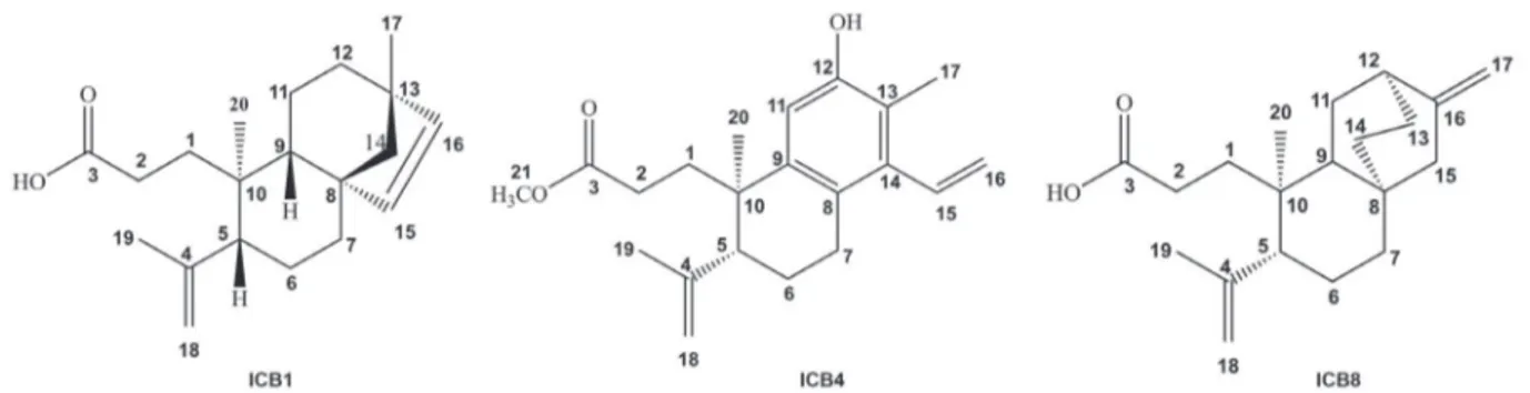
The compound ICB1, a colorless resin displayed an HRESIMS ion at m/z 303.2321 [M + H]+ (calcd. for
C20H31O2, 303.2324) or 301.2157 [M – H]- (calcd. for C20H29O2, 301.2168) in its HRESIMS spectra, [α]D21 +18.2° (c 0.11, CHCl3). Its IR spectrum showed characteristic
absorptions of O–H stretching of carboxylic acid (3400 to 2400 cm-1) and also C=O stretching at 1705 cm-1. It also showed absorptions at 1635 cm-1 of C=C stretching and 745 cm-1 of out-of-plane =C–H bending. The 1H NMR spectrum of compound ICB1 (Table 2) revealed the presence of protons of a cis and of a terminal carbon-carbon
double bonds at dH 5.72 (d, J 5.0 Hz, H-15), 5.49 (d,
J 5.0 Hz, H-16), 4.87 (s, H-18a) and 4.68 (s, H-18b). Three
singlets related to methyl protons at dH 0.77 (H-20), 1.01 (H-17) and 1.75 (H-19) were also observed. The latter one is characteristic of an isopropenyl group, while the other two seem to be attached to quaternary carbons. The
13C NMR-BB (broad-band) spectrum of compound ICB1
(Table 2) presented 20 spectral lines with the signal at
dC 180.8 (C-3) referring to an acid carboxyl, the signals at
dC 147.7 (C-4), 113.8 (C-18) and 23.9 (C-19) corresponding to the isopropenyl group, as well as two other olefinic carbons at dC 134.8 (C-15) and 137.0 (C-16). Comparison of the BB and distortionless enhancement of polarization
transfer (DEPT) 135° 13C NMR spectra permitted to
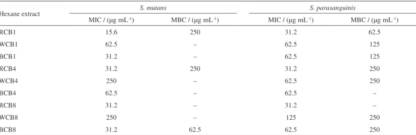
identify the hydrogenation pattern of each carbon of compound ICB1, confirming its molecular formula C20H30O2, indicating 6 double bond equivalents (DBE’s), Table1. MIC and MBC values of the hexane extracts from roots, heartwood and trunk bark of CB1, CB4 and CB8 on S. mutans and S. parasanguinis
Hexane extract S. mutans S. parasanguinis
MIC / (µg mL-1) MBC / (µg mL-1) MIC / (µg mL-1) MBC / (µg mL-1)
RCB1 15.6 250 31.2 62.5
WCB1 62.5 – 62.5 125
BCB1 31.2 – 62.5 125
RCB4 31.2 250 31.2 250
WCB4 250 – 62.5 250
BCB4 62.5 – 62.5 –
RCB8 31.2 – 31.2 –
WCB8 250 – 125 250
BCB8 31.2 62.5 62.5 250
from which three were related to three rings, two carbon-carbon double bonds and a carboxyl. The 1H,1H-COSY (correlation spectroscopy) spectrum analysis evidenced the correlations for the olefinic protons at dH 5.72 (d, J 5.0 Hz, H-15) and 5.49 (d, J 5.0 Hz, H-16). The presence of the
isopropenyl group was confirmed through the correlation of the protons of the methyl carbon at dH 1.75 (s, H-19) with the protons of the methylidene carbon at dH 4.87 (s, H-18a) and dH 4.68 (s, H-18b). The two dimensional 1H,13C-HMBC (heteronuclear multiple bond correlation)
spectrum revealed the presence of the isopropenyl group attached to carbon C-5, which was confirmed through the correlations of the terminal vinyl protons at dH 4.87 (s, H-18a) and 4.68 (s, H-18b) with the carbons at dC 147.7 (C-4), 51.3 (C-5) and 23.9 (C-19). The presence of the C-17 methyl linked to carbon C-13 was confirmed through the correlations observed for the protons of C-17 at dH 1.01 (s, 3H), with the carbons at dC 44.1 (C-13), 61.3 (C-14), 33.3 (C-12), and 137.0 (C-16). The cis olefinic system (C-15,
C-16) was ratified through the HMBC correlations of the protons at dH 5.72 (d, J 5.0 Hz, H-15) and 5.49 (d, J 5.0 Hz, H-16) with the carbons at dC 44.1 (C-13), 61.3 (C-14) and 49.1 (C-8). The relative stereochemistry was defined based on the 1H,1H-NOESY (nuclear Overhauser effect spectroscopy) spectrum by the dipolar coupling correlation of the proton at dH 1.99 (H-5) with the proton at dH 1.14 (H-9). The correlations for the protons of methyl C-20 at
dH 0.77 (s, 3H) with the protons at dH 5.72 (d, J 5.0 Hz, H-15), 1.81 (d, J 12.8 Hz, H-11a) and 4.68 (s, H-18b)
allowed the determination of the relative configurations at the stereogenic centers C-8, C-10 and C-13. It is possible to conclude that the compound ICB1 is the unknown diterpene 3,4-seco-beieren-3-oic acid (Figure 1). Compound ICB1 is
the “3,4-seco” counterpart of the ent-Beyer-15-en-18-oic
acid, previously isolated from C. sonderianus.31
The 1H NMR spectrum of compound ICB4 (Table 2) revealed the presence of olefinic protons of a vinyl moiety at
dH 6.73 (dd, J 11.4, 18.0 Hz, H-15), 5.57 (dd, J 2.0, 11.4 Hz, H-16a), and 5.26 (dd, J 2.0, 18.0 Hz, H-16b), in addition to
signals at 4.99 (s, H-18a) and 4.84 (s, H-18b), characteristic of the methylidene protons of a terminal carbon-carbon double bond, as observed for ICB4. There were also three singlets related to methyl protons at dH 1.24 (s, H-20), 1.77 (s, H-19) and 2.52 (s, H-17), and another singlet related to a methoxyl at dH 3.56. The 13C NMR-BB spectrum of ICB4 (Table 2) presented 21 spectral lines, from which the signal at dC 174.8 (C-3) was assigned to an ester carbonyl and the signals at dC 155.8 (C-12), 139.7 (C-14), 142.0 (C-9), 126.3 (C-8), 121.6 (C-13), and 112.5 (C-11), to the carbons of a penta-substituted benzene ring. In addition, four olefinic carbons at dc 147.9 (C-4) and 114.9 (C-18) of the terminal
double bond, 137.0 (C-15) and 119.9 (C-16), characteristic of the vinyl moiety, were also characterized. Comparison of the BB and the DEPT 135° 13C NMR spectra permitted to identify the hydrogenation pattern of ICB4, confirming the molecular formula C21H28O3, presenting 6 DBE’s. Its IR spectrum showed an intense absorption characteristic of C=O at 1730 cm-1, compatible with an ester carbonyl. A broad band centered at 3420 cm-1, characteristic of O–H; an absorption at 3050 cm-1, characteristic of benzene C–H stretching; besides absorptions at 1600 and 1480 cm-1 for the skeletal bands; at 1650 cm-1, with a shoulder, of the C=C stretching of alkenes, were also observed. Comparison of the 1H and 13C NMR-BB spectra with data from the literature,4 allowed to identify compound ICB4 as the methyl 12-hydroxy-3,4-seco
-cleistanta-8,11,13,15,4(18)-pentaene-3-oate, a diterpene, previously isolated from C. sonderianus (Figure 1).4
Compound ICB8 showed a 13C NMR-BB spectrum
with 20 spectral lines (Table 2). Comparison of the BB and DEPT 135° 13C NMR spectra permitted to establish the hydrogenation pattern of each carbon atom, as 2 methyls, 8 methylenes and 2 methylidenes, and 5 non-hydrogenated carbons, with the molecular formula C20H30O2, indicating
6 DBE’s. The 1H NMR spectrum of ICB8 (Table 2)
showed signals at dH 4.96 (s, H-18a), 4.89 (s, H-17a), 4.87 (s, H-18b) and 4.72 (s, H-17b) corresponding to terminal double bond protons, and 2 singlets for two methyl at
dH 1.81 (s, H-19) and 0.95 (s, H-20). Its IR spectrum showed characteristic absorptions for O–H stretching of carboxylic acid (3400 cm-1 to 2400 cm-1), and an intense absorption characteristic of C=O carboxyl stretching at 1705 cm-1. In addition, it showed absorptions at 1639 cm-1 of the C=C stretching of alkenes and the correspondent out-of-plane bending at 755 cm-1. The 1H and 13C NMR data (Table 2), suggested the structure of a tetracyclic diterpene with an ent-atisane backbone. According to the
analysis of the spectral data obtained, and comparison with the literature data, it was possible to suggest that compound ICB8 was indeed the ent-3,4-seco
atisa-4(18),16-dien-3-oic acid, previously isolated from Excoecaria
agallocha L.32 (Figure 1), isolated for the first time from C. blanchetianus.
Antimicrobial activity of extracts, fractions and diterpenes
The results of the antimicrobial assays performed with the hexane extracts from trunk bark, heartwood and roots of the three specimens of C. blanchetianus (CB1, CB4 and
CB8) are shown in Table 1.
Table2.1H and 13C NMR data for compounds ICB1, ICB4 and ICB8 isolated from the hexane extract from roots of the three individual specimens of
Croton blanchetianus
C ICB1
a ICB4b ICB8b
dC dH (mult., J in Hz) dC dH (mult., J in Hz) dC dH (mult., J in Hz)
1 36.4 1.57 (m, Ha)
1.36 (m, Hb)
36.1 2.22 (m, 2H) 34.9 1.95 (c, Ha)
1.79 (c, Hb)
2 28.6 2.39 (m, Ha)
2.30 (m, Hb)
30.3 2.45 (m, Ha)
2.16 (m, Hb)
29.9 2.57 (m, Ha)
2.44 (m, Hb)
3 180.8 – 174.8 – 176.9 –
4 147.7 – 147.8 – 148.3 –
5 51.3 1.99 (brd, 12.0, H) 47.3 2.49 (m, H) 51.2 2.07 (brd, 11.5, H)
6 20.8 1.43 (m, Ha)
1.33 (m, Hb)
25.8 1.83 (td, 3.9, 11.5, Ha) 1.77 (c, Hb)
25.4 1.72 (c, Ha) 1.31 (c, Hb)
7 32.6 1.55 (m, 2H) 29.2 2.81 (dt, 3.9, 15.0, Ha)
2.56 (c, Hb)
38.6 1.29 (brd, 11.0, Ha) 1.11 (brt, 11.0, Hb)
8 49.1 – 126.3 – 33.9 –
9 44.1 1.14 (d, 11.8, H) 142.0 – 44.4 1.39 (c, H)
10 39.3 – 41.7 – 40.3 –
11 26.2 1.81 (d, 11.8, Ha)
1.40 (m, Hb)
112.5 7.20 (s, H) 28.8 1.89 (m, Ha)
1.42 (m, Hb)
12 33.3 1.31 (m, Ha)
1.26 (m, Hb)
155.8 – 37.1 2.23 (brs, H)
13 43.8 – 121.6 – 27.7 1.60 (m, Ha)
1.51 (m, Hb)
14 61.3 1.49 (d, 9.2, Ha)
1.04 (d, 9.2, Hb)
139.7 – 28.9 1.62 (m, Ha)
0.97 (c, Hb)
15 134.8 5.72 (d, 5.0, H) 137.0 6.73 (dd, 11.4, 18.0, H) 48.7 2.03 (brd, 13.8, Ha)
1.92 (brd, 13.8, Hb)
16 137.0 5.49 (d, 5.0, H) 119.9 5.57 (dd, 2.0, 11.4, Ha)
5.26 (dd, 2.0, 18.0, Hb)
152.7 –
17 25.0 1.01 (s, 3H) 14.3 2.52 (s, 3H) 105.7 4.89 (s, Ha)
4.72 (s, Hb)
18 113.8 4.87 (s, Ha)
4.68 (s, Hb)
114.9 4.99 (s, Ha) 4.84 (s, Hb)
114.0 4.96 (s, Ha) 4.87 (s, Hb)
19 23.9 1.75 (s, 3H) 23.5 1.77 (s, 3H) 24.3 1.81 (s, 3H)
20 18.7 0.77 (s, 3H) 28.6 1.24 (s, 3H) 18.3 0.95 (s, 3H)
O–CH3 – – 51.8 3.56 (s, 3H) – –
a125 MHz, CDCl
3; b125 MHz, C5D5N; C: number of carbons in the compounds. ICBx: isolated from C. blanchetianus, x = 1 for specimen 1, x = 4 for
specimen 4, x = 8 for specimen 8.
extracts presented a bactericidal and bacteriostatic effect for both bacteria.
Considering the lowest inhibitory concentrations of the microbial growth, the hexane extracts from the roots were selected to perform the fractionation. In order to identify the compounds responsible for the antimicrobial action, the hexane extracts from roots were separated in acid and neutral fractions and then submitted to antibacterial assay.
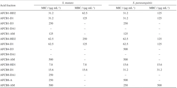
Acid and neutral fractions showed antimicrobial activity, however, the acid fractions presented the lowest values of concentration with effective action. The antibacterial results for all acid fractions are presented in Table 3.
Fraction AFCB8 showed a more effective bactericidal and bacteriostatic effect, with the lowest concentrations, 7.8 and 15.6 µg mL-1, against both bacteria. The antibacterial results for the neutral fractions are presented in Table S3 (SI section).
After several fractionation steps, three diterpenes (ICB1, ICB4 and ICB8) were isolated and their antibacterial activity was determined (Table 4).
ICB1 presented the same MIC and MBC values against
S. mutans (410 µM), while against S. parasanguinis showed
values of 206 and 410 µM, respectively. The AFCB1 fraction, from which ICB1 was obtained, presented a four-fold inhibitory effect of ICB1 against S. mutans and
two-fold increase against S. parasanguinis. However, MBC
values remained the same for both bacteria.
Regarding to ICB4, the compound was obtained from the neutral fraction NFCB4-D2. ICB4 presented MIC and MBC values of 95 µM against S. mutans, and 23.7 µM
against S. parasanguinis. ICB4 showed lower MIC values
compared to NFCB4-D2. Moreover, NFCB4-D2 showed no bactericidal activity.
ICB8, isolated from the acid fraction AFCB8-D1, showed MIC and MBC values of 103 µM against
S. mutans. On the other hand, AFCB8-D1 showed MIC
and MBC values lower compared to ICB8. The MIC Table3. MIC and MBC values of the acid fractions of CB1, CB4 and CB8 against S. mutans and S. parasanguinis
Acid fraction S. mutans S. parasanguinis
MIC / (µg mL-1) MBC / (µg mL-1) MIC / (µg mL-1) MBC / (µg mL-1)
AFCB1-HD2 31.2 62.5 31.2 125
AFCB1-D1 31.2 125 31.2 125
AFCB1-D3 250 – 250 –
AFCB1-DA1 – – – –
AFCB1-AM 125 – 125 –
AFCB4-HD2 62.5 250 62.5 125
AFCB4-D1 62.5 125 62.5 125
AFCB4-D3 – – 500 –
AFCB4-DA1 – – – –
AFCB4-AM 500 – 500 –
AFCB8-HD1 7.8 7.8 15.6 15.6
AFCB8-D1 15.6 15.6 31.2 31.2
AFCB8-DA1 250 – – –
AFCB8-A 250 – 500 –
AFCB8-AM 500 – 250 500
AFCBx: acid fractions of C. blanchetianus, x = 1 for specimen 1, x = 4 for specimen 4, and x = 8 for specimen 8; HD: hexane/CH2Cl2 50%; D: CH2Cl2;
DA: CH2Cl2/EtOAc 25%; AM: EtOAc/MeOH; HD1: first fraction collected using hexane/CH2Cl2 50%; HD2: second fraction collected using
hexane/CH2Cl2 50%; D1: first fraction collected using CH2Cl2; D3: third fraction collected using CH2Cl2; DA1: first fraction collected using CH2Cl2/EtOAc
25%. MIC: minimum inhibitory concentration; MBC: minimum bactericidal concentration.
Table4. MIC and MBC values of ICB1, ICB4, ICB8 diterpenes and
chlorhexidine gluconate (positive control) against S. mutans and S. parasanguinis
Compound S. mutans S. parasanguinis
MIC / µM MBC / µM MIC / µM MBC / µM
ICB1 410 410 206 410
ICB4 95 95 23.7 23.7
ICB8 103 103 103 206
Chlorhexidine 2.1 18.4 1.1 2.1
values of AFCB8-D1 and ICB8 remained the same against
S. parasanguinis, while MBC values of AFCB8-D1 were
lower. Chlorhexidine gluconate was used as a positive control, presenting values ranging from 1.1 to 18.4 µM.
In this study, it was observed that all the hexane extracts from trunk bark, heartwood and roots of C. blanchetianus
presented antimicrobial activity against S. mutans and S. parasanguinis, with concentrations ranging from 250
to 15.6 µg mL-1. Our results corroborate with the study of Silva et al.,33 where C. sonderianus Mull. ethanol extract inhibited dental caries-related bacteria growth, including
S. mitis ATCC 9811. The authors emphasize the possibility
of using this extract for oral pathologies treatment.34
The root hexane extract of C. sonderianus showed
antibacterial activity against B. subtilis, S. aureus and Mycobacterium smegmatis, and antifungal activity against Candida albicans, Trichophyton mentagrophytes, and Helminthosporium sp. The reported action was attributed to
the diterpenes hardwickiic acid and 3,4-secotrachylobanoic
acid.34 The EtOAc extract from C. macrostachyus trunk bark inhibited the growth of Salmonella typhi, E. coli, Klebsiella pneumoniae, Enterobacter aerogenes, and Listeria monocytogenes, with MIC values ranging from
125 to 250 mg mL-1, being considered as a promising antimicrobial agent against important human pathogens.35
In this work, acid and neutral fractions were separated and it was observed that the acid fractions presented antimicrobial activity in smaller concentrations when compared to the neutral fractions (see Table S3, SI section). A previous study31 with the acid and neutral fractions of C. sonderianus showed that acid fraction had
better antimicrobial activity against B. subtilis, S. aureus
and Pseudomonas aeruginosa when compared to neutral
fractions. Moreover, the authors isolated the compound
ent-Beyer-15-en-18-oic acid that presented antimicrobial
activity against B. subtilis.31
Croton species have been widely studied in relation to
their volatile and non-volatile constituents. Many species are producers of a large number of substances belonging to the alkaloids, phenylpropanoids and terpenoids classes.36 The diterpenes isolated from C. blanchetianus showed clearly antimicrobial activity against S. mutans
and S. parasanguinis. The diterpene class exhibits
significant antimicrobial activity against Gram-negative, Gram-positive bacteria and yeasts.37,38 In addition, it is suggested that diterpenes interact in a non-specific way with the bacterial cell membrane, causing disruption of its structure.38 A casbane diterpene isolated from
Croton nepetifolius Baill. showed potential antibacterial
activity against several streptococci species related to caries, with MIC values ranging from 250 to 62.5 µg mL
-1. In addition, the ability to inhibit the formation of oral
biofilms has been verified, therefore, being considered by the authors as a promising molecule on oral pathogens related to dental caries.39 Thus, the diterpenes isolated in this work have large potential for microbial therapy, however, toxicity tests are still required to determine the therapeutic index allowing the safety use.
Conclusions
In summary, in this study three 3,4-seco-diterpenes were
isolated from the hexane extracts of roots of three individual specimens of C. blanchetianus Baill., including a novel
diterpene 3,4-seco-beieren-3-oic acid (ICB1). Furthermore,
the acid and neutral fractions, and the isolated compounds, showed a potential activity against two oral streptococci.
Supplementary Information
Supplementary data of the compounds ICB1, ICB4 and ICB8 (NMR, IR and HRMS spectra, Figures S1-S25) and Tables S1-S3 are available free of charge at http://jbcs.sbq.org.br as PDF file.
Acknowledgments
This study was supported by grants from the Conselho Nacional de Desenvolvimento Científico e Tecnológico (CNPq), Coordenação de Aperfeiçoamento de Pessoal de Nível Superior (CAPES) and Fundação Cearense de Apoio ao Desenvolvimento Científico e Tecnológico (FUNCAP). EHT and ERS are senior investigators of CNPq and members of the Brazilian Academy of Sciences.
References
1. Salatino, A.; Salatino, M. L. F.; Negri, G.; J. Braz. Chem. Soc.
2007, 18, 11.
2. Palmeira Júnior, S. F.; Alves, V. L.; Moura, F. S.; Vieira, L. F. A.; Rev. Bras. Farmacogn.2006, 16, 397.
3. Lima, L. R.; Pirani, J. R.; Biota Neotrop.2008, 8, 2.
4. http://www.theplantlist.org/tpl1.1/record/kew-50556, accessed in November 2017.
5. http://www.theplantlist.org/tpl1.1/record/kew-50817, accessed in November 2017.
6. http://www.theplantlist.org/tpl1.1/record/kew-336462, accessed in November 2017.
7. Craveiro, A. A.; Silveira, E. R.; Braz Filho, R.; Mascarenhas, I. P.; Phytochemistry1981, 20, 852.
9. Silveira, E. R.; McChesney, J. D.; Phytochemistry1994, 36, 1457.
10. Gomes, A. P. S.; Sales, M. F.; Melo, A. L.; Acta Bot. Bras.2010, 24, 905.
11. Oliveira, I. M. M.; Santos, H. S.; Sena Jr., D. M.; Cruz, B. G.; Teixeira, A. M. R.; Freire, P. T. C.; Braz-Filho, R.; Sousa, J. W.; Albuquerque, M. R. J. R.; Bandeira, P. N.; Bernardino, A. C. S. S.; Gusmão, G. O. M.; Bento, R. R. F.; J. Mol. Struct.
2015, 1099, 226.
12. Corlay, N.; Delang, L.; Girard-Valenciennes, E.; Neyts, J.; Clerc, P.; Smadja, J.; Guéritte, F.; Leyssen, P.; Litaudon, M.; Fitoterapia2014, 97, 87.
13. Cordeiro, K. W.; Pinto, L. A.; Formagio, A. S.; Andrade, S. A. F.; Kassuya, C. A. L.; Freitas, K. C.; J. Ethnopharmacol.2012,
143, 331.
14. Lin, H. C.; Kuo, Y. L.; Lee, W. J.; Yap, H. Y.; Biomed. Res. Int.
2016, DOI 10.1155/2016/3237586.
15. Kalayou, S.; Haileselassie, M.; Gebre-Egziabher, G.; Tiku, T.; Sahle, S.; Tadelle, H.; Ghezu, M.; Asian Pac. J. Trop. Biomed.
2012, 2, 516.
16. Brighenti, F. L.; Salvador, M. J.; Delbem, A. C.; Delbem, Á. C.; Oliveira, M. A.; Soares, C. P.; Freitas, L. S.; Koga-Ito, C. Y.; Caries Res.2014, 48, 353.
17. Panda, S. K.; Mohanta, Z. K.; Padhi, L.; Park, Y. H.; Mohanta, T. K.; Bae, H.; Molecules2016, 14, 21.
18. Nascimento, A. M.; Maria-Ferreira, D.; de Souza, E. F.; Sassaki, G. L.; Iacomini, M.; Werner, M. F.; Ciprane, T. R.; Int. J. Biol. Macromol.2017, 95, 153.
19. Ximenes, R. M.; de Morais, N. L.; Cassundé, N. M.; Jorge, R. J.; dos Santos, S. M.; Magalhães, L. P.; Silva, M. R.; Viana, G. S. B.; Araújo, R. M.; de Sena, K. X.; de Albuquerque, J. F.; Martins, R. D.; J. Nat. Med.2013, 67, 758.
20. Kathiravan, V.; Ravi, S.; Ashokkumar, S.; Velmurugan, S.; Elumalai, K.; Khatiwada, C. P.; Spectrochim. Acta, Part A2015,
139, 200.
21. Jang, W. S.; Jyoti, M. A.; Kim, S.; Nam, K. W.; Ha, T. K.; Oh, W. K.; Song, H. Y.; J. Nat. Med.2016, 1, 127.
22. Thuong, P. T.; Khoi, N. M.; Ohta, S.; Shiota, S.; Kanta, H.; Takeuchi, K.; Ito, F.; Anti-Cancer Agents Med. Chem.2014, 14, 1051.
23. Lima, G. S.; Castro-Pinto, D. B.; Machado, G. C.; Maciel, M. A.; Phytomedicine2015, 22, 1133.
24. Angélico, E. C.; Costa, J. G. M.; Rodrigues, O. G.; Lima, E. Q.; Medeiros, R. S.; BioFar2011, 5, 44.
25. Alves, G. S.; Alves, J. M. F.; Martins, L. R. A.; Sousa, J. S.; Souto, J. S.; RVADS2014, 9, 50.
26. Santos, F. A.; Jeferson, F. A.; Santos, C. C.; Silveira, E. R.; Rao, V. S. N.; Life Sci.2005, 77, 2953.
27. Lima, G.; de Souza, T.; Freire, G. P.; Farias, D.; Cunha, A.; Ricardo, N.; de Morais, S.; Carvalho, A.; Parasitol. Res.2013,
112, 1953.
28. Silva, F. S.; Albuquerque, U. P.; Costa Júnior, L. M.; Lima, A. S.; Nascimento, A. L. B.; Monteiro, J. M.; J. Ethnopharmacol.
2014, 155, 1332.
29. Bjarnsholt, G. F. D. M.; da Costa, A. C. V.; Garino, F.; Medeiros, R. S.; Madruga, M. S.; Neto, V. Q.; Braz. J. Microbiol.2013,
44, 1189.
30. NCCLS, Approved Standard M7-A6; Methods for Dilution Antimicrobial Susceptibility Tests for Bacteria that Grow
Aerobically; NCCLS: Wayne, PA, 2003.
31. McChesney, J. D.; Clark, A. M.; Silveira, E. R.; Pharm. Res.
1991, 8, 1243.
32. Li, Y.; Liu, J.; Yu, S.; Proksch, P.; Gu, J.; Lin, W.; Phytochemistry
2010, 71, 2124.
33. Silva, V. A.; Oliveira, C. R. M.; Freitas, A. F. R.; Costa, M. R. M.; Pessoa, H. L. F.; Pereira, M. S. V.; Rev. Odontol. UNESP
2011, 40, 69.
34. McChesney, J. D.; Clark, A. M.; Silveira, E. R.; J. Nat. Prod.
1991, 54, 1625.
35. Obey, J. K.; Wright, A.; Orjala, J.; Kauhanen, J.; Kauhanen, C. T.; J. Pathog.2016, DOI 10.1155/2016/1453428.
36. Randau, K. P.; Florêncio, D. C.; Ferreira, C. P.; Xavier, H. S.; Rev. Bras. Farmacogn.2004, 14, 89.
37. Vasconcelos, M. A.; Arruda, F. V. S.; Santos, H. S.; Rodrigues, A. S.; Bandeira, P. N.; Albuquerque, M. R. J. R.; Cavada, B. S.; Teixeira, E. H.; Henriques, M.; Pereira, M. O.; Ind. Crops Prod.2014, 61, 499.
38. Carneiro, V. A.; Santos, H. S.; Arruda, F. V. S.; Bandeira, P. N.; Albuquerque, M. R. J. R.; Pereira, M. O.; Henriques, M.; Cavada, B. S.; Teixeira, E. H.; Molecules2011, 16, 190. 39. Sá, N. C.; Cavalcante, T. T. A.; Araújo, A. X.; dos Santos, H.
S.; Albuquerque, M. R. J.; Bandeira, P. N.; da Cunha, R. M.; Cavada, B. S.; Teixeira, E. H.; Arch. Oral Biol.2012, 57, 550.
Submitted: July 19, 2017