Article
Printed in Brazil - ©2018 Sociedade Brasileira de Química*e-mail: [email protected]
This paper was prepared in honor of the late Professor Angelo da Cunha Pinto
Synthesis of New
trans
-Dehydrocrotonin Nitrogenated Derivatives and their
Cytotoxic and DNA-Topoisomerase I Inhibitory Activities
Andressa Esteves-Souza,a Kenia Pissinate,a Maria A. M. Macielb,c and
Aurea Echevarria*,a
aDepartamento de Química, Universidade Federal Rural do Rio de Janeiro,
23851-970 Seropédica-RJ, Brazil
bDepartamento de Química, Universidade Federal do Rio Grande do Norte,
59072-970 Natal-RN, Brazil
cPrograma de Pós-graduação em Biotecnologia, Universidade Potiguar,
Laureate International Universities, Campus Salgado Filho, 59060-000 Natal-RN, Brazil
A new series of 19-nor-clerodane diterpene derivatives was synthesized from the natural
trans-dehydrocrotonin obtained from stem barks of Croton cajucara (Euphorbiaceae), a native medicinal plant of the Brazilian Amazon. The new derivatives were obtained by changes in the ketone moiety of trans-dehydrocrotonin leading to nitrogenated derivatives which are: three substituted hydrazine diterpenes, oxime, and methyloxime. The cytotoxic effect of the diterpene derivatives was evaluated by MTT (3-(4,5-dimethylthiazol-2-yl)-2,5-diphenyltetrazolium bromide) assay against Ehrlich carcinoma and K562 human leukemia cells. The cytotoxic activity of the hydrazine and oxime semi-synthetic derivatives was better than the one of the natural product trans-dehydrocrotonin. Moreover, all diterpenes were tested for their DNA topoisomerase I inhibitory activity, and the most effective one, in general, was observed to the phenyl-hydrazone derivative. Results indicated that the topoisomerase I inhibitory effect is correlated with the cytotoxic activity.
Keywords: Croton cajucara, diterpenes, trans-dehydrocrotonin, cytotoxic activity, DNA-topoisomerase I
Introduction
Medicinal plants are the dominant form of medicine in most countries, and the World Health Organization estimates that around 80% of the world population in developing countries relies on traditional plant medicines for primary healthcare needs.1
Many Euphorbiaceae plants are well known in different parts of the world as toxic and/or medicinal, and Croton is a large genus of this family widely distributed in tropical and subtropical regions of both hemispheres. Croton cajucara
Benth, commonly known as “sacaca”, is a medicinal plant largely grown in the Brazilian Amazon. Both stem bark and leaves are extensively used in the form of tea or pills for the treatment of several diseases.2,3 It is widely known in the Amazonian traditional phytotherapy for the treatment
of diarrhea and gastrointestinal disorders,3 liver diseases and weight loss,4 diabetes5 and high blood cholesterol levels.6 The terpenoid classes are the predominant special metabolite constituents of this genus, and the clerodane diterpenes, the prevalent type of Croton.3,7-10
Several clerodane diterpenes isolated from different species had been tested for their biological activities, and presented potentially useful medicinal properties, such as antimicrobial,11-14 anti-inflammatory,15,16 antiprotozoal,17-21 and antitumoral.22-26
The trans-dehydrocrotonin (1) is a nor-clerodane diterpene isolated from Croton cajucara as major component.27 Experimental laboratory animals and in vitro studies have shown that 1 has insecticidal,28 antigenotoxic,29 antiulcerogenic,3,30-33 anti-inflammatory,34 anti-leishmanial35 and antitumor effects.36-40
1 (Figure 1). Not only did we consider the fact that 1 is the principal and abundant component of C. cajucara,27 but also the previous in vivo and in vitro results of anti-tumor activities.36,37 Clerodane 2, a natural component of C. cajucara stem bark, was also evaluated against representative neoplastic cell lines.
In the present work, we report the synthesis of five new
trans-dehydrocrotonin nitrogenated derivatives, and their
in vitro evaluation of cytotoxic activity. The new derivatives were prepared from clerodane 1, previously isolated from the stem bark of Croton cajucara,3,27 and they were also evaluated against Ehrlich carcinoma and K562 human leukemia cells. Furthermore, the inhibitory action of the new derivatives, as well as the isolated natural compounds 1
and 2, was evaluated for DNA-topoisomerase I inhibition (Topo I).
Results and Discussion
Synthesis and characterization
Clerodane 1 was isolatedfrom the stem bark of methanol extract of Croton cajucara, as previously described.3,27 The synthesis of the diterpene derivatives involved reactions of the C-2 carbonyl group in the A ring. The structure of the clerodane skeleton was maintained to assure its natural feature in an attempt to preserve or enhance the cytotoxic activities previously observed.37,38 Thus, to construct a sensible series of trans-dehydrocrotonin derivatives for SAR purpose, some new semi-synthetic derivatives were prepared.
Those derivatives were performed including nitrogenated moieties at the ketone carbon (C-2) of A ring. The new compounds were the hydrazone derivatives, such as the unsubstituted hydrazone (3), methyl-hydrazone (4) and phenyl-hydrazone (5), as well as the oxime(6), and the methylated oxime (7). The synthetic route of the semi-synthetic clerodanes 3-7 is outlined in Scheme 1.
Derivatives 3, 4 and 5 were obtained from the treatment of 1 using hydrazine hydrate, methyl hydrazine and phenyl hydrazine, respectively, at room temperature, at different Figure 1.trans-Dehydrocrotonin (1) and trans-crotonin (2) clerodane
diterpenes isolated from stem bark of Croton cajuacara.
time reactions (6, 21 and 24 h, respectively), in ethanol as solvent, following the previously reported procedure.41 The corresponding hydrazones were obtained in 80, 60 and 75% yield for 3, 4 and 5, respectively. The oxime derivative (6) was prepared from 1 using hydroxylamine hydrochloride in basified ethanol with NaOH, for 10 min under reflux, thus yielding 80%. The reaction between 6 and methyl iodide, and Ag2O, at low temperature,42 furnished the corresponding methylated oxime (7)in 60% yield. All the compounds were purified by silica gel chromatographic column using CHCl3, hexane and ethyl acetate as eluents. These new semi-synthetic diterpene derivatives 3-7 were characterized by infrared, 1H and 13C nuclear magnetic resonance (NMR) spectroscopy, and then briefly discussed.
T h e i n f r a r e d s p e c t r a s h ow e d t h e a b s e n c e of carbonyl moiety signal at 1666 cm-1 observed to
trans-dehydrocrotonin27 and, the presence of absorption in the range of 1622-1599 cm-1 assigned to C=N group of hydrazone derivatives, 3, 4 and 5. 1H NMR spectra of 3, 4 and 5 presented a similar pattern, detaching the chemical shifts in the range of d 6.51-6.98, and signals corresponding to aromatic hydrogens of phenyl-hydrazone derivative (5) were observed. The N−H signal was observed in the range of d 5.00-6.00. The 13C NMR spectra of 3, 4 and 5 presented coherent changes to respect 1 in accordance to the moiety nature attached to C-2. Typical values of imino function were observed for C-2 in the range of d 151.9-152.0, and smaller variations in the carbons C-1, C-3 and C-4, as expected.
The infrared spectra of 6 and 7 derivatives showed absorptions of 1635 and 1610 cm-1, respectively, assigned to C=N moiety, instead of C=O absorption of trans-dehydrocrotonin (1666 cm-1).27 The 1H NMR chemical shifts observed for 6, the oxime derivative, and for methyl-oxime (7) were coherent with the structure proposal, in which H-3 appeared in d 6.13 for 7, presenting deshielding effect when compared to 6 (d 5.89), due to the stereo-spatial position of the methyl group. The signal corresponding to the hydroxyl-oxime of 6 appeared in d 8.05 as a wide singlet. 13C NMR chemical shifts were in accordance to the proposal structures; d 156.2 and 158.7 were attributed to C-2 at 6 and 7, respectively; and, shielding effect (γ effect) was observed at C-3, with ∆d 6.5 and ∆d 10.0 when compared to 1, for 6 and 7, respectively.
Cytotoxic effects
The cytotoxic action of special metabolites isolated from C. cajucara Benth stem bark extracts against tumor cells36,37 led us to continue studying the possible action of the new semi-synthetic derivatives of trans-dehydrocrotonin
diterpene (1). The antiproliferative effect of 1-7 against the ascitic Ehrlich carcinoma and human leukemia K562 cells was evaluated. Assays were performed using the MTT method43 with quercetin and vincristine as positive controls for Ehrlich and K562 leukemia, respectively.
In vitro assays using Ehrlich carcinoma cells with 4, 5,
6 and 7 nitrogenated derivatives showed a more significant antiproliferative activity when compared to 1,the major active component of C. cajucara, previously evaluated by Grynberg et al.,36 with dose dependent responses over 48 h culture period. IC50 values 45.78 ± 4.35, 16.78 ± 1.42, 21.88 ± 1.96 and 43.43 ± 3.96 µmol L-1 were also compared to quercetin (IC50 = 44 µmol L-1), as shown in Table 1. Interestingly, the unsubstituted hydrazone derivative 3
did not present any significant activity until the maximum concentration of 50 µmol L-1 used in the assays, thus suggesting a positive contribution of hydrophobic effect.
The inhibitory effect on K562 cells in vitro, after 96 h in culture, was not observed for 1 nor 2 until the dose of 50 µmol L-1.37 However, the new nitrogenated derivatives and the unsaturated alcohol presented cytotoxic effect against K562, also with dose dependent responses, with IC50 = 24.74 ± 2.3 (3), 7.85 ± 1.49 (4), 13.08 ± 1.12 (5), and 40.72 ± 5.63 µmol L-1 (7), as shown in Table 1.
DNA-topoisomerase I inhibitory effect
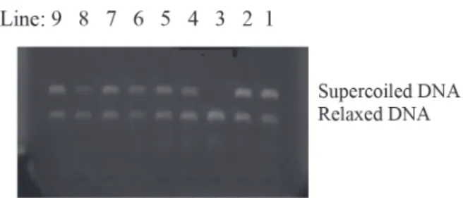
The conversion of supercoiled plasmid DNA to relaxed DNA by human topoisomerase I (Topo 1) was examined in the presence of 1, 2 and the new semi-synthetic diterpene derivatives. The activity of the compounds on Topo I was observed through relaxation assays using pBR322 plasmid DNA. Campothecin, a well-known Topo 1 enzyme inhibitor, was used as a positive control.44,45
Table 1. IC50 values for nor-clerodane diterpene derivatives against Ehrlich
carcinoma and K562 leukemia cells
Derivative IC50 ± SD / (µmol L
-1)
Ehrlich K562
1 > 50a > 50
2 > 50b > 50
3 > 50 24.74 ± 2.3
4 45.78 ± 4.35 7.85 ± 1.49
5 16.78 ± 1.42 13.08 ± 1.12
6 21.88 ± 1.96 > 50
7 43.43 ± 3.96 40.72 ± 5.63
Quercetin 44.0 ± 3.9 −
Vincristine − 0.060 ± 0.001
a1, IC
Results were observed through the alteration of the electrophoretic mobility of pBR322 plasmid DNA, combining Topo I action and the drugs at 200 µmol L-1. After this development, the results were analyzed with ethidium bromide in UV light, and recorded by photographing with a digital camera. As shown in Figure 2, the mobility of supercoiled closed circular double-stranded plasmid DNA increased on Topo I mediated relaxation, when subjected to electrophoresis with ethidium bromide (line 3). In the presence of 200 µmol L-1 campothecin (positive control), the relaxation effect was not observed (line 1). The relaxation inhibitory effect was also observed in the presence of Topo I with 1, 3, 4, 5, 6, and 7. When the assays were performed at 20 µmol L-1, only 4 did not show inhibitory effect on Topo I. These results must justify the cytotoxic activities observed.
Conclusions
In summary, new clerodane diterpene derivatives were synthesized from trans-dehydrocrotonin, the major bioactive metabolite of the Amazonian medicinal plant
Croton cajucara, by simple processes and with good yields. In vitro studies of cytotoxic activities against Ehrlich carcinoma and K562 leukemia cells showed that the nitrogenated derivatives were more active than the natural products 1 and 2, especially phenyl and methyl-hydrazone. The study of a possible mechanism of action showed the strong inhibitory effect of the nitrogenated derivatives, except 4, on DNA-topoisomerase I, in the relaxation assay.
Experimental
Chemistry
Reagents and apparatus
The melting points were determined on a Quimis hot
stage instrument. Infrared spectra (KBr pellets or CHCl3) were recorded on a PerkinElmer 1605 spectrometer and expressed in cm-1. The microanalyses were carried out with a Carlo Erba EA-1110 CHNS-O Elemental Analyser. 1H and 13C NMR spectra were obtained on a Bruker AC 200 spectrometer (200 and 50.3 MHz), using CDCl3 or DMSO-d6 as solvent, and TMS as internal standard. Elemental analyses were performed on a PerkinElmer 2400 CHN in the Laboratory of Environmental Science at the State University of Northern Rio de Janeiro (UENF). Thin layer chromatography (TLC) analyses were performed on silica gel 60 F254 plates. All the reagents were purchased from Merck or Sigma-Aldrich.
Vegetal material and the isolation of 1 and 2
Plant material, Croton cajucara Benth was collected in Jacundá-PA (Amazon region, Brazil), and botanically authenticated by Dr Nelson A. Rosa, Museu Paraense Emílio Goeldi. A voucher specimen No. 247 is deposited at the Herbarium of the same museum. The methanolic extraction from the powdered bark of Croton cajucara
and the isolation of 1 and 2 was carried out as previously reported.3,27
General method for the preparation of hydrazone derivatives (3-5)
For the solution of 1 (0.30 mmol) in ethanol (1 mL), 1.2 mmol of corresponding hydrazine was added; the reaction mixture was stirred for 6 h at room temperature for hydrazine hydrate, and for 21 and 24 h under reflux for methyl and phenyl-hydrazine, respectively. At these times, the hydrazine excess had been evaporated, and the crude products were purified by flash chromatography on silica gel (EtOAc:hexane, 10:90) for 4 and 5, and by recrystallization from EtOH and powdered charcoal for 3.
2-Hydrazone-dehydrocrotonin (3)
80% yield; mp 119-121 °C; IR (KBr) νmax / cm-1 3433, 2927-2876, 1695, 1378; 1H NMR (200 MHz, CDCl
3) d 1.11 (3H), 1.66 (1H, β), 1.84 (1H, α), 1.94 (3H, br s), 2.15 (1H,
α), 2.22 (1H), 2.35-2.39 (1H), 2.48 (1H, β), 3.14 (1H, br s), 3.44 (2H), 5.40 (1H), 5.86 (1H), 2.35-2.39 (1H), 6.38 (1H, s), 7.42 (2H); 13C NMR (50 MHz, CDCl
3) d 17.4, 21.6, 27.5, 28.5, 30.1, 38.8, 40.9, 41.6, 51.8, 45.5, 72.0, 108.8, 124.1, 125.2, 140.2, 143.8, 151.9, 160.1, 177.4. Anal. calcd. for C19H24N2O3: C, 69.49; H, 7.37; N, 8.53. Found: C, 69.73; H, 7.28; N, 8.72.
2-Methylhydrazone-dehydrocrotonin (4)
60% yield; mp 135-136 °C; IR (KBr) νmax / cm-1 3296, Figure 2. Effect of trans-dehydrocrotonin derivatives in 200 µmol L-1
on Topo I. Line 1: 0.25 µg DNA pBR322 + 1U Topo I + campothecin 200 µmol L-1; line 2: 0.25 µg DNA pBR322; line 3: 0.25 µg DNA pBR322
2960-2864, 1746, 1318; 1H NMR (200 MHz, DMSO-d 6) d 1.10 (3H), 1.59 (1H, β), 1.71 (2H), 1.81 (1H, α), 2.11 (1H, α), 2.20 (1H), 2.47 (1H, m), 2.57 (1H, β), 2.77 (3H), 2.86 (1H, br s), 5.58 (2H, br s), 5.86 (1H), 6.75 (1H, s), 7.79 (1H), 7.93 (1H); 13C NMR (50 MHz, CDCl
3) d 17.6, 20.8, 25.5, 28.3, 30.2, 38.0, 40.9, 41.0, 44.1, 51.6, 71.7, 108.9, 124.1, 125.4, 140.6, 144.4, 177.4. Anal. calcd. for C20H26N2O3: C, 70.15; H, 7.65; N, 8.18. Found: C, 70.37; H, 7.43; N, 8.24.
2-Phenylhydrazone-dehydrocrotonin (5)
75% yield; mp 171-172 °C; IR (KBr) νmax / cm-1 3328, 2958-2858, 1743, 1322; 1H NMR (200 MHz, CDCl
3) d 1.13 (3H, d, J 5.1 Hz), 1.62 (1H, β), 1.70 (2H), 1.77 (2H), 1.84 (3H, br s), 1.94 (1H, α), 2.18 (1H, α, d, J 3.3 Hz), 2.24-2.27 (1H), 2.57 (1H, br s, β), 2.33-2.70 (1H, dd, J 14.0, 6.3 Hz), 2.83 (1H, br s), 5.51 (1H), 5.59 (1H, sl), 6.48 (1H), 6.83 (2H, dd, J 0.7, 8 Hz), 6.96 (1H, d, J 6.3 Hz), 6.98 (2H, d,
J 0.7 Hz), 7.58 (1H), 7.59 (1H); 13C NMR (50 MHz, CDCl 3) d17.4, 21.4, 26.0, 27.6, 30.0, 39.6, 40.4, 41.7, 45.9, 51.5, 72.3, 108.0, 113.2, 120.3, 122.3, 126.6, 129.1, 139.1, 144.3, 152.0, 153.1, 166.1, 177.4.Anal. calcd. for C25H28N2O3: C, 74.23; H, 6.98; N, 6.93. Found: C, 74.37; H, 6.72; N, 7.04.
The preparation of 2-oxime-dehydrocrotonin (6)
For the solution of 1 (0.5 mmol), HONH2-HCl (0.8 mmol), 2 mL of ethanol in 0.4 mL of water was added under stirring NaOH (2.75 mmol). The mixture had been refluxed for 10 min, and when it reached the room temperature, it was added to an acid solution (0.3 mL HCl and 2 mL water). After that, the precipitate was filtered, washed with water and dried at room temperature. 80% yield; mp 197-198 °C; IR (KBr) νmax / cm-1 3279, 2941-2860, 1751, 1635, 1463-1436, 1351, 1184; 1H NMR (200 MHz, CDCl3) d 1.87 (1H, α), 1.12 (3H, d, J 5.1 Hz), 1.55 (1H), 1.63 (1H, β), 1.74 (1H), 1.82 (3H, br s), 2.15 (1H, α), 2.25 (2H), 2.44 (2H, t, J 8.3 Hz), 2.92 (1H, β), 3.44 (1H, d, J 5.1 Hz), 5.40 (1H, t, J 8.3 Hz), 5.89 (1H), 6.50 (1H, s), 7.43 (1H), 7.49 (1H), 8.05 (1H, br s); 13C NMR (50 MHz, CDCl3) d17.6, 21.3, 28.5, 30.2, 38.4, 41.8, 46.3, 51.8, 72.1, 108.2, 119.2, 125.1, 140.0, 144.2, 153.2, 156.2, 177.4. Anal. calcd. for C19H23NO4: C, 69.28; H, 7.04; N, 4.25. Found: C, 69.41; H, 6.98; N, 4.32.
The preparation of 2-O-methyloxime-dehydrocrotonin (7)
For the solution of 3 (0.15 mmol) in CH2Cl2 (2 mL), Ag2O (0.22 mmol) and CH3I (0.75 mmol) were slowly added. The solution was stirred at room temperature for 3 h. After that, CH2Cl2 was evaporated, and the residue
was purified by flash chromatography on silica gel (hexane:AcOEt, 90:10), thus providing product 7 as an oil in 60% yield. IR (CHCl3) νmax / cm1 2929-2860, 1749, 1610, 1319; 1H NMR (200 MHz, CDCl
3) d 1.23 (3H, br s), 1.66 (1H, β), 1.91 (1H, α), 2.00 (2H), 2.17 (1H, m, α), 2.44 (1H, t, J 7.6 Hz), 2.93 (1H, m, β), 3.17 (1H, br s), 5.05 (3H, br s), 5.42 (1H, br s), 6.13 (1H, br s), 6.53 (1H), 7.44 (1H), 7.49 (1H); 13C NMR (50 MHz, CDCl
3) d 17.6, 22.0, 28.0, 30.0, 39.4, 39.7, 40.3, 44.0, 45.1, 51.6, 64.7, 72.3, 108.1, 116.6, 126.7, 140.0, 144.5, 158.6, 166.3, 177.2. Anal. calcd. for C20H25NO4: C, 69.95; H, 7.34; N, 4.08. Found: C, 70.09; H, 7.21; N, 4.
Biological assays
Materials
The dye MTT (3-(4,5-dimethylthiazol-2-yl)-2,5-diphenyltetrazolium bromide) was obtained from Sigma Co. (USA); quercetine and vincristine sulfate, used as positive control in cytotoxic assays, were obtained from Sigma-Aldrich Co. (USA) and Merck Co. (Germany), respectively.
Cell culture
Ehrlich carcinoma cells were maintained for 12-14 days in Swiss mice. The tumor cell cultures were initiated from mouse Ehrlich ascites with at least one in vitro
passage prior to use. K562 cells were maintained in RPMI 1640 medium, containing 10% fetal calf serum (FCS), 100 U mL-1 penicillin G and 100 µg mL-1 streptomycin. The cell cultures were incubated at 37 °C in a 5% CO2 humidified atmosphere.
Drugs and cytotoxic assay
out in triplicate. After 48 h (Ehrlich) and 96 h (K562), at 37°C under 5% CO2, the cultures were incubated with MTT (5 mg mL-1) for 3 h. The formazan produced by live cells were solubilized with acidic isopropanol, and the absorbance was measured at 570 nm. IC50 values were obtained by linear regression analysis of the absorbance (percent) versus the log of drug concentration. The data are expressed as means ± standard deviation (SD) of 3 independent experiments. Statistical significance was assessed by the Student’s t-test, p < 0.05 was considered significant difference.
DNA-topoisomerase I assay
Topo I inhibition was determined by relaxation assay, which was carried out as described in the TopoGEN screening kit. For Topo I, one unit of the enzyme was utilized to relax 0.125 µg of the supercoiled ϕ×174 plasmid DNA. The reaction mixture (10 µL) contained the drug, DNA, assay buffer, 1U of Topo I and water. The mixture was incubated at 37 °C for 30 min, and the reaction was finalized by the addition of 1 µL of dye solution containing 25% bromophenol blue, 50% glycerol and 10% SDS. Reaction products were loaded onto a 1% agarose gel, containing ethidium bromide. Electrophoresis was carried out in tris-acetate-EDTA, pH 8.5, at 15 V, for 3.5 h; and then, it was photographed with a digital camera by illumination.
Supplementary Information
Supplementary data (1H and 13C NMR spectra) are available free of charge at http://jbcs.sbq.org.br as PDF file.
Acknowledgments
The authors thank the Coordenação de Aperfeiçoamento de Pessoal de Nível Superior (CAPES), Conselho Nacional de Desenvolvimento Científico e Tecnológico (CNPq) and Fundação de Amaparo à Pesquisa do Estado do Rio de Janeiro (FAPERJ) for financial support and the fellowships.
References
1. Sampson, J. H.; Phillipson, J. D.; Bowery, N. G.; O’Neill, M. J.; Houston, J. G.; Lewis, J. A.; Phytother.Res.2000, 14, 24. 2. Salatino, A.; Salatino, M. L. F.; Negri, G.; J. Braz. Chem. Soc.
2007, 18, 11.
3. Maciel, M. A. M.; Pinto, A. C.; Arruda, A. C.; Pamplona, S. G. S. R.; Vanderlinde, F. A.; Lapa, A. J.; Cólus, I. M. S.; Echevarria, A.; Grynberg, N. F.; Farias, R. A. F.; Luna, A. M. C.; Rao, V. S. N.; J. Ethnopharmacol.2000, 70, 41.
4. Grassi-Kassisse, D. M.; Wolf-Nunes, V.; Miotto, A. M.; Farias-Silva, E.; Souza-Brito, A. R. M.; Nunes, D. S.; Spadari-Bratfisch, R. C.; J. Pharm. Pharmacol. 2003, 55, 253. 5. Rodrigues, G.; Marcolin, E.; Bona, S.; Porawski, M.; Lehmann,
M.; Marroni, N. P.; Arq. Gastroenterol.2010, 47, 301. 6. Silva, R. M.; Santos, F. A.; Maciel, M. A. M.; Pinto, A. C.; Rao,
V. S. N.; Planta Med. 2001, 67, 763.
7. Huang, W.; Li, G.; Wu, Y.; Ge, W.; Chung, H. Y.; Ye, W.; Li, Y.; Wang, G.; Heterocycles2014, 89, 1585.
8. Yang, L.; Zhang, Y.; Wu, Z.; Chen, N.; Jiang, S.; Li, Y.; Wang, G.; Chem. Lett.2016, 45, 1235.
9. Li, R.; Morris-Natschke, S. L.; Lee, K.; Nat. Prod. Rep.2016, 33, 1166.
10. Liu, C. P.; Xu, J. B.; Zhao, J. X.; Xu, C. H.; Dong, L.; Ding, J.; Yue, J. M.; J. Nat. Prod. 2014, 77, 1013.
11. Gupta, V. K.; Tiwari, N.; Gupta, P.; Verma, S.; Pal, A.; Srivastava, S. K.; Darokar, M. P.; Phytomedicine 2016, 23, 654.
12. Cavin, A. L.; Hay, A. E.; Marston, A.; Stoeckli-Evans, H.; Diallo, D.; Hostettmann, K.; J. Nat. Prod. 2006, 69, 768. 13. Huang, Z.; Jiang, M. Y.; Zhou, Z. Y.; Xu, D.; Z. Naturforsch.
B: J. Chem. Sci.2010, 65, 83.
14. Bayor, M. T.; Ayim, J. S. K.; Marston, G.; Phillips, R. M.; Shnyder, S. D.; Wheelhouse, R. T.; Wright, C. W.; Nat. Prod. Commun. 2008, 3, 1875.
15. Wu, T.; Cheng, Y.; Chen, C.; Ng, L.; Chou, L.; Huang, L.; Chen, Y.; Kuo, S.; El-Shazly, M.; Wu, Y.; Chang, F.; Liaw, C.; Molecules 2014, 19, 2049.
16. Dai, S. J.; Liang, D. D.; Ren, Y.; Liu, K.; Shen, L.; Chem. Pharm. Bull.2008, 56, 207.
17. M a m bu , L . ; G r e l l i e r, P. ; F l o r e n t , L . ; J oy e a u , R . ; Ramanitrahasimbola, D.; Rasoanaivo, P.; Frappier, F.; Phytochemsitry 2006, 67, 444.
18. Harinantenaina, L.; Takahara, Y.; Nishizawa, T.; Kohchi, C.; Soma, G. I.; Asakawa, Y.; Chem. Pharm. Bull. 2006, 54, 1046. 19. Munro, T. A.; Duncan, K. K.; Xu, W.; Wang, Y.; Liu-Chen, L.
Y.; Carlezon Jr, W. A.; Cohen, B. M.; Beguin, C. B.; Bioorg. Med. Chem. 2008, 16,1279.
20. Chang, H. L.; Chang, F. R.; Chen, J. S.; Wang, Y. P.; Wu, Y. H.; Wang, C. C.; Wu, Y. C.; Hwang, T. L.; Eur. J. Pharmacol.
2008, 586, 332.
21. Bautista, E.; Toscano, A.; Calzada, F.; Díaz, E.; Yepez-Mulia, L.; Ortega, A.; J. Nat. Prod. 2013, 76, 1970.
22. Qiu, M.; Cao, D.; Gao, Y.; Li, S.; Zhu, J.; Yang, B.; Zhou, L.; Zhou, Y.; Jin, J.; Zhao, Z.; Fitoterapia 2016, 108, 81. 23. Maciel, M. A. M.; Pinto, A. C.; Brabo, S. N.; Silva, M. N.;
Phytochemistry1998, 49, 823.
25. Jullian, V.; Bounduelle, C.; Valentin, L.; Acebey, L.; Duigou, A. G.; Prevostb, M. F.; Sauvain, M.; Bioorg. Med. Chem.2005, 15, 5065.
26. Williams, R. B.; Norris, A.; Miller, J. S.; Birkinshaw, C.; Ratovoson, F.; Andriantsiferana, R.; Rasamison, V. E.; Kingston, D. G. I.; J. Nat. Prod. 2007, 70, 206.
27. Kubo, I.; Asaka, Y.; Shibata, K.; Phytochemistry1991, 30, 2546. 28. Poersch, A.; Santos, F. V.; Maciel, M. A. M.; Câmara, J. K. P.;
Dantas, T. N. C.; Cólus, I. M. S.; Mutat. Res.2007, 629, 14. 29. Souza-Brito, A. R. M.; Rodriguez, J. A.; Hiruma-Lima, C. A.;
Haun, M.; Nunes, D. S.; Planta Med. 1998, 64, 126.
30. Hiruma-Lima, C. A.; Spadari-Bratfisch, R. C.; Grassi-Kassisse, D. M.; Souza-Brito, A. R. M.; Planta Med.1999, 65, 325. 31. Rodriguez, J. A.; Hiruma-Lima, C. A.; Souza-Brito, A. R. M.;
Hum. Exp. Toxicol. 2004, 23, 455.
32. Hiruma-Lima, C. A.; Tona, W.; Gracioso, J. S.; Almeida, A. B. A.; Batista, L. M.; Magri, L.; Paula, A. C. B.; Soares, F. R.; Nunes, D. S.; Souza-Brito, A. R. M.; Biol. Pharm. Bull. 2002, 25, 425.
33. Carvalho, J. C. T.; Silva, M. F. C.; Maciel, M. A. M.; Pinto, A. C.; Nunes, D. S.; Lima, R. M.; Bastos, J. K.; Sarti, S. J.; Planta Med.1996, 62, 402.
34. Silva, R. M.; Oliveira, F. M.; Cunha, K. M. A.; Maia, J. L.; Maciel, M. A. M.; Pinto, A. C.; Nascimento, N. R. F.; Santos, F. A.; Rao, V. S. N.; Vasc. Pharmacol.2005, 43, 11.
35. Lima, G. S.; Castro-Pinto, D. B.; Machado, G. C.; Maciel, M. A. M.; Echevarria, A.; Phytomedicine 2015, 22, 1133. 36. Grynberg, N. F.; Echevarria, A.; Lima, J. E.; Pamplona, S. S.
R.; Pinto, A. C.; Maciel, M. A.; Planta Med.1999, 65, 687. 37. Maciel, M. A. M.; Martins, J. R.; Pinto, A. C.; Kaiser, C. R.;
Esteves-Souza, A.; Echevarria, A.; J. Braz. Chem. Soc. 2007, 18, 391.
38. Corrêa, D. H. A.; Melo, P. D. S.; Carvalho, C. A. A.; Azevedo, M. B. M.; Durán, N.; Haun, M.; Eur. J. Pharmacol. 2005, 510, 17.
39. Freire, A. C. G.; Assis, C. F.; Frick, A. O.; Melo, P. D. S.; Haun, M.; Aoyama, H.; Durán, N.; Sauer, M. M.; Kallás, E. G.; Ferreira, C. V.; Leuk. Res. 2003, 27, 823.
40. Frungillo, L.; Martins, D.; Teixeira, S.; Anazetti, M. C.; Melo, P. D. S.; Durán, N.; J. Pharm. Sci. 2009, 98, 4796.
41. Newkome, G. R.; Fishel, D. L.; J. Org. Chem. 1966, 31, 677. 42. Brehm, M.; Gӧckel, V. H.; Jarglis, P.; Lichtenthaler, F. W.;
Tetrahedron: Asymmetry2008, 19, 358. 43. Mosmann, T.; J. Immunol. Methods1983, 65, 55.
44. Pommier, Y.; Pourquier, P.; Fan, Y.; Strumberg, D.; Biochim. Biophys. Acta1998, 1400, 83.
45. Kim, D. H.; Lee, N.; Mini-Rev. Med. Chem. 2002, 2, 611.
Submitted: March 27, 2017
Published online: June 27, 2017