Article
J. Braz. Chem. Soc., Vol. 28, No. 6, 1014-1022, 2017. Printed in Brazil - ©2017 Sociedade Brasileira de Química 0103 - 5053 $6.00+0.00
*e-mail: [email protected]
Miscellaneous Diterpenes from the Aerial Parts of
Plectranthus ornatus
Codd
Fábio N. Ávila,a Francisco C. L. Pinto,a Thiciana S. Sousa,a Maria Conceição M. Torres,a
Leticia V. Costa-Lotufo,b,c Danilo D. Rocha,b Mayron A. de Vasconcelos,d
Nairley Cardoso-Sá,d Edson H. Teixeira,d Maria Rose Jane R. Albuquerque,e
Edilberto R. Silveiraa and Otília D. L. Pessoa*,a
aDepartamento de Química Orgânica e Inorgânica, Universidade Federal do Ceará,
60021-970 Fortaleza-CE, Brazil
bDepartamento de Fisiologia e Farmacologia, Universidade Federal do Ceará,
60430-270 Fortaleza-CE, Brazil
cDepartamento de Farmacologia, Universidade de São Paulo, 05508-900 São Paulo-SP, Brazil
dLaboratório Integrado de Biomoléculas, Departamento de Patologia e Medicina Legal,
Universidade Federal do Ceará, 60441-750 Fortaleza-CE, Brazil
eCoordenação de Química, Centro de Ciências Exatas e Tecnológicas,
Universidade Estadual Vale do Acaraú, 62040-340 Sobral-CE, Brazil
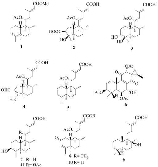
Five new diterpenes derivatives named as ornatin A, B, C, D and E, in addition to six known related diterpenes were isolated from the aerial parts of cultivated specimens of Plectranthus ornatus.The structures were elucidated using a combination of 1D/2D nuclear magnetic resonance (NMR) spectroscopy, high-resolution electrospray ionization mass spectrometry (HRESIMS) and comparison with published NMR data of analogous compounds. All isolated compounds were assayed against four human cancer cell lines, and Gram-positive and Gram-negative bacteria strains. None of them showed any cytotoxic activity, but ornatin C, D, E and three related diterpenes displayed marginal bactericidal or bacteriostatic effects against the Gram-positive strains.
Keywords:Plectranthus ornatus, diterpenes, ent-clerodanes, bacteriostatic
Introduction
The genus Plectranthus L’Her (Lamiaceae, subfamily Nepetoideae, tribe Ocimeae, subtribe Plectranthinae)
comprises approximately 300 species largely distributed over the African, Asian and Australian continents.1
Plectranthus is a large genus well known by its diversity
of ethnobotanical uses, being especially indicated to treat digestive disorders, skin diseases, infections and respiratory problems.2 In general, are rich source
of essential oils, terpenoids and phenol compounds.3
Pharmacological properties such as anti-inflammatory,4
antioxidant,5 antimicrobial,6 anti-tumoral7 and diuretic8
have been demonstrated for several isolated compounds from Plectranthus, corroborating with its larger medicinal
uses.
Plectranthus ornatus Codd (syn. Coleus comosus Hochst.
ex Gurke) is a perennial and aromatic herb, widespread around the new world.9 In the northeast of Brazil, P. ornatus,
popularly known as “malva santa” or “boldo miúdo”, is cultivated as a very important medicinal plant, indicated as analgesic and particularly to treat gastric disorders.10 Several
antimicrobial labdanes, ent-clerodanes, and halimane-type
diterpenoids have been previously isolated from P. ornatus.11
As part of a multidisciplinary program, where the efforts are devoted to study medicinal plants or congeners, in order to unveil their pharmacological or biological properties, herein we describe the isolation and characterization of five new diterpenes derivatives together with six known compounds from the aerial parts of cultivated specimens of P. ornatus, as
Experimental
General experimental procedures
Optical rotations were measured on a PerkinElmer 341 digital polarimeter. Infrared (IR) spectra were obtained on a PerkinElmer FT-IR spectrum 1000 spectrometer. Accurate mass spectra were acquired on a liquid chromatography-mass spectrometry ion-trap and time-of-flight (LCMS-IT-TOF, Shimadzu) spectrometer. Nuclear magnetic resonance (NMR) spectra were performed either on Bruker DPX-300 or DRX-500 spectrometers. Open column chromatography (CC) were carried out with silica gel (60 or 230 mesh, Merck) and Sephadex LH-20 (Phenomenex), while thin layer chromatography (TLC) were conducted on precoated silica gel aluminum sheets (60 F254, 0.20 mm,
Merck). Semi preparative Gemini-Phenomenex C-18 column (150 × 10 mm) was used on an ultra-fast liquid chromatography (UFLC, Shimadzu) system equipped with a SPD-M20A diode array UV-Vis detector. High performance liquid chromatography (HPLC) procedures were carried out
using UV PDA detection 210-400 nm, injection volume 200 µL, and flow rate of 4.72 mL min-1.
Plant material
Plectranthus ornatus was harvested in May 2012
at the medicinal plant garden Francisco José de Abreu Matos, Universidade Federal do Ceará (UFC). The plant authentication was performed by Dr Maria Iracema B. Loiola from the Herbario Prisco Bezerra (EAC), UFC, where a voucher (No. 56806) was deposited.
Extraction and isolation
The air-dried and powdered aerial parts of P. ornatus
(1.50 kg, 7.5% of fresh weight) was extracted with
n-hexane followed by EtOH (3 × 13 L), at room
temperature, to give the respective crude extracts after the solvents evaporation under reduced pressure. The
n-hexane extract (30.00 g, 0.22% of dry weight) was
subjected to a chromatography column (CC) over silica gel
eluted with binary mixture of n-hexane/EtOAc (100:0, 7:3,
1:1, 3:7, 0:100) to yield ten fractions (FA-FJ). FC (2.8 g) was subjected to silica gel CC and eluted with increasing amounts of n-hexane in EtOAc to yield compound 1
(50.7 mg), which was obtained by elution with n
-hexane-EtOAc 8:2. FG (573.6 mg) was chromatographed on a Sephadex LH-20 CC and eluted with MeOH to yield four subfractions (FG.1-FG.4). Subfraction FG.3 (350.6 mg) was subjected to CC silica gel column eluted with
n-hexane-EtOAc (95:5, 90:10, 80:20, 75:25, 70:30, 60:40,
1:1) to afford seven subfractions (FG.3-1-FG.3-7) from which FG.3-1 (256.6 mg) was further purified by HPLC using a semi preparative C-18 column and a solvent system constituted of H2O (TFA 0.1%)/MeOH (30:70) to
100% MeOH over 15 min, followed by 10 min of 100% MeOH, to give 2 (12.0 mg, tR = 7.85) and 3 (23.1 mg,
tR = 9.35). FH (2.2 g) was subjected to repeated CC
over silica gel, always using n-hexane-EtOAc gradient
mixtures to afford two main fractions FH1 (167.8 mg) and FH2 (146.5 mg). Both were subjected to HPLC using a semi preparative C-18 column and in the mobile phase, a gradient consisting of H2O (TFA 0.1%)/MeOH (35:65) to
100% MeOH over 20 min, followed by 10 min of 100% MeOH. From FH1 were isolated compounds 6 (24.0 mg, tR = 9.43) and 7 (4.5 mg, tR = 14.47), while from FH2 were
obtained compounds 8 (12.9 mg, tR = 6.82) and 9 (6.7 mg,
tR =11.64). FI (792.0 g) was subjected to Sephadex LH-20
by elution with MeOH to afford three main fractions, FI1-FI3. FI2 (351.8 mg) was rechromatographed on Sephadex LH-20 CC eluting with MeOH to yield subfractions FI2.1 and FI2.2. FI2.2 (276.6 mg) was further subjected to HPLC using a semipreparative C-18 column and a gradient of H2O (TFA 0.1%)/MeOH (30:70) to
90% MeOH over 15 min, followed by 10 min of 100% MeOH, to afford 4 (13.4 mg, tR = 9.03), 5 (16.4 mg,
tR = 11.45), 10 (24.0 mg, tR = 8.09) and 11 (17.0 mg,
tR = 9.50).
Ornatin A (1)
Colorless resin: [α]D22 –43.96° (c 0.1, CH2Cl2);
UV (MeOH) λmax / nm 215; IR (ATR, attenuated total
reflectance) ν / cm-1 1717, 1647, 1436, 1369, 1235, 1152; 1H and 13C NMR (CDCl
3), see Tables 1 and 2, respectively;
HRESIMS (high-resolution electrospray ionization mass spectrometry) m/z, observed: 399.2508; C23H36O4
[M + Na]+ requires 399.2506.
Ornatin B (2)
Colorless resin: [α]D22−34.44° (c 0.06, CH2Cl2); UV
(MeOH) λmax / nm 218; IR (KBr) ν / cm-1 3444, 2926, 1694,
1635, 1380, 1235; 1H and 13C NMR (MeOD), see Tables 1
and 2, respectively; HRESIMS m/z, observed:433.2197;
C22H34O7 [M + Na]+ requires 433.2202.
Ornatin C (3)
Colorless resin; [α]D22 −48.06° (c 0.1, MeOH); UV
(MeOH) λmax / nm217; IR (KBr) ν / cm-1 3436, 2934,
2867, 1711, 1643, 1439, 1244; 1H and 13C NMR (CDCl 3),
see Tables 1 and 2, respectively; HRESIMS m/z, observed:
419.2403;C22H36O6 [M + Na]+ requires 419.2404.
Ornatin D (4)
Greenish oil; [α]D22−47.50° (c 0.1, CH2Cl2); UV (MeOH)
λmax / nm 219; IR (ATR) ν / cm-1 3452, 2926, 2876, 1734,
1691, 1641, 1238, 1149, 1020; 1H and 13C NMR (CDCl 3),
see Tables 1 and 2, respectively; HRESIMS m/z, observed:
399.2114; C22H32O5 [M + Na]+ requires 399.2142.
Ornatin E (5)
Colorless resin; [α]D22−47.50° (c 0.1, CH2Cl2); UV
(MeOH) λmax / nm 218; IR (ATR) ν / cm-1 3396, 2925, 1736,
1688, 1641, 1435, 1237, 1207; 1H and 13C NMR (CDCl 3),
see Tables 1 and 2, respectively; HRESIMS m/z, observed:
399.2495; C22H32O4 [M + K]+ requires399.1938.
Cytotoxicity evaluation - MTT assay
Cytotoxicity was evaluated against four different human cancer cell lines provided by the National Cancer Institute U.S. (Bethesda, MD): HCT-116 (colon adenocarcinoma), HL-60 (leukemia), OVCAR-8 (ovarian carcinoma) and SF-295 (glioblastoma). Cells were maintained in RPMI 1640 medium supplemented with 10% fetal bovine serum (v/v), 2 mmol L-1 glutamine, 100 U mL-1
penicillin, 100 µg mL-1 streptomycin at 37 °C under a
5% CO2 atmosphere. Cytotoxicity was also evaluated
against peripheral blood mononuclear cells (PBMC) obtained from the peripheral blood of healthy volunteers after centrifugation on a Ficoll gradient. Cells were removed, washed with phosphate buffered saline (PBS) and resuspended in a RPMI 1640 medium supplemented with 20% of fetal bovine serum, 100 U mL-1 penicillin
and 100 µg mL-1 of streptomycin to a final concentration
of 3 × 10⁵ cell mL-1. Phytohemagglutinin (3%) was
added to induce cell proliferation. Compounds 1-11 were tested at concentrations ranging from 0.05 to 25 µmol L-1
during 72 h. The effect on cell proliferation was evaluated
in vitro using the MTT
[3-(4,5-dimethyl-2-thiazolyl)-2,5-diphenyl-2H-tetrazolium bromide] assay, as described
by Mosmann.12 IC
50 (the concentration that inhibits growth
(Sigma Aldrich Co.) was used as positive control (0.01 to 5 µmol L-1), while the vehicle used to dilute the substances,
DMSO at a final concentration of 0.5%, was used as negative control.
Antibacterial assay
The antibacterial activity of the diterpenes was evaluated against four bacteria: Staphylococcus aureus ATCC 25923, S. epidermidis ATCC 12128, Pseudomonas aeruginosa
ATCC 9027 and Escherichia coli ATCC 11303. Before
experimental procedures, each bacterial species was grown in tryptic soy agar (TSA, Himedia, USA) for 24 h at 37 °C. The cells were inoculated in tryptic soy broth (TSB, Himedia, USA) and incubated for 18 h at 37 °C under constant agitation. Subsequently, cell’s concentration of each bacterium was adjusted to 1 × 10⁶ cell mL-1 using
spectrophotometry and calibration curves previously determined. The minimal inhibitory concentration (MIC) and minimal bactericidal concentration (MBC) were determined by the broth microdilution method according with the guidelines from the National Committee for Clinical Laboratory Standards, M7-A6 (NCCLS, 2003),13 with
some modifications. Briefly, different concentrations of the compounds (7.8 to 500 µg mL-1) were prepared in TSB (with
4% of dimethyl sulfoxide) and aliquots of 100 µL of each compound were mixed with bacterial suspensions (100 µL) in the 96-well polystyrene plates. The plates were incubated at 37 °C during 24 h under constant agitation. The MIC value was established as the lowest concentration of compound able to inhibit the visible growth of microorganism. MBC value was determined by transferring 10 µL from each well without visible growth into TSA plates. MBC was considered the lowest concentration that completely inhibited microbial growth in the plates.
Results and Discussion
The chromatographic fractionation of the n-hexane
extract from the aerial parts of P. ornatus was performed,
leading to the isolation of five new diterpenes which were designated as ornatins A-E (1-5), as well as the known diterpenes: coleon R (6),14ent-3α-hydroxycleroda-4(18),
13E-dien-15-oic acid (7),15 plectrornatin A (8),16
labd-13-en-8β-hydroxy-15-oic acid (9),17 11-acetoxy-2-oxo-
ent-cleroda-3,13E-dien-15-oic acid (10)18 and ent-11R
-acetoxy-2-oxocleroda-3,13E-dien-15-oic acid (11).16
Compound 1, isolated as an colorless resin, [α]D22
–43.96° (c 0.1, CH2Cl2), had its molecular formula
assigned as C23H36O4 based on the [M + Na]+ ion peak at
m/z 399.2508 (calcd. 399.2506, D +0.50 ppm) in the HRESI
mass spectrum. The 1H NMR spectrum showed signals to
olefinic protons at dH 5.64 (s, 1H, H-14) and 5.19 (s, 1H,
H-3), to an oxymethine proton at dH 5.44 (d, 1H, J 10.9 Hz,
H-11) and a methoxyl at dH 3.66. In addition, were observed
six methyl signals, two of which attached to sp2 carbons at dH 2.14 (s, 3H, Me-16) and 1.57 (s, 3H, Me-18), another one
of an acetoxyl group at dH 1.99 (s, 3H, C-11’), two others
attached to quaternary carbons (dH 0.76, Me-20) and dH 1.04
(s, 3H, Me-19), and the last one attached to a tertiary carbon
dH 0.97 (d, 3H, J 6.1 Hz, Me-17). The 13C NMR spectrum
showed 23 carbon signals, assigned through the composite pulse decoupling (CPD), distortionless enhancement by polarization transfer (DEPT 135) and heteronuclear single quantum correlation (HSQC) spectra, into seven methyls, six methylenes, five monohydrogenated, including two methine, one oxymethine and two sp2 carbons, and six
non-hydrogenated carbons including four sp2 hybridized
carbons at dC 170.8 (C-11’), 166.7 (C-15), 144.5 (C-4)
and 158.1 (C-13) and two quaternary carbons at dC 38.7
(C-5) and 43.4 (C-9). The 1H and 13C NMR spectral data
(Tables 1 and 2) of 1 were similar to those reported for the diterpenes 11-acetoxyneoclerodane, which were previously isolated of the P. ornatus.19 The only difference between
these two compounds was the replacement of the carboxyl function at C-15 (dC 171.7) for a methyl ester (dC 166.7,
51.0 / dH 3.66). The double bond C-13/C-14 was defined
as E based on chemical shifts (see Table 1) in agreement
with previous diterpenes isolated of P. ornatus.16 As can
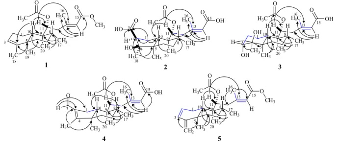
be seen in the Experimental section, no MeOH has been used during any extraction or chromatographic procedures, thus, eliminating the possibility of 1 being an artifact. The final structure of 1 was confirmed by the heteronuclear multiple bond correlation (HMBC) spectrum through the selected long-range correlations depicted by arrows in Figure 2, while its relative stereochemistry was established by the nuclear Overhauser spectrum (NOESY) (see Supplementary Information). The trans-configuration for
the A and B rings of the decaline moiety was confirmed by the dipolar coupling effect (NOE) between H-8/H-10 and Me-19/Me-20 both set in a 1,3-diaxial relationship. The observed NOE’s for Me-20 with Me-17 and Me-11’ (methyl of the acetoxyl group) confirmed the α-position inferred for both AcO-11’ and Me-17 (Figure 3), also in agreement with the stereochemistry reported previously for 11R* diterpenes from P. ornatus.16,18 Thus, compound 1
was assigned as the ent-clerodane diterpene designated as
ornatin A, in allusion to the species.
Compound 2, obtained as a colorless resin, [α]D22
−34.44° (c 0.06, CH2Cl2), had its molecular formula
determined as C22H34O7 based on the [M + Na]+ ion peak
the HRESIMS. Its 1H NMR spectrum displayed signals for
an olefinic proton at dH 5.66 (s, 1H, H-14), an oxymethine
proton at dH 5.10 (d, 1H, J 10.3 Hz, H-11), and six methyl
groups, including one attached to a sp2 carbon at d
H 2.20 (s,
3H, H-16) and another one of an acetoxyl group at dH 2.02
(s, 3H, H-11’). The 13C NMR spectrum exhibited 22 carbon
signals, which in accordance with the DEPT 135 and HSQC spectra were defined as six methyls, four methylenes, five monohydrogenated and seven non-hydrogenated carbons, including an olefinic carbon dC 160.6 (C-13), two rid
carboxyls dC 180.3 (C-3) and dC 171.6 (C-15), one of which
α,β-unsaturated, and an acetoxyl moiety dC 170.9 (C-11’).
Comparison of the 1H and 13C NMR data of 2 with those of 1
showed that these compounds share a partial similarity, but differing markedly in the A ring. In the HMBC spectrum, the long range correlation for the methylene protons
dH 2.15 (m, 1H, H-1a) with the methine carbons at dC 51.4
(C-2) and 45.7 (C-10), and with the carboxyl at dC 180.3
(C-3), as well as correlation of the dH 1.35 (s, 3H, Me-18)
with the quaternary carbon at dC 49.5 (C-5) and with an
oxygenated non-hydrogenated carbon at 82.7 (C-4) allowed to suggest a rearranged five membered ring bearing a carboxyl group at C-2 and a hydroxyl at C-4. Finally, the relative stereochemistry of 2 was established by the NOESY spectrum which showed dipolar couplings for H-2/Me-18 and Me-18/Me-19 indicating a β-position to the carboxyl acid at C-2, while the NOE’s of H-8/H-10 and Me-19/ Me-20 supported a trans fusion of the A/B rings, as depicted
in Figure 3. The stereochemistry of C-11 was suggested as R* in accordance with previously diterpenes isolated
from P. ornatus.16,18 Thus, the structure of compound 2 was
established as a new rearranged (4→2)-abeo-clerodane,
named as ornatin B.
Compound 3, a colorless resin, [α]D22−48.06° (c 0.1,
MeOH), had its molecular formula C22H36O6 inferred
from the [M + Na]+ ion peak at m/z 419.2403 (calcd. for
C22H36O6Na, 419.2404, D−0.24 ppm) as showed through
HRESIMS. The 1H NMR spectrum showed signals for
an olefinic proton at dH 5.64 (H-14, s), two oxymethine
protons at dH 5.40 (d, 1H, J 11.2 Hz, H-11) and 3.50 (br t,
1H, J 2.3 Hz, H-3) and signals to six methyl groups, one
of which attached to a sp2 carbon at d
H 2.20 (s, 3H, H-16)
and another one of an acetoxyl group at dH 1.99 (s, 3H,
H-11’). The 13C NMR spectrum showed signals for 22
carbon atoms, accounting for six methyls, five methylenes, five methines including one olefinic dC 119.6 (C-14)and
one oxymethine carbons dC 77.6 (C-3). The 13C NMR and
DEPT spectra displayed six non-hydrogenated carbons, including one olefinic carbon dC 158.6 (C-13),an acetoxyl dC 172.8 (C-11’) and a conjugated acid carboxyl dC 169.7
(C-15). The 13C NMR data of 3 were similar to those of 1,
except by the appearance of two carbon signals at dC 77.6
and dC 76.9 corresponding to the hydroxylation of C-3 and
C-4, instead the carbon-carbon double bond. Based on the HSQC spectrum the carbon signal at dC 77.6 exhibited
correlation with the proton signal at dH 3.50 (br t, J 2.3 Hz,
H-3). Comparing these data with those of 3β,4β and 3α,4β -dihydroxylated ent-clerodane diterpenes from literature,20,21
the structure of compound 3 was established as the 3α,4β
derivative which was named as ornatin C.
Compound 4, a greenish oil, [α]D22−47.50° (c 0.1,
CH2Cl2), had its molecular formula C23H38O6 determined by
HRESIMS through theion peak [M + Na]+ at m/z 399.2114
(calcd. for C22H32O5Na,399.2142, D +1.75 ppm). The 1H NMR spectrum showed signals for an aldehyde proton
at dH 9.93 (s, 1H, H-3), an olefinic proton at dH 5.64 (s,
1H, H-14), an oxymethine proton at dH 5.12 (dd, 1H, J 9.6,
2.6 Hz, H-11), and six methyl groups, two of which attached to sp2 carbons at d
H 2.12 (s, 3H, H-16) and 1.99 (s, 3H,
H-18), and another one of an acetyl group at dH 1.96 (s,
3H, Me-11’). The 13C NMR spectrum exhibited 22 carbon
signals, classified into six methyls, four methylenes, four methines and seven non-hydrogenated carbons, including two acetoxy carbonyl dC 170.8 (C-11’), dC 171.4 (C-15) and
an aldehyde carbonyl dC 188.9 (C-3). Two α,β-conjugated
systems [dC 159.6 (C-4), 137.9 (C-2), 188.9 (C-3)] and
[dC 170.7 (C-13), 118.1 (C-14), 171.4 (C-15)]were
assigned as established by the DEPT 135, HSQC and HMBC spectra. Complete 1H and 13C assignments of 4
were made using a combination of 1D and 2D-NMR data and comparison with the NMR data of 2 (Tables 1 and 2), where the difference between these compounds were the presence of a double bond at C2/C3 and an aldehyde function at C-2, instead of a carboxyl acid. This proposition was supported by the HMBC spectrum, which exhibited correlations for the aldehyde proton dH 9.93 (H-3) with the
carbon signals at dC 137.9 (C-2) and dC 28.1 (C-1), as well
as correlations of dH 1.99 (s, 3H, Me-18) with dC 137.9
(C-2) and dC 51.7 (C-5). It is worthy of notice that both
structures of 2 and 4 were also supported by comparison of the 1H and 13C NMR data of these with those reported
to the rearranged (4→2)-abeo-clerodane diterpenes.22
The relative stereochemistry of 4, as depicted in Figure 3, was defined by NOESY and literature data,16,18 which was
similar to compound 2. Thus, the structure of compound 4, a new (4→2)-abeo-clerodane diterpene, was established
and named as ornatin D.
Compound 5, a colorless oil, had itsmolecular formula C22H32O4 determined from theion peak [M + K]+ at
m/z 399.2495 in the HRESIMS. Its IR spectrum exhibited
UV absorption bands at 3393 and 1691 cm-1 for conjugated
1H NMR spectrum showed signals for olefinic protons
at dH 6.05 (d, 1H, J 9.5 Hz, H-3), 5.80 (m, 1H, H-2),
5.66 (s, 1H, H-14), 4.80 (s, 1H, H-18a) and 4.66 (s, 1H, H-18b), the latter two corresponding to vinyl protons of an exocyclic double bond. In addition, it displayed signals for an oxymethine proton at dH 5.33 (d, 1H, J 10.3 Hz, H-11)
and five methyl groups, including a methyl attached to an olefinic carbon at dH 2.11 (s, 3H, Me-16) and a methyl of
an acetoxyl group at dH 2.01 (s, 3H, C-11’). The 13C NMR
spectrum exhibited signals to 22 carbons atoms assigned as five methyls, five methylenes, six methines and six non-hydrogenated carbons, including four carbon-carbon double bonds and two carboxyl carbons at dC 171.0 (C-15)
and dC 170.8 (C-11’). The 1H and 13C NMR data of 5
were similar to those reported initially to 1-4, showing differences only for the A ring. A system comprising an exocyclic double bond of conjugated diene located at A ring was confirmed through the long range correlations of the allylic protons dH 2.40 (m, 2H, H-1) with dC 128.1
(C-2) and dC 129.1 (C-3) and correlations of the
exo-methylene protons dH 4.80; 4.66 (s, 1H-18ab) with C-3
and C-5 (Figure 2). Finally, the relative stereochemistry of 5 was confirmed by NOESY spectrum, similar to those of compounds 1-4. Based on the aforementioned data, the complete structure of 5 was established as a new
ent-clerodane diterpene which was named ornatin E.
Following their identification, compounds 1-11 were assessed for cytotoxicity in four human cancer cell lines, HCT-116, HL-60, OVCAR-8 and SF-295. All compounds showed to be non-cytotoxic at tested concentrations (IC50 values > 25 µM, data not shown). The anthracycline,
doxorubicin, used as positive control, presented IC50 values
ranging from 0.02 µM in HCT-116 cells to 0.33 µM in SF-295 cells.
Regarding the antibacterial activity, compounds 1 through 11 were evaluated by MIC and MBC assay against clinically relevant bacteria. The results showed that some diterpenes were bacteriostatic or bactericidal against Gram-positivebacteria. Compounds 3, 4 and 6 showed MIC for S. aureus at concentrations of 250 µg mL-1 and
7 and 8 at 500 µg mL-1. Furthermore, S. epidermidis
was susceptible only to compound 5 at 500 µg mL-1.
Interestingly, only compound 3 showed bactericidal effects on S. aureus at 500 µg mL-1. On the other hand,
no compounds were effective against Gram-negative bacteria. The antibacterial results found here corroborates with other studies reporting on the antimicrobial activity of diterpenes.23 Moreover, the effective concentration
of the diterpenes used in this study were similar to those found for other works.24 According Urzúa et al.25
the antimicrobial activity of diterpenes occurs by destabilization of the plasma membrane caused by the interaction with the lipid bilayer fatty acids present in its structure. Moreover, some studies showed that diterpenes exhibit antibacterial activity only against Gram-positive bacteria, but not against Gram-negative bacteria. Probably, the outer membrane of the Gram-negative bacteria decreases the permeability of the diterpenes and increases resistance to the action of these compounds.
Conclusion
Five new diterpenes, along with six known ones, were isolated from the aerial parts of Plecthranthus ornatus.
Diterpenes are the most characteristic compounds of the genus Plecthranthus, especially those having the abietane
and labdane skeletons. However, previous work on P. ornatus
have reported it, instead, as a prolific source of clerodane
Figure 3. Diagnostic NOESY correlations for compounds 1-5.
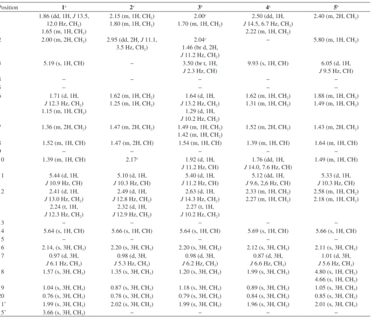
Table 1. 1H NMR data for compounds 1, 2, 4 and 5 (CDCl
3), and 3 (MeOD)
Position 1a 2a 3b 4a 5b
1 1.86 (dd, 1H, J 13.5, 12.0 Hz, CH2)
1.65 (m, 1H, CH2)
2.15 (m, 1H, CH2)
1.80 (m, 1H, CH2)
2.00c
1.70 (m, 1H, CH2)
2.50 (dd, 1H, J 14.5, 6.7 Hz, CH2)
2.22 (m, 1H, CH2)
2.40 (m, 2H, CH2)
2 2.00 (m, 2H, CH2) 2.95 (dd, 2H, J 11.1,
3.5 Hz, CH2)
2.04c
1.46 (br d, 2H, J 11.2 Hz, CH2)
− 5.80 (m, 1H, CH2)
3 5.19 (s, 1H, CH) − 3.50 (br t, 1H,
J 2.3 Hz, CH)
9.93 (s, 1H, CH) 6.05 (d, 1H, J 9.5 Hz, CH)
4 − − − − −
5 − − − −
6 1.71 (d, 1H,
J 12.3 Hz, CH2)
1.15 (m, 1H, CH2)
1.62 (m, 1H, CH2)
1.25 (m, 1H, CH2)
1.64 (d, 1H, J 13.2 Hz, CH2)
1.29 (d, 1H, J 10.2 Hz, CH2)
1.62 (m, 1H, CH2)
1.31 (m, 1H, CH2)
1.88 (m, 1H, CH2)
1.49 (m, 1H, CH2)
7 1.36 (m, 2H, CH2) 1.47 (m, 2H, CH2) 1.49 (m, 1H, CH2)
1.42 (m, 1H, CH2)
1.52 (m, 2H, CH2) 1.43 (m, 2H, CH2)
8 1.52 (m, 1H, CH) 1.47 (m, 2H, CH) 1.54 (m, 1H, CH) 1.39 (m, 1H, CH) 1.64 (m, 1H, CH)
9 − − − − −
10 1.39 (m, 1H, CH) 2.17c 1.92 (d, 1H,
J 11.2 Hz, CH)
1.76 (dd, 1H, J 14.0, 7.6 Hz, CH)
1.49 (m, 1H, CH)
11 5.44 (d, 1H,
J 10.9 Hz, CH)
5.10 (d, 1H, J 10.3 Hz, CH)
5.40 (d, 1H, J 11.2 Hz, CH)
5.12 (dd, 1H, J 9.6, 2,6 Hz, CH)
5.33 (d, 1H, J 10.3 Hz, CH)
12 2.41 (d, 1H,
J 13.0 Hz, CH2)
2.24 (t, 1H, J 12.3 Hz, CH2)
2.49 (d, 1H, J 12.8 Hz, CH2)
2.32 (d, 1H, J 12.9 Hz, CH2)
2.63 (d, 1H, J 14.3 Hz, CH2)
2.27 (t, 1H, J 10.2 Hz, CH2)
2.33 (m, 1H, CH2)
2.27 (m, 1H, CH2)
2.58 (m, 1H, CH2)
2.18 (m, 1H, CH2)
13 − − − − −
14 5.64 (s, 1H, CH) 5.66 (s, 1H, CH) 5.64 (s, 1H, CH) 5.69 (s, 1H, CH) 5.66 (s, 1H, CH)
15 − − − − −
16 2.14, (s, 3H, CH3) 2.20 (s, 3H, CH3) 2.20 (s, 3H, CH3) 2.12 (s, 3H, CH3) 2.11 (s, 3H, CH3)
17 0.97 (d, 3H,
J 6.1 Hz, CH3)
0.98 (d, 3H, J 5.3 Hz, CH3)
0.98 (d, 3H, J 6.2 Hz, CH3)
0.87 (d, 3H, J 6.6 Hz, CH3)
1.01 (d, 3H, J 5.6 Hz, CH3)
18 1.57 (s, 3H, CH3) 1.35 (s, 3H, CH3) 1.20 (s, 3H, CH3) 1.99 (s, 3H, CH3) 4.80 (s, 1H, CH2)
4.66 (s, 1H, CH2)
19 1.04 (s, 3H, CH3) 0.87 (s, 3H, CH3) 1.18 (s, 3H, CH3) 0.89 (s, 3H, CH3) 1.05 (s, 3H, CH3)
20 0.76 (s, 3H, CH3) 0.78 (s, 3H, CH3) 0.79 (s, 3H, CH3) 0.84 (s, 3H, CH3) 0.85 (s, 3H, CH3)
11’ 1.99 (s, 3H, CH3) 2.02 (s, 3H, CH3) 1.99 (s, 3H, CH3) 1.96 (s, 3H, CH3) 2.01 (s, 3H, CH3)
15’ 3.66 (s, 3H, CH3) − − − −
and halimane diterpenes which are not well common to the genus. The results reported in the present work indeed corroborate the tendency of P. ornatus to behave differently
of the other species belonging to Plecthranthus. Nine (1-5,
7, 8, 10, 11), out of eleven diterpenes reported here belong to the clerodane class, while the two others belong to the labdane (9) and abietane (6) classes, respectively. Two of the isolated ent-clerodanes (2, 4) present an unusual
(4→2)-abeo-clerodane rearranged, while no halimane has
been isolated in the present work. Unfortunately, none of the eleven isolated compounds showed any cytotoxic action, and the antimicrobial activity was too weak in order to be considered effective.
Supplementary Information
Supplementary information (Figures S1-S42) is available free of charge at http://jbcs.sbq.org.br as a PDF file.
Acknowledgments
This work was supported by grants from Conselho Nacional de Desenvolvimento Científico e Tecnológico (CNPq), Coordenação de Pessoal e Nível Superior (CAPES) and Programa de Núcleo de Excelência (PRONEX).
References
1. Alasbahi, R. H.; Melzig, M. F.; Planta Med. 2010, 76, 653; Rice, L. J.; Brits, G. J.; Potgieter, C. J.; Van Staden, J.; S. Afr. J. Bot. 2011, 77, 947.
2. Lukhoba, C. W.; Simmonds, M. S. J.; Paton, A. J.; J. Ethnopharmacol. 2006, 103, 1.
3. Waldia, S.; Joshi, B. C.; Pathak, U.; Joshi, M. C.; Chem. Biodiversity 2011, 8, 244.
4. Chen, Y. S.; Yu, H. M.; Shie, J. J.; Cheng, T. J. R.; Wu, C. Y.; Fang, J. M.; Wong, C. H.; Bioorg. Med. Chem. 2014, 22, 1766; Napagoda, M.; Gerstmeier, J.; Wesely, S.; Popella, S.; Lorenz, S.; Scheubert, K.; Svatos, S.; Werz, O.; J. Ethnopharmacol.
2014, 151, 800.
5. Albuquerque, R. L.; Kentopff, M. R.; Machado, M. I. L.; Silva, M. G. V.; Matos, F. J. A.; Quim. Nova2007, 30, 1882. 6. Khare, R. S.; Banerjee, S.; Kundu, K.; Int. J. Pharma Bio Sci.
2011, 2, 488.
7. Xing, X.; Wu, H.; Wang, X.; Huang, Y.; Li, Q.; Li, C.; Yang, Y.; Liu, J.; J. Chemother.2008, 20, 238.
8. Patel, R.; Mahobia, N. K.; Gendle, R.; Kaushik, B.; Singh, S.; Pharmacogn. Res.2010, 2, 86.
9. Ascensão, L.; Mota, L.; Castro, M. M.; Ann. Bot. (Oxford, U. K.)1999, 84, 437.
10. Dellar, J. E.; Cole, M. D.; Waterman, P. G.; Phytochemistry
1996, 41, 735; Passinho-Soares, H.; Felix, D.; Kaplan, M. A.; Margis-Pinheiro, M.; Margis, R.; Planta Med. 2006, 72, 929.
11. Abdel-Mogib, M.; Albar, H. A.; Batterjee, S. M.; Molecules
2002, 7, 271.
12. Mosmann, T.; J. Immunol. Methods1983, 65, 55.
13. National Committee for Clinical Laboratory Standards; Document M7-A6; Methods for Dilution Antimicrobial Susceptibility Tests for Bacteria that Grow Aerobically; NCCLS: Wayne, PA, USA, 2003.
14. Arihara, S.; Ruedi, P.; Eugster, C. H.; Helv. Chim. Acta1975, 58, 343.
15. Wijethne, E. M. K.; Silva, L. B.; Tezuka, Y.; Kikuchi, T.; Phytochemistry1995, 39, 443.
16. Oliveira, P. M.; Ferreira, A. A.; Silveira, D.; Alves, R. B.; Rodrigues, G. V.; Emerenciano, V. P.; Raslan, D. R.; J. Nat. Prod. 2005, 68, 588.
17. Pacheco, A. G.; Oliveira, P. M.; Piló-Veloso, D.; Alcantara, A. F. C.; Molecules 2009, 14, 1245.
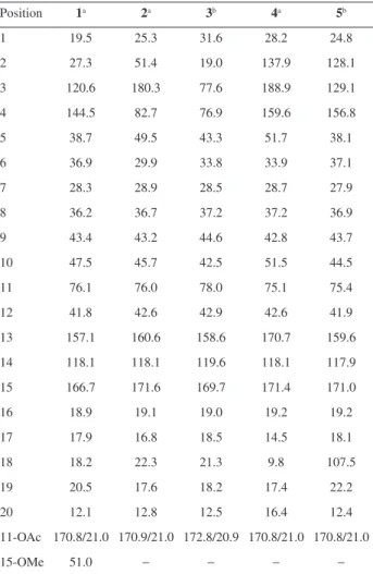
Table 2.13C NMR chemical shift (d) of compounds 1, 2, 4 and 5 (CDCl 3),
and 3 (MeOD)
Position 1a 2a 3b 4a 5b
1 19.5 25.3 31.6 28.2 24.8
2 27.3 51.4 19.0 137.9 128.1
3 120.6 180.3 77.6 188.9 129.1
4 144.5 82.7 76.9 159.6 156.8
5 38.7 49.5 43.3 51.7 38.1
6 36.9 29.9 33.8 33.9 37.1
7 28.3 28.9 28.5 28.7 27.9
8 36.2 36.7 37.2 37.2 36.9
9 43.4 43.2 44.6 42.8 43.7
10 47.5 45.7 42.5 51.5 44.5
11 76.1 76.0 78.0 75.1 75.4
12 41.8 42.6 42.9 42.6 41.9
13 157.1 160.6 158.6 170.7 159.6
14 118.1 118.1 119.6 118.1 117.9
15 166.7 171.6 169.7 171.4 171.0
16 18.9 19.1 19.0 19.2 19.2
17 17.9 16.8 18.5 14.5 18.1
18 18.2 22.3 21.3 9.8 107.5
19 20.5 17.6 18.2 17.4 22.2
20 12.1 12.8 12.5 16.4 12.4
11-OAc 170.8/21.0 170.9/21.0 172.8/20.9 170.8/21.0 170.8/21.0
15-OMe 51.0 − − − −
18. Rijo, P.; Rodriguez, B.; Duarte, A.; Simões, M. F.; Nat. Prod. J. 2011, 1, 57.
19. Rijo, P.; Gaspar-Marques, C.; Simões, M. F.; Jimeno, M. L.; Rodriguez, B.; Biochem. Syst. Ecol.2007, 35, 215.
20. Tsichritzis, F.; Jakupovic, J.; Phytochemistry1990, 29, 3173. 21. Yang, S.-M.; Wu, S.-H.; Qin, X.-D.; Luo, X.-D.; Wu, D.-G.;
Helv. Chim. Acta2004, 87, 1279.
22. Tung-Ho, W.; Yung-Yi, C.; Chao-Jung, C.; Lean-Teik, N.; Li-Chen, C.; Li-Jiau, H.; Yung-Husan, C.; Sheng-Chu, K.; Mohamed, E.; Yang-Chang, W.; Fang-Rong, C.; Chih-Chuang, L.; Molecules2014, 19, 2049.
23. Carneiro, V. A.; Santos, H. S.; Arruda, F. V. S.; Bandeira, P. N.; Albuquerque, M. R. J. R.; Pereira, M. O.; Henriques, M.; Cavada, B. S.; Teixeira, E. H.; Molecules2011, 16, 190; Cardoso Sá, N.; Cavalcante, T. T.; Araújo, A. X.; dos Santos, H. S.;
Albuquerque, M. R.; Bandeira, P. N.; da Cunha, R. M.; Cavada, B. S.; Teixeira, E. H.; Arch. Oral Biol. 2012, 57, 550; Urzúa, A.; Jara, F.; Tojo, E.; Wilkens, M.; Mendoza, L.; Rezende, M. C.; J. Ethnopharmacol. 2006, 103, 297.
24. Vasconcelos, M. A.; Arruda, F. V. S.; Santos, H. S.; Rodrigues, A. S.; Bandeira, P. N.; Albuquerque, M. R. J. R.; Cavada, B. S.; Teixeira, E. H.; Henriques, M.;Pereira,M. O.; Ind. Crops Prod. 2014, 61, 509.
25. Urzúa, A.; Rezende, M. C.; Mascayano, C.; Vásquez, L.; Molecules2008, 13, 882.
Submitted: June 30, 2016
Published online: September 14, 2016