Gut of Patients with Inflammatory Bowel Disease
Eleonora Franze`1, Roberta Caruso1, Carmine Stolfi1, Massimiliano Sarra1, Maria Laura Cupi1, Flavio Caprioli2,3, Ivan Monteleone1, Francesca Zorzi1, Daniela De Nitto1, Alfredo Colantoni1, Livia Biancone1, Francesco Pallone1, Giovanni Monteleone1*
1Department of Systems Medicine, University ‘‘TOR VERGATA’’ of Rome, Rome, Italy,2Unit of Gastroenterology and Pathology Unit, Fondazione Istituto di Ricovero e Cura a Carattere Scientifico Ca` Granda Ospedale Maggiore Policlinico, Milan, Italy,3Department of Pathophysiology and Transplantation, University of Milan, Milan, Italy
Abstract
Monocytes/macrophages displaying different markers of activation/differentiation infiltrate the inflamed gut of patients with inflammatory bowel diseases (IBD), but the role that each monocyte/macrophage subpopulation plays in the pathogenesis of IBD is not fully understood. The hemoglobin scavenger receptor CD163, a specific marker of monocytes/ macrophages, has been associated with either anti-inflammatory or inflammatory functions of macrophages in several pathologies. In this study we examined the tissue distribution and function of CD163-expressing monocytes/macrophages in IBD. CD163 RNA and protein expression was more pronounced in IBD in comparison to normal controls, with no significant difference between Crohn’s disease and Ulcerative colitis. In IBD, over-expression of CD163 was restricted to areas with active inflammation and not influenced by current therapy. Immunohistochemical analysis confirmed the accumulation of CD163-expressing cells in IBD, mostly around and inside blood vessels, thus suggesting that these cells are partly recruited from the systemic circulation. Indeed, FACS analysis of circulating mononuclear cells showed that the fractions of CD163-positive monocytes were increased in IBD patients as compared to controls. Functionally, interleukin-6 up-regulated CD163 expression in lamina propria mononuclear cells and mucosal explants of normal subjects. In IBD blood and mucosal cell cultures, cross-linking of CD163 with a specific monoclonal anti-CD163 antibody enhanced tumor necrosis factor-asynthesis. These findings indicate that IBD mucosa is abundantly infiltrated with CD163-positive cells, which could contribute to amplify the inflammatory cytokine response.
Citation:Franze` E, Caruso R, Stolfi C, Sarra M, Cupi ML, et al. (2013) Lesional Accumulation of CD163-Expressing Cells in the Gut of Patients with Inflammatory Bowel Disease. PLoS ONE 8(7): e69839. doi:10.1371/journal.pone.0069839
Editor:Massimo Federici, University of Tor Vergata, Italy
ReceivedApril 2, 2013;AcceptedJune 12, 2013;PublishedJuly 26, 2013
Copyright:ß2013 Franze` et al. This is an open-access article distributed under the terms of the Creative Commons Attribution License, which permits unrestricted use, distribution, and reproduction in any medium, provided the original author and source are credited.
Funding:This work was supported by ‘‘Fondazione Umberto Di Mario ONLUS’’ (Rome, Italy), Giuliani Spa (Milan, Italy), funding for the IPODD consortium under Grant Agreement 202020 of the Seventh Research Framework Program of the European Union. The funders had no role in study design, data collection and analysis, decision to publish, or preparation of the manuscript.
Competing Interests:This work was supported by ‘‘Fondazione Umberto Di Mario ONLUS (Rome, Italy), Giuliani Spa (Milan, Italy), funding for the IPODD consortium under Grant Agreement 202020 of the Seventh Research Framework Program of the European Union. This does not alter the authors’ adherence to all PLOS ONE policies on sharing data and materials.
* E-mail: [email protected]
Introduction
The gastrointestinal mucosa is the largest body surface to interface with the external environment and the largest reservoir of macrophages in the body [1,2]. Preferentially located in the subepithelial lamina propria, intestinal macrophages play a key role in the maintenance of mucosal homeostasis and progression of tissue destructive immune responses. In the normal intestine, macrophages express low levels of microbe-associated molecular pattern receptors, including the lipopolysaccharide (LPS) recep-tors, CD14 and toll-like receptor (TLR)-4, produce anti-inflam-matory molecules [e.g. (interleukin)-10, IL-25] and are able to engulf and kill microbes without eliciting inflammatory responses [3,4,5]. In contrast, during chronic inflammatory disorders, such as Crohn’s disease (CD) and Ulcerative colitis (UC), the major forms of inflammatory bowel disease (IBD) in human beings, macrophages express high levels of CD14, TLRs and co-stimulatory molecules and synthesize huge amounts of pro-inflammatory mediators in response to bacterial stimulation [2,6]. Phenotypic analysis of surface-cell markers has contributed
to show that distinct macrophage subsets infiltrate the gut of patients with CD and patients with UC, even though the role that each macrophage subpopulation plays in the pathogenesis of IBD is not fully understood [7,8].
pro-inflammatory cytokines (i.e. TNF-a, IL–1b and IL-6) [12,13]. CD163 can also facilitate Gram-positive and -negative bacteria-induced proinflammatory cytokine response [14]. Consistently, up-regulation of CD163 has been documented in many inflam-matory pathologies [15,16,17,18]. On the other hand, there is evidence that CD163-expressing macrophages produce counter-regulatory molecules, which are involved in the resolution of many inflammatory processes [19]. In addition to the full-length moiety, a soluble form of CD163 (sCD163), generated by shedding of the cell-surface protein by matrix metalloproteinases, [20,21] is elevated in many inflammatory conditions. sCD163 inhibits activation of human T lymphocytes thereby contributing to suppression of inflammatory responses [22].
Previous studies have evaluated CD163 in IBD with conflicting results [23,24,25] probably depending on the methodology adopted to assess the expression of the scavenger. Our study was aimed at further characterizing the tissue distribution of CD163 in IBD and ascertaining whether CD163-delivered signals are either inflammatory or anti-inflammatory in the gut. Using several techniques, we here show that: CD163-expressing cells are abundant in the inflamed intestine of patients with CD and patients with UC; IL-6, a cytokine over-produced in IBD, positively regulates CD163 expression in normal lamina propria mononuclear cells (LPMC) and mucosal explants; CD163 triggers inflammatory signals. Overall these data suggest a novel mecha-nism by which mucosal inflammation is amplified and perpetuated in IBD.
Materials and Methods
Ethics Statement
Each patient who took part in the study gave written informed consent and the study protocol was approved by the local Ethics Committees (Tor Vergata University Hospital, Rome).
Patients and samples
Biopsies were taken from the inflamed mucosa of 12 patients with colonic CD, 7 patients with ileocolonic CD, 1 patient with ileal CD and 25 patients with UC undergoing colonoscopy for a clinically active disease at the Gastrointestinal Unit of Tor Vergata University (Rome, Italy) or Fondazione IRCCS Ca` Granda, Ospedale Maggiore Policlinico (Milan, Italy). Paired biopsies were also taken from the inflamed and uninflamed mucosa of 3 patients with ileocolonic CD and 6 patients with UC. Eleven patients (2 colonic CD, 2 ileocolonic CD and 7 UC) were taking no drug and biopsies were collected at the time of initial diagnosis. Moreover, biopsies were taken from 18 patients (7 colonic CD, 2 ileocolic CD, and 9 UC) receiving mesalamine, 10 patients (1 colonic CD, 2 ileocolonic CD, and 7 UC) taking steroids and 6 patients (2 colonic CD, 1 ileocolonic CD, 1 ileal CD and 2 UC) on immunosup-pressive drugs. In all these patients, endoscopy was performed for a clinical relapse of the disease.
Additionally, surgical specimens were taken from 15 patients with colonic CD and 19 patients with UC undergoing surgery for a chronic active disease poorly responsive to medical treatment and from 10 patients with ileal CD undergoing surgery due to stricturing disease. In 3 out of 19 UC patients, surgical specimens were available from both involved and uninvolved mucosa. Clinical characteristic of IBD patients are shown in table 1.
Controls (CTR) included biopsies taken from unaffected colonic mucosa of 22 subjects and unaffected ileal mucosa of 7 subjects undergoing colonoscopy for colorectal cancer screening. Addi-tional controls were mucosal specimens taken from
macroscopi-cally and microscopimacroscopi-cally unaffected colonic areas of 24 patients undergoing surgery for colon cancer.
Autologous peripheral blood samples were obtained by standard venipucture from 9 UC patients, 27 CD patients and 9 CTR.
RNA extraction, cDNA preparation and real-time PCR
Total RNA was extracted using TRIzol reagent (Invitrogen, Milan, Italy). A constant amount of RNA (1mg/sample) was retro-transcribed into complementary DNA (cDNA) and then 1ml of cDNA/sample was amplified using the following conditions: denaturation 1 minute at 95uC; annealing 30 seconds at 62uC for TNF-a and at 60uC forb-Actin, followed by 30 seconds of extension at 72uC. Primers sequence was as follows: TNF-a: forward, 59-AGGCGGTGCTTGTTCCTCAG-39; reverse, 59 -GGCTACAGGCTTGTCACTCG -39; b-actin: forward, 59 -AAGATGACCCAGATCATGTTTGAGACC-39; reverse, 59 -AGCCAGTCCAGACGCAGGAT-39) was used as internal con-trol gene.
CD163 was evaluated using a commercial TaqMan probe (Applied Biosystems, Foster City, CA). RNA expression was calculated relative to the housekeepingb-Actin gene on the base of theDDCt algorithm.
Total protein extraction and Western blotting
Colonic mucosal explants of CTR, UC patients and CD patients were lysed on ice in buffer containing 10 mM HEPES (pH 7.9), 10 mM KCl, 0.1 mM EDTA, 0.2 mM EGTA and 0.5% Nonidet P40 supplemented with 1 mM dithiothreitol, 10 mg/ml aprotinin, 10 mg/ml leupeptin, 1 mM phenylmethylsulfonyl fluoride, 1 mM Na3VO4 and 1 mM NaF. Lysates were clarified
Table 1.Clinical characteristic of Crohn’s disease patients and ulcerative colitis patients.
CD n = 72 UC n = 53
Gender, male: n (%) 38 (52.7) 28 (52.8)
Age: median (range) 33 (22–65) 50 (20–82)
CD location: n (%)
Terminal ileum 24 (33.3) Pre-anastomotic ileum 5 (6.9)
Ileo-colon 13 (18.1)
Colon 30 (41.7)
UC extent: n (%)
Proctitis 7 (13.3)
Left-side colitis 19 (35.8) Extensive colitis 27 (50.9)
Current therapy: n (%)
None 4 (5.5) 7 (13.3)
Sistemic CS 32 (44.4) 29 (54.7) Budesonide 3 (4.2)
Mesalamine 21 (29.2) 11 (20.7)
ISS 8 (11.1) 3 (5.6)
Anti-TNF 2 (2.8) 1 (1.9)
Sistemic CS+ISS 2 (2.8) 1 (1.9)
Sistemic CS+Anti-TNF 1 (1.9)
Abbreviations:CD, Crohn’s disease, UC, ulcerative colitis, CS, corticosteroids, ISS, immunosuppressive drugs, TNF, tumor necrosis factor.
by centrifugation at 4uC, 12.0006g for 30 minutes, and separated
on 8% sodium dodecyl sulphate-poliacrilamide gel electrophoresis. CD163 was detected using a mouse anti-human CD163 (AbD Serotec Endeavour House, Langford Lane, Kidlington, UK) followed by a horseradish peroxidase–conjugated rabbit anti-mouse IgG monoclonal antibody (Dako, Milan, Italy). The reaction was detected with a sensitive enhanced chemilumines-cence kit (Pierce, Rockford, IL). After the analysis of CD163, blots were stripped and incubated with a mouse anti-human b-Actin antibody (Sigma-Aldrich, Milan, Italy) as internal loading control, followed by a goat anti-mouse antibody conjugated to horseradish peroxidase.
Immunohistochemistry
Immunohistochemistry was performed on formalin-fixed, par-affin-embedded sections of CTR and IBD patients. The sections were deparaffinized and dehydrated through xylene and ethanol and the antigen retrieval was performed in citrate buffer (pH 6.0) for 20 minutes in microwave. Immunohistochemical staining was performed using a mouse monoclonal antibody directed against human CD163 (Biocare Medical, Concord, CA) or human CD68 (Dako) at room temperature for 1 hour followed by a biotin-free HRP-polymer detection technology with 3,39diaminobenzidine (DAB) as a chromogen (MACH 4 Universal HRP-Polymer Kit, Biocare Medical). The sections were counterstained with haema-toxylin, dehydrated and mounted. Isotype control IgG-stained sections were prepared under identical immunohistochemical conditions as described above, replacing the primary antibody with a purified mouse normal IgG control antibody (R&D Systems, Minneapolis, MN, USA). The CD163- and CD68-expressing cells were counted in at least 3 fields per section using IAS 2000 System (Delta Sistemi, Rome, Italy) and expressed as number of cells for high power field (hpf).
Ex vivoorgan cultures
Freshly obtained normal colonic mucosal samples were cultured as described elsewhere [26]. Briefly, samples were placed on iron grids with the mucosal face upward in the central well of an organ culture dish containing AQIX medium (Aqix Ltd, London, UK) supplemented with 1% L-glutamine penicillin (P) (100 U/ml), streptomycin (S) (100mg/ml) and gentamycin (G) (50mg/ml) (all from Lonza Vervies, Belgium) in the presence or absence of
TNF-a(20 ng/ml, R&D Systems) or IL-6 (50 ng/ml, R&D Systems). Dishes were then placed in a tight container with 95% O2, and 5% CO2 at 37uC, at 1 bar. After 24 hours, mucosal samples were used to evaluate CD163 expression by Western blotting.
Cell Isolation, Purification of HLA-DR-expressing LPMC and Culture
Human peripheral blood mononuclear cells (PBMC) were isolated from EDTA-stabilized blood samples of CTR and IBD patients by Ficoll gradients and used for flow cytometry analysis or to perform cell cultures in RPMI 1640 medium supplemented with 10% fetal bovine serum (FBS) and P/S/G.
LPMC were isolated from colonic specimens of CTR and IBD patients as described elsewhere [27] and resuspended in RPMI 1640 medium supplemented with 10% FBS and P/S/G. Moreover, LPMC isolated from surgical specimens of CTR and CD patients were depleted of CD3- e CD19-positive cells and then used to purify HLA-DRII-expressing cells by commercial kits (Miltenyi Biotec, Bergish Gladbach, Germany) according to the manufacturer’s instruction. To evaluate whether inflammatory stimuli can modulate CD163 expression, normal LPMC (or HLD-DR-expressing LPMC) were seeded at a concentration of 26106
cells/ml into 24-well culture dishes either left unstimulated or stimulated with TNF-a20 ng/ml, R&D Systems) or IL-6 (50 ng/ ml, R&D Systems) for 48 hours and then analyzed by Western blotting. To examine functional properties of CD163, IBD LPMC
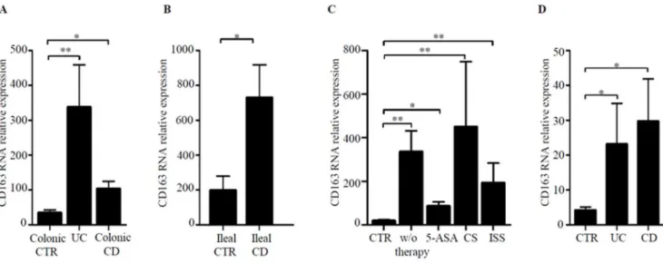
Figure 1. CD163 transcripts are increased in inflamed IBD mucosa.A. CD163 RNA expression was evaluated in colonic biopsies taken from 22 normal controls (CTR), 25 patients with Ulcerative Colitis (UC) and 19 patients with Crohn’s disease (CD) by real-time PCR and levels were normalized tob-actin. Data indicate mean6SEM of all samples; *p = 0.01; **p,0.0001. B. CD163 RNA expression was evaluated in ileal biopsies taken from 7 ileal CTR and 6 ileal CD patients by real-time PCR. Data indicate mean6SEM of all samples; *p = 0.03. C. Colonic biopsies taken from 22 CTR, 11 IBD patients (4 CD and 7 UC) receiving no therapy (w/o therapy), 18 IBD patients (9 CD and 9 UC) treated with mesalamine (5-ASA), 10 IBD patients (3 CD and 7 UC) treated with steroids (CS) and 5 IBD patients (3 CD and 2 UC) treated with immunomodulators (ISS) were analyzed for CD163 RNA expression by real-Time PCR. Levels are normalized tob-actin. Data are expressed as mean6SEM of all samples; *p = 0.01; **p,0.01. D. CD163 RNA expression was evaluated in LPMC from 6 normal controls (CTR), 4 patients with UC and 6 patients with CD by real-time PCR. Levels are normalized to b-actin. Data indicate mean6SEM of all samples; *p = 0.02.
(or IBD HLA-DR-expressing LPMC) and PBMC were seeded at a concentration of 16106cells/ml into 48-well culture dishes
pre-coated with 10mg/ml EDHU1-Ab (AbD Serotec, Du¨sseldorf,
Germany) or control Ab mouse IgG1 (R&D Systems) for 2 hours at 37uC and cultured for 6 and 48 hours. To examine whether CD163 expression correlates with TNF-a production, PBMC were isolated from IBD patients and used to purify CD14+
monocytes using isolation kit (Miltenyi Biotec, Bergish Gladbach, Germany) according to the manufacturer’s instruction. RNA was
then extracted from those cell samples and CD163 and TNF-a
RNA transcripts were evaluated by Real Time PCR.
TNF-aenzyme-linked immunosorbent assay
TNF-a was measured in supernatants of IBD LPMC and PBMC cultured for 48 hours as described above using a sensitive commercial enzyme-linked immunosorbent assay (ELISA) kit (R&D Systems) according to the manufacturer’s instructions.
Figure 2. CD163 protein expression is increased in IBD.A. Representative Western blots showing CD163 andbactin in total proteins extracted from mucosal samples of 2 CTR, 2 UC patients and 2 CD patients. Right panel shows the quantitative analysis of CD163/b-actin ratio in mucosal samples taken from 8 CTR, 8 UC patients and 8 CD patients as measured by densitometry scanning of Western blots. Values are expressed in arbitrary units (a.u.) and indicate mean6SEM of all samples; *p = 0.003; **p = 0.03. B. Representative photomicrographs (1006original magnification) of CD163-stained paraffin-embedded sections of surgical samples taken from 1 CTR, 1 patient with UC and 1 patient with CD. Isotype control antibody-stained section is also shown. Right panel shows the number of CD163-positive cells for high power field (hpf) in colonic sections taken from 3 CTR, 3 UC patients and 4 CD patients. Data are expressed as mean 6 SD; *p = 0.03; **p = 0.02. C Representative photomicrographs (2006original magnification) of CD163-stained paraffin-embedded sections of surgical samples taken from 1 patient with UC and 1 patient with CD. CD163-positive cells are evident inside and around vessels. Insets show higher magnification (4006) images.
doi:10.1371/journal.pone.0069839.g002
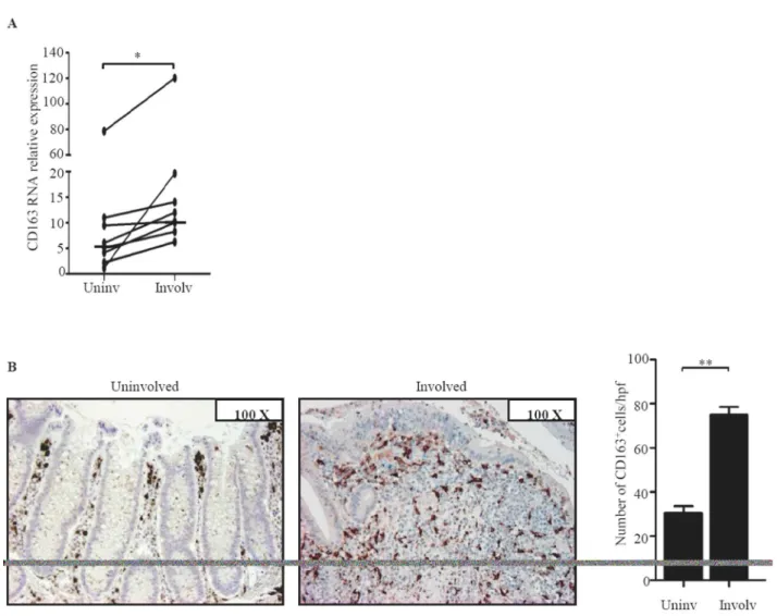
Figure 3. CD163 RNA and protein expression is increased in the inflamed areas of IBD.A. Paired biopsies taken from the involved (Involv) and uninvolved (Uninv) mucosa of 6 UC patients and 3 CD patients were analyzed for CD163 RNA expression by real-time PCR. Levels are normalized tob-actin; horizontal bars indicate the median values; *p = 0.003. B. Representative photomicrographs (original magnification 1006) of CD163-stained sections of colonic mucosal samples taken from involved and uninvolved mucosa of 1 UC patient. Right panel shows the number of CD163-positive cells per high power field (hpf) of colonic sections taken from the involved (Involv) and uninvolved (Uninv) mucosa of 3 UC patients. Data indicate the mean values6SEM of all samples; **p = 0.01.
Flow cytometry analysis
CTR and IBD PBMC were stained with CD163 PE (1:50 final dilution; eBioscience, San Diego, CA), CD14 FITC, (1:50 final dilution, Immunotools, Friesoythe; Germany), CD16 PerCP (1:50 final dilution; Invitrogen) or isotype control IgGs (BD Biosciences) and cell-surface fluorescence intensity was assessed using a FACSCalibur analyzer and analyzed using CellQuest software (BD Biosciences, Milan, Italy).
Statistical analysis
Differences between groups were compared using the Mann– Whitney U test and Wilcoxon test. Correlation between CD163 and TNF-a was examined using Spearman’s non parametric correlation.
Results
CD163 RNA transcripts are up-regulated in IBD
CD163 RNA expression was more pronounced in both CD and UC colonic samples in comparison to CTR (Fig. 1A). UC biopsies contained more CD163 transcripts than CD biopsies but the difference was not statistically significant (Fig. 1A). High CD163 RNA was also seen in ileal CD samples as compared to ileal CTR samples (Fig. 1B). To exclude the possibility that up-regulation of CD163 in IBD was secondary to current therapy, CD163 RNA transcripts were compared between patients receiving or not drugs. Figure 1C shows that CD163 RNA expression was not influenced by the ongoing treatments. Finally, we showed that CD163 transcripts were up-regulated in IBD LPMC as compared to control samples (Fig. 1D).
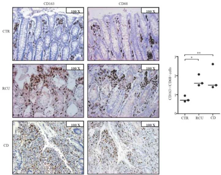
Figure 4. Increased CD163+cells/CD68+cells ratio in IBD tissue.A. Representative photomicrographs (1006original magnification) in serial paraffin-embedded sections of surgical samples taken from 1 CTR, 1 patient with UC and 1 patient with CD and stained with CD163 or CD68. Right panel shows the ratio of CD163+and CD68+cells counted in colonic sections taken from 3 CTR, 3 UC patients and 3 CD patients. Horizontal bars indicate the median values; *p = 0.03; **p = 0.02.
CD163-positive cells accumulate in the inflamed intestine of IBD patients
Western blotting analysis showed that CD163 was expressed in all IBD and CTR samples, but immunoreactivity corresponding to CD163 was more pronounced in IBD in comparison to CTR (Fig. 2A). Immunohistochemical analysis confirmed the abundant expression of CD163 in IBD and showed accumulation of these cells around the deep ulcers (Fig. 2B, right lower panel) and around and inside the blood vessels (Fig. 2C).
To examine whether, in IBD, CD163 is restricted to areas with mucosal lesions, paired biopsies were taken from both involved and uninvolved areas of IBD patients and examined for CD163 by real-time PCR and immunohistochemistry. CD163 RNA and protein expression was up-regulated in the inflamed samples as compared to uninvolved mucosal samples of the same IBD patients (Fig. 3). In contrast, expression of CD163 did not differ between uninvolved biopsies of IBD patients and normal CTR (not shown). Moreover, serial sections of IBD and control specimens stained with CD163 or CD68 showed that the number of CD68-expressing cells was higher than that of CD163-positive cells in control samples, while in both CD and UC the number of CD163-positive cells exceeded that of CD68-expressing macro-phages (Fig. 4). These data suggest that up-regulation of CD163 cells in IBD mucosa is not secondary to the increased macrophage infiltration.
CD163 -positive PBMC are increased in IBD
The fact that, in IBD tissue, CD163-expressing cells are located around and inside blood vessels (Fig. 2B–C) raises the possibility that these cells are in part recruited from the systemic circulation. Thus, in subsequent experiments, we evaluated the expression of CD163 in PBMC of IBD patients and CTR. The percentage of CD163-expressing PBMC was increased in IBD patients as compared to CTR (Fig. 5A). Further analysis revealed that the fractions of CD14+, CD16+and CD14+/CD16+cells expressing CD163 were increased in IBD PBMC in comparison to control PBMC (Fig. 5 B–D).
IL-6 enhances CD163 expression in normal colonic explants and LPMC
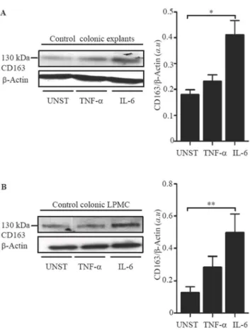
Next, we determined whether CD163 can be modulated by cytokines over-produced in IBD [28,29]. To this end, normal colonic explants and LPMC were stimulated with TNF-aand IL-6, two cytokines that up-regulate CD163 in other systems [28,29]. Western blot analysis showed that IL-6, but not by TNF-a
increased CD163 protein expression in both explants and LPMC (Fig. 6A–B). To examine whether increased expression of CD163 resulted from direct effects of IL-6 on normal intestinal macrophages, HLA-DR-expressing CD3- and CD19-negative LPMC were stimulated with IL-6 for 6 hours and then CD163 RNA was evaluated by real-time PCR. IL-6 enhanced CD163 RNA transcripts in all samples analyzed (1.260.2 in unstimulated cells vs 561.4 in IL-6-stimulated cells).
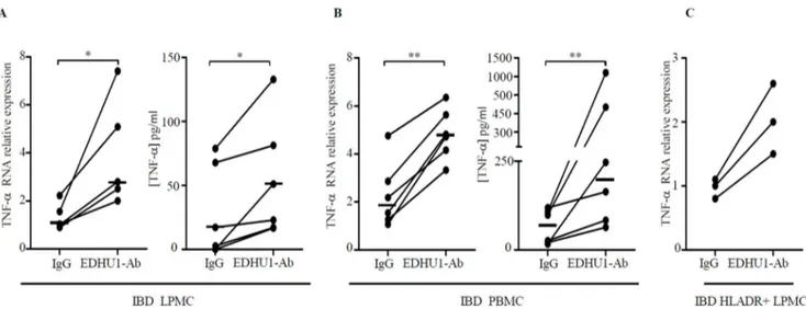
Cross-linking of CD163 with EDHU1-Ab increases TNF-a
production
In a final set of experiments we evaluated if CD163-delivered signals control cytokine expression. IBD LPMC and PBMC were cultured in the presence or absence of a specific activating CD163 monoclonal antibody (EDHU1-Ab) or control IgG for 6–48 hours and TNF-a RNA and protein were then examined by real-time PCR and ELISA respectively. Cross-linking of CD163 with EDHU1-Ab increased TNF-a RNA and protein expression and this was evident in both LPMC and PBMC (Fig. 7A–B). Consistently, stimulation of CD HLA-DR-expressing CD3- and CD19-negative LPMC with EDHU1-Ab increased TNF-aRNA expression (Fig. 7C). Although these data indicate that CD163 cross-linking on monocytes/macrophages enhances TNF-a ex-pression, no correlation was seen between CD163 and TNF-a
transcripts in CD14+ monocytes isolated from blood of IBD patients (not shown), perhaps due to the multiple signals controlling TNF-aproduction in monocytes/macrophages.
Discussion
The present study was undertaken to evaluate the expression and function of CD163 in IBD. CD163 is a multifunctional receptor, which can bind multiple ligands and activate various
Figure 5. CD163-expressing cells are increased in the blood of IBD patients.A. The Histograms show the percentage of CD163-expressing cells in total PBMC (A), CD14+
cells (B), CD16+
cells (C) and CD14+ CD16+
cells (D) of 23 IBD patients (5 UC patients, 18 CD patients) and 9 CTR. Cells were isolated from the blood of IBD patients and controls and examined by flow-cytometry as indicated in material and methods. Data indicate the mean values6SEM of all samples; *p,0.001 **p = 0.001.
intracellular pathways, thereby controlling many biological processes. Initial studies showed that CD163 identifies a specific subsets of alternatively activated macrophages [30], which exert anti-inflammatory effects following interaction and internalization of the complex Hb-Hp [10]. This function of CD163 was supported by the demonstration that CD163-expressing macro-phages constitute the predominant macrophage population during the late or resolution phase of many inflammatory reactions [31,32] and CD163 expression is strongly induced by anti-inflammatory mediators, such as glucocorticoids and IL-10 [33,34]. More recently, it has been demonstrated that CD163 can also bind both Gram-negative and Gram-positive bacteria and activate a number of intracellular events, which lead to production of pro-inflammatory cytokines [14]. Therefore, it is plausible that CD163 may have a dual role in the control of immune responses perhaps depending on the microenvironment in which CD163-expressing cells are located.
We here show that IBD-related inflammation is marked by enhanced CD163 expression at both RNA and protein level. High CD163 is restricted to mucosal areas with active inflammation and
not influenced by current therapy. While this study was ongoing, Bain and co-workers showed that CD14-positive LPMC isolated from the inflamed ileum of CD patients express low levels of CD163 as compared to control cells [25]. We have attempted to reproduce such results using the same LPMC isolation and flow-cytometry procedures adopted by Bain. However, CD163 was undetectable on LPMC of both IBD patients and controls using 3 different commercial flow-cytometry antibodies, including that used by Bain and co-workers. The reason for this apparent discrepancy remains unknown. The fact that analysis of this receptor in our samples was performed using a very sensitive technique (i.e. real-time PCR) and data were confirmed by Western blotting and immunohistochemistry suggests however that CD163 is over-expressed in the inflamed mucosa of IBD patients. This hypothesis is supported by a previous immunostain-ing study showimmunostain-ing enhanced accumulation of CD163+ macro-phages in the intestinal mucosa of patients with CD and patients with spondyloarthropathy [23]. CD163 was detectable in PBMC by flow-cytometry and positivity was not affected by exposure of cells to reagents used to isolate LPMC (e.g. EDTA, collagenase). Thus, it is unlikely the difficulty to detect CD163 in LPMC by flow-cytometry is secondary to cleavage of the receptor during the cell preparations.
The relevance of the increased number of CD163+cells in IBD tissue is highlighted by further observations. First, the increase of CD163+ cells reflects an increase of a specific macrophage subpopulation rather than a global increase of macrophages, since the number of CD68+cells was similar or even decreased to that of CD163+ cells in IBD samples. Second, the up-regulation of CD163+ cells in patients with CD and patients with UC could reflect a similar pathophysiological mechanism, which accounts for the accumulation of this particular cell subset in the two IBD. Third, CD163 was enhanced in normal LPMC by IL-6, a cytokine that is over-produced in both CD and UC and supposed play a key role in the pathogenesis of IBD-associated tissue-damaging immune response. Fourth, cross-linking of CD163 with a specific activating antibody leads to enhanced production of TNF-a in cultures of IBD LPMC and HLADR-expressing LPMC.
Overall our data are in line with previous studies documenting high expression of CD163 in pathological conditions. For example, Yawalkar and Fuentes-Duculan demonstrated that CD163-positive macrophages infiltrate heavily the lesional skin of psoriatic patients and express bioactive forms of pro-inflamma-tory cytokines, such IL-12 and IL-23 [15,16]. Consistent with this is the demonstration that CD163-positive macrophages infiltrating the fibrous tissue in facet joints of patients with ankylosing spondylitis are a major source of inflammatory cytokines, such IL-23 [35]. Wakusawa et al described a patient with Necrobiosis lipoidica associated with Insulin-dependent type 1 diabetes mellitus, in which granulomas contained mostly pro-inflammatory CD163-positive macrophages [36]. Increased expression of CD163 has been also seen in skin macrophages and peripheral blood monocytes of patients with systemic sclerosis, an autoim-mune disease characterized by local inflammatory infiltrates and widespread fibrosis [37]. CD163-expressing macrophages are also overrepresented in the colon of patients with diverticulitis [38] and in the kidney of patients with IgA nephropathy [39].
In conclusion, data of the present study show that CD163+cells are abundant in the inflamed gut of patients with IBD and suggest a role for these cells in the amplification and perpetuation of the ongoing mucosal inflammation in IBD.
Figure 6. IL-6 enhances CD163 expressions in normal colonic explants and LPMC.A. Representative Western blots showing CD163 andb-actin in total proteins extracted from normal colonic explants treated with or without (unstimulated = UNST) TNF-a(20 ng/ml) or IL-6 (50 ng/ml) for 24 hours. Right panel shows the quantitative analysis of CD163/b-actin ratio in normal colonic explants. Values are expressed in arbitrary units (a.u.) and indicate mean 6 SEM of four separate experiments; *p = 0.04. B. Representative Western blots showing CD163 andb-actin in total proteins extracted from normal LPMC treated as above for 48 hours. Right panel shows the quantitative analysis of CD163/b-actin ratio in LPMC protein extracts. Values are expressed in arbitrary units (a.u.) and indicate mean 6 SEM of three separate experiments; **p = 0.01.
Author Contributions
Conceived and designed the experiments: GM. Performed the experi-ments: EF RC CS MS MLC FC IM FZ DDN AC. Analyzed the data: EF
RC CS MS MLC FC IM FZ DDN AC. Contributed reagents/materials/ analysis tools: EF RC CS MS MLC FC IM FZ DDN AC LB. Wrote the paper: EF GM LB FP.
References
1. Brandtzaeg P (1989) Overview of the mucosal immune system. Curr Top Microbiol Immunol 146: 13–25.
2. Smith PD, Ochsenbauer-Jambor C, Smythies LE (2005) Intestinal macrophages: unique effector cells of the innate immune system. Immunol Rev 206: 149–159. 3. Smythies LE, Sellers M, Clements RH, Mosteller-Barnum M, Meng G, et al. (2005) Human intestinal macrophages display profound inflammatory anergy despite avid phagocytic and bacteriocidal activity. J Clin Invest 115: 66–75. 4. Caruso R, Sarra M, Stolfi C, Rizzo A, Fina D, et al. (2009) Interleukin-25
inhibits interleukin-12 production and Th1 cell-driven inflammation in the gut. Gastroenterology 136: 2270–2279.
5. Ulevitch RJ, Tobias PS (1995) Receptor-dependent mechanisms of cell stimulation by bacterial endotoxin. Annu Rev Immunol 13: 437–457. 6. Rugtveit J, Nilsen EM, Bakka A, Carlsen H, Brandtzaeg P, et al. (1997) Cytokine
profiles differ in newly recruited and resident subsets of mucosal macrophages from inflammatory bowel disease. Gastroenterology 112: 1493–1505. 7. Rugtveit J, Brandtzaeg P, Halstensen TS, Fausa O, Scott H (1994) Increased
macrophage subset in inflammatory bowel disease: apparent recruitment from peripheral blood monocytes. Gut 35: 669–674.
8. Hunter MM, Wang A, Parhar KS, Johnston MJ, Van Rooijen N, et al. (2010) In vitro-derived alternatively activated macrophages reduce colonic inflammation in mice. Gastroenterology 138: 1395–1405.
9. Fabriek BO, Dijkstra CD, van den Berg TK (2005) The macrophage scavenger receptor CD163. Immunobiology 210: 153–160.
10. Kristiansen M, Graversen JH, Jacobsen C, Sonne O, Hoffman HJ, et al. (2001) Identification of the haemoglobin scavenger receptor. Nature 409: 198–201. 11. Van Gorp H, Delputte PL, Nauwynck HJ (2010) Scavenger receptor CD163, a
Jack-of-all-trades and potential target for cell-directed therapy. Mol Immunol 47: 1650–1660.
12. Van den Heuvel MM, Tensen CP, van As JH, Van den Berg TK, Fluitsma DM, et al. (1999) Regulation of CD 163 on human macrophages: cross-linking of CD163 induces signaling and activation. J Leukoc Biol 66: 858–866. 13. Ritter M, Buechler C, Kapinsky M, Schmitz G (2001) Interaction of CD163
with the regulatory subunit of casein kinase II (CKII) and dependence of CD163 signaling on CKII and protein kinase C. Eur J Immunol31: 999–1009. 14. Fabriek BO, van Bruggen R, Deng DM, Ligtenberg AJ, Nazmi K, et al. (2009)
The macrophage scavenger receptor CD163 functions as an innate immune sensor for bacteria. Blood 113: 887–892.
15. Yawalkar N, Tscharner GG, Hunger RE, Hassan AS (2009) Increased expression of IL-12p70 and IL-23 by multiple dendritic cell and macrophage subsets in plaque psoriasis. J Dermatol Sci 54: 99–105.
16. Fuentes-Duculan J, Suarez-Farinas M, Zaba LC, Nograles KE, Pierson KC, et al. (2010) A subpopulation of CD163-positive macrophages is classically activated in psoriasis. J Invest Dermatol 130: 2412–2422.
17. Sugaya M, Miyagaki T, Ohmatsu H, Suga H, Kai H, et al. (2012) Association of the numbers of CD163(+) cells in lesional skin and serum levels of soluble CD163 with disease progression of cutaneous T cell lymphoma. J Dermatol Sci 68: 45–51.
18. Baeten D, Demetter P, Cuvelier CA, Kruithof E, Van Damme N, et al. (2002) Macrophages expressing the scavenger receptor CD163: a link between immune alterations of the gut and synovial inflammation in spondyloarthropathy. J Pathol 196: 343–350.
19. Philippidis P, Mason JC, Evans BJ, Nadra I, Taylor KM, et al. (2004) Hemoglobin scavenger receptor CD163 mediates interleukin-10 release and heme oxygenase-1 synthesis: antiinflammatory monocyte-macrophage responses in vitro, in resolving skin blisters in vivo, and after cardiopulmonary bypass surgery. Circ Res 94: 119–126.
20. Fabriek BO, Moller HJ, Vloet RP, van Winsen LM, Hanemaaijer R, et al. (2007) Proteolytic shedding of the macrophage scavenger receptor CD163 in multiple sclerosis. J Neuroimmunol 187: 179–186.
21. Etzerodt A, Maniecki MB, Moller K, Moller HJ, Moestrup SK (2010) Tumor necrosis factor alpha-converting enzyme (TACE/ADAM17) mediates ectodo-main shedding of the scavenger receptor CD163. J Leukoc Biol 88: 1201–1205. 22. Frings W, Dreier J, Sorg C (2002) Only the soluble form of the scavenger receptor CD163 acts inhibitory on phorbol ester-activated T-lymphocytes, whereas membrane-bound protein has no effect. FEBS Lett 526: 93–96. 23. Demetter P, De Vos M, Van Huysse JA, Baeten D, Ferdinande L, et al. (2005)
Colon mucosa of patients both with spondyloarthritis and Crohn’s disease is enriched with macrophages expressing the scavenger receptor CD163. Ann Rheum Dis 64: 321–324.
24. Yuan PQ, Wu SV, Elliott J, Anton PA, Chatzaki E, et al. (2012) Expression of corticotropin releasing factor receptor type 1 (CRF(1)) in the human gastrointestinal tract and upregulation in the colonic mucosa in patients with ulcerative colitis. Peptides 38: 62–69.
25. Bain CC, Scott CL, Uronen-Hansson H, Gudjonsson S, Jansson O, et al. (2012) Resident and pro-inflammatory macrophages in the colon represent alternative context-dependent fates of the same Ly6C(hi) monocyte precursors. Mucosal Immunol.
26. Monteleone G, Kumberova A, Croft NM, McKenzie C, Steer HW, et al. (2001) Blocking Smad7 restores TGF-beta1 signaling in chronic inflammatory bowel disease. J Clin Invest 108: 601–609.
Figure 7. Cross-linking of CD163 with the EDHU1-Ab enhances TNF-aexpression in LPMC, PBMC and in purified HLADR-expressing LPMC of IBD patients.A–B. Left panels. TNF-aRNA expression was evaluated in LPMC of 4 CD patients and 1 UC patients (A) and in PBMC of 3 CD patients and 3 UC patients (B) treated with EDHU1-Ab or control IgG for 6 hours by real-time PCR. Levels are normalized tob-actin; horizontal bars indicate the median values; *p = 0.03; **p = 0.01. Right panels. TNF-asecretion was measured in supernatants of IBD LPMC and PBMC treated as described above for 48 hours. Horizontal bars indicate the median values; *p = 0.03; **p = 0.01. C. TNF-aRNA expression was evaluated by real-time PCR in HLA-DR-expressing, CD3- and CD19-negative LPMC of 3 patients with CD and treated with EDHU1-Ab or control IgG for 6 hours. Levels are normalized tob-actin.
27. Monteleone G, Biancone L, Marasco R, Morrone G, Marasco O, et al. (1997) Interleukin 12 is expressed and actively released by Crohn’s disease intestinal lamina propria mononuclear cells. Gastroenterology 112: 1169–1178. 28. Reinecker HC, Steffen M, Witthoeft T, Pflueger I, Schreiber S, et al. (1993)
Enhanced Secretion of Tumor-Necrosis-Factor-Alpha, Il-6, and Il-1-Beta by Isolated Lamina Propria Mononuclear-Cells from Patients with Ulcerative-Colitis and Crohns-Disease. Clinical and Experimental Immunology 94: 174– 181.
29. Buechler C, Ritter M, Orso E, Langmann T, Klucken J, et al. (2000) Regulation of scavenger receptor CD163 expression in human monocytes and macrophages by pro- and antiinflammatory stimuli. J Leukoc Biol 67: 97–103.
30. Gordon S (2003) Alternative activation of macrophages. Nat Rev Immunol 3: 23–35.
31. Zwadlo G, Voegeli R, Schulze Osthoff K, Sorg C (1987) A monoclonal antibody to a novel differentiation antigen on human macrophages associated with the down-regulatory phase of the inflammatory process. Exp Cell Biol 55: 295–304. 32. Verschure PJ, Van Noorden CJ, Dijkstra CD (1989) Macrophages and dendritic cells during the early stages of antigen-induced arthritis in rats: immunohisto-chemical analysis of cryostat sections of the whole knee joint. Scand J Immunol 29: 371–381.
33. Morganelli PM, Guyre PM (1988) IFN-gamma plus glucocorticoids stimulate the expression of a newly identified human mononuclear phagocyte-specific antigen. J Immunol 140: 2296–2304.
34. Sulahian TH, Hogger P, Wahner AE, Wardwell K, Goulding NJ, et al. (2000) Human monocytes express CD163, which is upregulated by IL-10 and identical to p155. Cytokine 12: 1312–1321.
35. Appel H, Maier R, Bleil J, Hempfing A, Loddenkemper C, et al. (2013) In situ analysis of IL-23 and IL-12 positive cells in the spine of patients with ankylosing spondylitis. Arthritis Rheum.
36. Wakusawa C, Fujimura T, Kambayashi Y, Furudate S, Hashimoto A, et al. (2012) Pigmented Necrobiosis Lipoidica Accompanied by Insulin-dependent Diabetes Mellitus Induces CD163 Proinflammatory Macrophages and Interleu-kin-17-producing Cells. Acta Derm Venereol.
37. Vogel DY, Vereyken EJ, Glim JE, Heijnen PD, Moeton M, et al. (2013) Macrophages in inflammatory multiple sclerosis lesions have an intermediate activation status. J Neuroinflammation 10: 35.
38. von Rahden BH, Kircher S, Thiery S, Landmann D, Jurowich CF, et al. (2011) Association of steroid use with complicated sigmoid diverticulitis: potential role of activated CD68+/CD163+macrophages. Langenbecks Arch Surg 396: 759– 768.