J of Evolution of Med and Dent Sci/ eISSN- 2278-4802, pISSN- 2278-4748/ Vol. 4/ Issue 23/ Mar 19, 2015 Page 3947
ROLE OF MAGNETIC RESONANCE IMAGING IN SPINAL TRAUMA WITH
SURGICAL CORRELATION
M. Vijaya Kumari1, Suneetha2, V. L. Eshwari3, Mythri Priyadarshini Kodali4, Sudha Bindu5, Ashwini
Jyothi6, Archana7, Ramachandraiah8
HOW TO CITE THIS ARTICLE:
M. Vijaya Kumari, Suneetha, V. L. Eshwari, Mythri Priyadarshini Kodali, Sudha Bindu, Ashwini Jyothi, Archana, Ramachandraiah. Role of Magnetic Resonance Imaging in Spinal Trauma with Surgical Correlation. Journal of Evolution of Medical and Dental Sciences 2015; Vol. 4, Issue 23, March 19; Page: 3947-3960,
DOI: 10.14260/jemds/2015/569
ABSTRACT: Spinal trauma is a common cause of disability. The common causes of spinal trauma are blunt injuries – motor vehicle accidents, falls, sport injuries, assaults. MRI plays a crucial role in evaluating and detecting spinal trauma specially subtle bone marrow, soft tissue and spinal cord abnormalities. Many advantages of MRI such as high contrast resolution, absence of bony artifacts, multiplanar capability and choice of various pulse sequences make it possible to diagnose spinal trauma more accurately. Information about neural and extra-neural injuries requiring surgical interventions can be obtained. AIMS: Role of MRI in spinal trauma with surgical correlation.
MATERIAL AND METHODS: Prospective evaluation of 85 patients with history of spinal injury in hemodynamically stable patients on 1.5 Tesla MRI and surgical correlation. RESULTS: Age of patients ranged from 11-80 years with mean age 45 years. Cervical spine is most commonly involved and RTA being most common cause of spinal injury. Cord compression, haemorrhage are most common presentation in MRI. While MRI is less sensitive in detecting posterior element fractures, over estimates ligament injuries and shows highest sensitivity for intervertebral disc injury. In our study, we have seen one case of pseudomeningocele formation with brachial plexus injury and two cases of vertebral artery thrombosis. CONCLUSION: Magnetic Resonance Imaging is the only tool available for depicting the changes within the cord, ligaments and paraspinal soft tissues which helps in the management of the patients and in predicting the prognosis of recovery.
KEYWORDS: MRI, Spinal trauma, Cervical trauma, Cord compression, Cord haemorrhage, Cord transection, Pseudomeningocele, Vertebral artery thrombosis.
INTRODUCTION: Spinal trauma is a common cause of disability. The common causes of spinal trauma are blunt injuries – motor vehicle accidents, falls, sport injuries, assaults. Penetrating injuries like stab wounds or gunshot wounds are rare causes of spinal trauma.
Diagnostic imaging, particularly MRI plays a crucial role in evaluating and detecting spinal trauma. Subtle bone marrow, soft tissue and spinal cord abnormalities which may not be apparent on other imaging modalities, can be readily detected on MRI. Early detection often leads to prompt and accurate diagnosis, expeditious management.
J of Evolution of Med and Dent Sci/ eISSN- 2278-4802, pISSN- 2278-4748/ Vol. 4/ Issue 23/ Mar 19, 2015 Page 3948 MRI is an excellent diagnostic modality for evaluation of spinal trauma as the MRI findings correlated well with surgical findings and also with degree of weakness and prognosis. The purpose of this study was to evaluate this correlation.
MATERIALS AND METHODS: We prospectively evaluated 85 patients with a history of spinal injury and who had undergone MR imaging of the spine from 2011 to 2013 were included in the study after informed consent. Inclusion criteria: All patients with traumatic spinal injuries were included in the study. Exclusion criteria: Patients who are hemodynamically unstable, with metallic implants, pacemakers.
In all the patients’ mode of injury, findings on neurological examination and neurological status at the time of admission (American Spinal Injury Association –ASIA score) were noted.
MRI was performed on a 1.5 Tesla electromagnet (General Electrical Medical Systems) without contrast administration (contrast administration was not a part of standard protocol). The pulse sequences included T1WI, T2WI using spin echo and gradient echo techniques and Short Tau Inversion Recovery sequences. Axial and sagittal T2WI, sagittal T1WI, Sagittal and coronal STIR images were obtained. Images were obtained with an interslice gap of 5.2mm, slice thickness of 4mm and a matrix size of 512 x 512. On MRI, signal changes in vertebrae, ligaments, spinal cord and soft tissues were noted:
In patients who were operated, the preoperative findings were noted. The neurological status of the patient at the time of discharge was noted.
MR imaging findings were compared with surgical findings and prognosis of the patient based on ASIA score at the time of admission and discharge.
OBSERVATIONS AND RESULTS: Eighty five patients who presented to our hospital with the history of spinal trauma and underwent MRI of the spine in a time period from December 2011 to September 2013 were included in study.
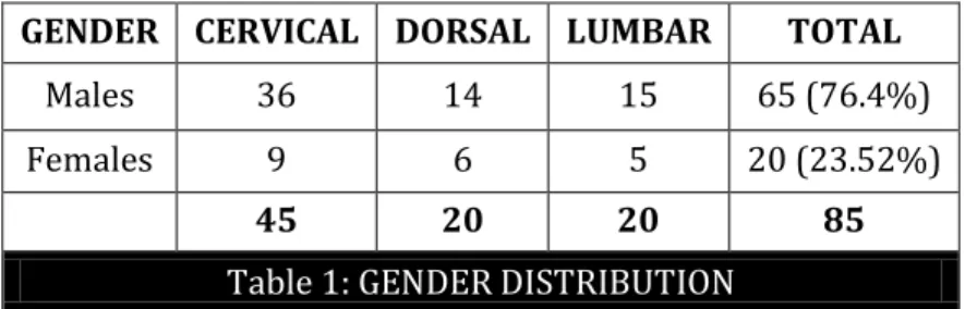
Of the 85 patients, 65 were males and 20 were female patients. The below table shows the gender and spinal distribution of the injury. (Table 1)
GENDER CERVICAL DORSAL LUMBAR TOTAL
Males 36 14 15 65 (76.4%)
Females 9 6 5 20 (23.52%)
45 20 20 85
Table 1: GENDER DISTRIBUTION
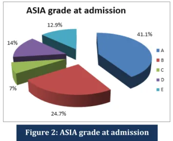
J of Evolution of Med and Dent Sci/ eISSN- 2278-4802, pISSN- 2278-4748/ Vol. 4/ Issue 23/ Mar 19, 2015 Page 3949 The neurological status of the patients at the time of admission and discharge was graded according to the ASIA classification. The commonest grade at presentation was A in the cervical and dorsal regions and E in the lumbar region. (Figure 2)
Of the 85 patients, 45 had injury to the cervical spine, 20 to the dorsal spine and 20 to the lumbar spine. The commonest level of involvement was C5/C6 in cervical spine; D11/D12 in dorsal spine and L1 vertebra in lumbar spine. (Table 2)
REGION NO. OF CASES
CERVICAL 45 (52.9%) DORSAL 20 (23.5%) LUMBAR 20 (23.5%) Table 2: REGION DISTRIBUTION
Figure 1: MODES OF INJURY
J of Evolution of Med and Dent Sci/ eISSN- 2278-4802, pISSN- 2278-4748/ Vol. 4/ Issue 23/ Mar 19, 2015 Page 3950 In the cervical spine, subluxation with bilateral facet dislocation was the commonest pattern of skeletal injury which is unstable and needs surgical intervention; whereas in the dorso-lumbar spine, wedge compression fracture was the commonest pattern. (Table 3, 4, 5)
Hyper-flexion sprain 5 Flexion compression fracture 3 Flexion tear-drop fracture 1 Bilateral facet dislocation 15 Unilateral facet dislocation 6
Burst fracture 1 Posterior element fractures 5
Table 3: SKELETAL INJURIES IN CERVICAL SPINE
Wedge compression fracture 7 Burst fracture 6 Posterior element fractures 2 Subluxations/dislocations 4 Table 4: SKELETAL INJURIES IN DORSAL SPINE
Wedge compression fracture 11 Burst fracture 7 Posterior element fractures 3 Table 5: SKELETAL INJURIES IN LUMBAR SPINE
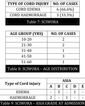
Cord Injury-The various types of cord injuries noted, were SCIWORA (spinal cord injury without radiographic abnormality), edema, haemorrhage, transection, compression, syrinx. Spinal cord compression and haemorrhage were the common pattern of cord injury in our study. (Table 6)
TYPE CERVICAL DORSAL LUMBAR TOTAL
SCIWORA 9 - - 9 (7.8%)
EDEMA 12 1 - 13 (11.3%)
HEMORRHAGE 25 13 3 41 (35.6%)
TRANSECTION 3 2 - 5 (4.3%)
COMPRESSION 24 14 7 45 (39.1%)
SYRINX 1* 1 2 (1.73%)
115
Table 6: CORD INJURIES
*Syrinx involving cervical and dorsal cord
J of Evolution of Med and Dent Sci/ eISSN- 2278-4802, pISSN- 2278-4748/ Vol. 4/ Issue 23/ Mar 19, 2015 Page 3951
TYPE OF CORD INJURY NO. OF CASES
CORD EDEMA 6 (66.6%) CORD HAEMORRAGE 3 (33.3%)
Table 7: SCIWORA
AGE GROUP (YRS) NO. OF CASES
10-20 2
21-30 2
31-40 1
41-50 3
51-60 1
Table 8: SCIWORA – AGE DISTRIBUTION
Type of Cord injury ASIA
A B C D E
EDEMA - 5 - 1 - HAEMORRAGE 2 - 1 - - Table 9: SCIWORA – ASIA GRADE AT ADMISSION
CORD EDEMA: This was noted in 13 cases – 12 in the cervical and one in the dorsal region. (Table 10, 11)
MODE OF INJURY NO. OF CASES
RTA 8
Fall from a height 3
Assault 2
Table 10: CORD EDEMA – MODE OF INJURY
ASIA GRADE AT ADMISSION NO. OF CASES
A 4
B 7
C -
D 2
E -
Table 11: CORD EDEMA – ASIA GRADE AT ADMISSION
J of Evolution of Med and Dent Sci/ eISSN- 2278-4802, pISSN- 2278-4748/ Vol. 4/ Issue 23/ Mar 19, 2015 Page 3952
MODE OF INJURY NO. OF CASES TOTAL
CERVICAL DORSAL LUMBAR
RTA 16 8 - 24
Fall from a height 5 5 3 13
Fall of a heavy weight 2 - - 2
Assault 2 - - 2
25 13 3 41
Table 12: MODE OF INJURY – CORD HAEMORRHAGE
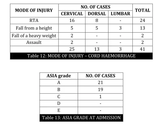
ASIA grade NO. OF CASES
A 21
B 19
C 1
D -
E -
Table 13: ASIA GRADE AT ADMISSION
Seven of these patients had died during the hospital stay. In the remaining, most of the patients had not shown significant improvement in the neurological status at the time of discharge.
CORD COMPRESSION: Compression of the cord to a varying degree by bone, herniated disc or epidural haematoma was noted in 45 cases. This was the commonest cord injury in our study. In most of the cases, it was associated with cord haemorrhage and poor neurological scores at admission. (Table 14, 15)
MODE OF INJURY NO. OF CASES TOTAL
CERVICAL DORSAL LUMBAR
RTA 17 8 - 25
Fall from a height 3 6 7 16
Fall of a heavy weight 3 - - 3
Assault 1 - - 1
24 14 7 45
J of Evolution of Med and Dent Sci/ eISSN- 2278-4802, pISSN- 2278-4748/ Vol. 4/ Issue 23/ Mar 19, 2015 Page 3953
ASIA GRADE NO. OF CASES
A 21
B 20
C 2
D 2
E -
45
Table 15: ASIA GRADE AT ADMISSION
CORD TRANSECTION: Transection was noted in 5 cases – 3 in the cervical and 2 in the dorsal region. (Table 16)
MODE OF INJURY
REGION
CERVICAL DORSAL LUMBAR
RTA 2 2 -
Fall from a
height - - -
Fall of a heavy
weight 1 - -
3 2 -
Table 16: MODE OF INJURY
LIGAMENTOUS INJURIES: In our study, ligament injuries were commonly noted in cervical spine. Of the ligaments involved, posterior ligaments (Interspinous, Supraspinous, Ligamentum flavum) were commonly involved than Anterior and Posterior longitudinal ligaments. (ALL & PLL).(table 17)
LIGAMENT REGION TOTAL
CERVICAL DORSAL LUMBAR
ALL 18 2 1 21
PLL 14 2 - 16
POSTERIOR
LIGAMENTS 22 3 1 26
Table 17: LIGAMENTOUS INJURIES
VASCULAR INJURIES: In two cases, loss of flow void was noted in right vertebral artery. In one case, thrombosis was confirmed with Doppler; in another case with MRA.
J of Evolution of Med and Dent Sci/ eISSN- 2278-4802, pISSN- 2278-4748/ Vol. 4/ Issue 23/ Mar 19, 2015 Page 3954
TREATMENT: Of the 85 patients, 56 (65.8%) were operated and 29 (34.11%) were treated conservatively. Of the operated cases, 28 in cervical, 15 in dorsal and 13 were in lumbar region.(fig3)
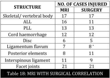
CORRELATION OF MR IMAGING FINDINGS WITH SURGICAL FINDINGS: Approach to spine surgery can be anterior, posterior or combined. The operative approach determined the structures visualized during surgery. For the anterior approach, the anterior longitudinal ligament, vertebral body, disc, posterior longitudinal ligament are adequately visualized. For the posterior approach, posterior osseous elements, ligamentum flavum, interspinous ligaments were adequately visualized. Depending upon the approach, surgical information was available for either the anterior or posterior structures.
In our hospital, anterior approach was commonly used for cervical spine and posterior approach was commonly used for dorsolumbar spine. But the approach varies depending on the predominant spinal column injured.
Of the 56 cases operated, 20 through anterior approach and 36 through posterior approach. In anterior approach, vertebral body fractures or subluxations were noted in 17 patients; ALL injury in 11 patients; PLL in 13patients; disc injury in 5 patients; cord haemorrhage in 12 patients.
In posterior approach, injury to Ligamentum flavum was noted in 8 patients; Interspinous ligament injury was noted in 9 cases; posterior element osseous injury was noted in 11 patients; facet joint dislocation was noted in 7 patients. (Table 20)
STRUCTURE NO. OF CASES INJURED
MRI SURGERY
Skeletal/ vertebral body 17 17
ALL 16 11
PLL 13 13
Cord haemorrhage 12 12
Disc 6 5
Ligamentum flavum 7 8 *
Posterior elements 8 11 Interspinous ligament 11 9
Facet joints 21 21 Table 18: MRI WITH SURGICAL CORRELATION
J of Evolution of Med and Dent Sci/ eISSN- 2278-4802, pISSN- 2278-4748/ Vol. 4/ Issue 23/ Mar 19, 2015 Page 3955 Positive predictive value of MRI in detecting the skeletal injuries was 100%.
Positive predictive value of MRI in detecting the ligamentous injuries: Anterior longitudinal ligament – 68.75%
Posterior longitudinal ligament – 100% Ligamentum flavum – 85.7%
Interspinous ligaments -81.8%
Positive predictive value of MRI in detecting the disc injuries – 83.3%.
Sensitivity of MRI** Present study Zhuge et al(99) (2013)
ALL injury 100% 100%
PLL injury 100% 80%
Ligamentum flavum injury 75% 80% Interspinous ligament injury 100% 100%
Intervertebral disc injury 100% 100%
** specificity couldn’t be calculated as all the patients were not operated. Figure 4: MRI WITH SURGICAL
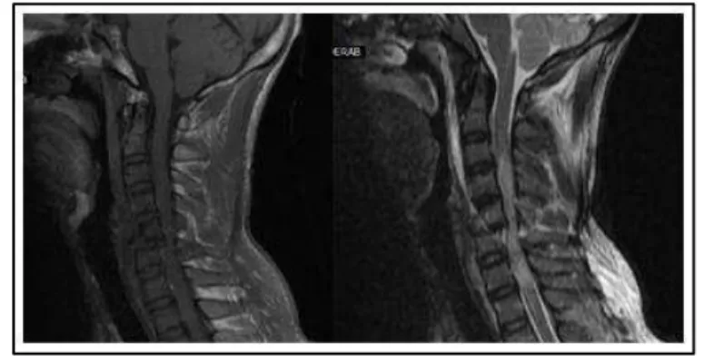
Figure 5: Mid sagittal T2W, T1W, STIR images-tear-drop fracture of C5 vertebral body with cord compression by retropulsed C5 & cord haemorrhage (C2-C7),
J of Evolution of Med and Dent Sci/ eISSN- 2278-4802, pISSN- 2278-4748/ Vol. 4/ Issue 23/ Mar 19, 2015 Page 3956
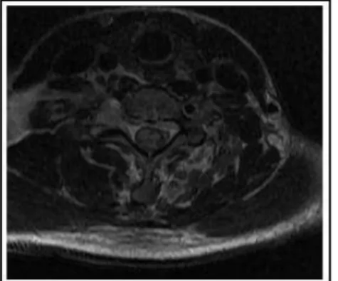
PUT ARROW AT LESION:
Figure 6: Acial T2WI – cord haemorrhage with flow void in vertebral artery
Figure 7 & 8: Midsagittal T2WI and T1WI – dislocation of C5 over C6 with cord transection and haemorrhage, disruption of ligaments
J of Evolution of Med and Dent Sci/ eISSN- 2278-4802, pISSN- 2278-4748/ Vol. 4/ Issue 23/ Mar 19, 2015 Page 3957
DISCUSSION: The purpose of MR imaging in spinal trauma are (a) to identify vertebral injury that compromises spinal canal (b) to identify ligamentous injuries (c) to assess the type and extent of cord injury (d) to assess the potential for surgical decompression and (e) to predict the prognosis for recovery.
Our study of MRI evaluation of spinal trauma consisted of 85 patients presenting with history of spinal trauma and underwent MR imaging. Of the 85 patients, 56 were operated and radiological and surgical correlations are compared.
Of the 85 patients, 65 were males and 20 were females accounting for 76.4% & 23.52% of the cases respectively. The greater incidence of spinal injury in males was also noted in study by Mahmood et al.1
The spinal injuries are commonly caused by blunt injuries and rarely by penetrating injuries. The various modes on injury noted in our study were RTA (road traffic accident), fall from a height, fall of a heavy object over neck and assault. The commonest causes were RTA and fall from a height accounting for 51.76% and 37.64% of the cases respectively.
The cervical spine was the most commonly involved (52.9%); the dorsal and lumbar spine (23.5%). D11/D12; C5/C6; L1 were the most commonly involved. The dorso-lumbar junction is more prone to injury due to the change in the curvature and centre of gravity.
Bilateral facet dislocation in cervical spine and wedge compression fracture in dorsolumbar spine, are commonest patterns of skeletal injury. Posterior element fractures were noted in 10 cases. One case of Jefferson’s fracture and tear drop fracture were noted. One case of osteoporotic and chance fracture are identified each.
SCIWORA was noted in 9 cases accounting for 7.8%of the cases. The types of injuries noted in these patients are cord edema (66.6%) and cord hemorrhage (33.3%). Siddhartha Sharma et al(89) noted Spinal Cord Injury Without Radiographic Abnormalities suspected cervical spine injury showing cord edema (58.3%) and haemorrhage (41.6%)in his study.
In our study, cord edema was seen in 13 cases – 12 in cervical and 1 in the dorsal region. The results were similar to study by Mahmood et al. 1
Cord hemorrhage was noted in 41 cases. Of them, 25 cases were in the cervical, 13 in the dorsal and 3 in the lumbar region. They showed no significant improvement in the neurological status at the time of discharge. The results were similar to Flanders et al 2, Silberstein et al. 3
Cord compression was the most common cord injury in our study noted in 45 cases. 40 % of the patients had shown improvement in the neurological status at the time of discharge. The results were almost similar to study by Silberstein et al.3
In our study, cord transection was noted in 5 cases – 3 in the cervical and 2 in the dorsal region, accounting for 4.3% of the total cord injuries. Three of them (cervical cord transection) expired and the remaining had shown no improvement in the neurological status at the time of discharge. Poor prognosis in cord transection was also reported by Silberstein et al.3
J of Evolution of Med and Dent Sci/ eISSN- 2278-4802, pISSN- 2278-4748/ Vol. 4/ Issue 23/ Mar 19, 2015 Page 3958 In two cases, loss of flow void in right vertebral artery was noted. One case was confirmed with MRA. Torina et al noted vertebral artery thrombosis in 83 patients out of 632 cases of cervical spinal trauma.4
We found that T2W sagittal images are sensitive for demonstrating the spinal injuries as reported by Anthony Bozzo et al.(5) STIR sequence best suited for detection of paraspinal soft tissue
injury.
In our study, 30 yr old male patient with history of RTA 15 days back presented weakness of upper limbs. On MRI, spinal cord was normal but there was nerve root avulsion injury with pseudomeningocele on right at C6/C7 level and edema of brachial plexus on left side. Tedesco et al reported 2 cases of cervical pseudomeningocele after brachial plexus injury.6
Of the 85 patients, 56 (65.8%) were operated. Of which 28 are cervical, 15 are dorsal and 13 were in lumbar region. In our hospital, anterior approach was commonly used for cervical spine and posterior approach for dorso-lumbar spine.
In anterior approach, vertebral body injury was noted in 17 cases which were confirmed on surgery (100% sensitivity, 100% PPV). Posterior element injury was noted in 8 cases on MRI whereas in surgery, posterior element fracture was noted in 11 cases (72.7% sensitivity, 100% PPV). Morais et al study of clinical application of MRI in acute spinal trauma, conclude that radiological findings are better visualized using MRI, except for the posterior elements are best on CT.7
On MRI, Anterior longitudinal ligament was involved in 16 cases, intraoperatively only in 11 cases (sensitivity 100%, PPV was 68.75%). On MRI, Posterior longitudinal ligament was involved in 13 cases which were confirmed on surgery (100% sensitivity and PPV). On MRI, Ligamentum flavum was involved in 7 cases, whereas in surgery Ligamentum flavum injury was noted in 6 cases (75% sensitivity, 85.7% PPV). Interspinous ligament injury was noted in 11 cases on MRI and 9 cases intraoperatively (sensitivity 100%, PPV 81.8%). Intervertebral disc injury was noted in 6 cases on MRI, 5 cases intraoperatively (sensitivity 100%, PPV 83.3%). These results were comparable to study by Zhuge et al.8
Though MRI overestimates anterior longitudinal ligament injury, Interspinous ligament injury and underestimates Ligamentum flavum injury; it has good sensitivity in depicting ligamentous injuries.
CONCLUSION: MRI features of 85 patients with spinal trauma were studied and imaging findings were correlated with neurological outcome based on ASIA score and with the surgical findings in feasible cases:
1. The age group ranged from 11yrs to 80yrs with a mean age of 45 yrs with male preponderance. 2. The cervical spine was the most commonly involved (in 52.9% of the cases); RTA and fall from
a height were the most common causes of spinal injury.
3. Cord compression and hemorrhage were the common presentations of cord injuries and cord edema had better prognosis compared to cord compression and hemorrhage; worse neurological outcome was seen with cord transection and hemorrhage.
J of Evolution of Med and Dent Sci/ eISSN- 2278-4802, pISSN- 2278-4748/ Vol. 4/ Issue 23/ Mar 19, 2015 Page 3959 5. We encountered 5 cases of cord transection, one case of nerve root avulsion with pseudomeningocele formation with brachial plexus injury and two cases of vertebral artery thrombosis.
6. MRI was able to detect intervertebral disc injury with 100% sensitivity.
7. Though MRI overestimated anterior longitudinal ligament injury, Interspinous ligament injury and underestimated Ligamentum Flavum injury; it has good sensitivity in depicting ligamentous injuries.
Magnetic Resonance Imaging is the only tool for identifying pathology within the cord, ligaments and para-spinal soft tissues wand helps in the management and prognostic assessment of the patients.
REFERENCES:
1. N. S. Mahmood, R Kadavigere, A K Ramesh and V R Rao. Magnetic resonance imaging in acute cervical spinal cord injury: a correlative study on spinal cord changes and 1 month motor recovery: Spinal Cord (2008) 46, 791–797.
2. Flanders AE, et al. Forecasting motor recovery after cervical spinal cord injury: value of MR imaging. Radiology 1996; 201(3): 649–655.
3. Silberstein M, T ress BM, Hennessy O. Prediction of neurologic outcome in acute spinal cord injury: the role of CT and MR. AJNR Am J Neuroradiol 1992; 13(6): 1597–608.
4. Torina PJ, Flanders AE, Carrino JA, et al. Incidence of vertebral artery thrombosis in cervical spine trauma: Correlation with severity of spinal cord injury. AJNR Am J Neuroradiol. 2005; 26: 2645-2651.
5. Anthony Bozzo, Judith Marcoux, Mohan Radhakrishna, Julie Pelletier, and Benoit Goule: The Role of Magnetic Resonance Imaging in the Management of Acute Spinal Cord Injury: J Neurotrauma. 2011 August; 28(8): 1401–1411.
6. Tedesco-Marchese, Antônio J.; Bor-Seng-Shu, Edson; Poetscher, Arthur W; Teixeira, Manoe: Giant Traumatic Cervical Pseudomeningocele After Brachial Plexus Injury: A Report of 2 Cases - l J. Neurosurgery Quarterly: August 2013 - Volume 23 - Issue 3 - p 206–209.
7. Morais DF, de Melo Neto JS, Meguins LC, Mussi SE, Filho JR, Tognola WA. Clinical applicability of magnetic resonance imaging in acute spinal cord trauma- Eur Spine J. 2014 July; 23(7): 1457-63. Epub 2013 Oct 4.
J of Evolution of Med and Dent Sci/ eISSN- 2278-4802, pISSN- 2278-4748/ Vol. 4/ Issue 23/ Mar 19, 2015 Page 3960
AUTHORS:
1. M. Vijaya Kumari 2. Suneetha 3. V. L. Eshwari
4. Mythri Priyadarshini Kodali 5. Sudha Bindu
6. Ashwini Jyothi 7. Archana
8. Ramachandraiah
PARTICULARS OF CONTRIBUTORS:
1. Associate Professor, Department of Radiology, Osmania General Hospital, Hyderabad.
2. Senior Resident, Department of Radiology, Osmania General Hospital, Hyderabad.
3. Professor, Department of Radiology, Osmania General Hospital, Hyderabad. 4. Senior Resident, Department of
Radiology, Osmania General Hospital, Hyderabad.
FINANCIAL OR OTHER
COMPETING INTERESTS: None
5. Assistant Professor, Department of Radiology, Osmania General Hospital, Hyderabad.
6. Assistant Professor, Department of Radiology, Osmania General Hospital, Hyderabad.
7. Assistant Professor, Department of Radiology, Osmania General Hospital, Hyderabad.
8. Assistant Professor, Department of Radiology, Osmania General Hospital, Hyderabad.
NAME ADDRESS EMAIL ID OF THE CORRESPONDING AUTHOR:
Dr. Mythri Priyadarshini Kodali, # 12-234, Adarsh Nagar, Bala Nagar, Hyderabad-500037. E-mail: [email protected]