artigo 305
ORIgInAL ARTICLE
The authors declare that they did not have any conflict of interests in producing this article.
1 – PhD and Master’s Degree in Orthopedics from Unifesp; Orthopedic Doctor and Head of Residency Service of the Hospital São Bento Cardioclínica S/A – Belo Horizonte, MG. 2 – PhD in General Pathology from UFMG; Master’s Degree in Rehabilitation from Unifesp; Resident Doctor in Orthopedics and Traumatology at the Hospital São Bento
Cardioclínica S/A – Belo Horizonte, MG.
3 – Orthopedic Doctor and Specialist in Knee Surgery at the Hospital São Bento Cardioclínica S/A – Belo Horizonte, MG. Work carried out at the Hospital São Bento Cardioclínica S/A – Belo Horizonte, MG.
Correspondence: Tania Clarete Fonseca Vieira Sales Sampaio – Rua Agena, 180 – Santa Lúcia – 30360-730 – Belo Horizonte, MG. E-mail: [email protected] Work received for publication: March 8, 2010; accepted for publication: May 4, 2010.
coMparaTiVe sTudy beTWeen radiological classificaTion
and Macro and Microscopic analysis on
osTeoarThriTis lesions of The knee
Carlos Antônio Garrido1, Tania Clarete Fonseca Vieira Sales Sampaio2, Frederico de Souza Ferreira3
Rev Bras Ortop. 2011;46(2):155-9
InTRODUCTIOn
Osteoarthritis (OA) is a progressive degenerative disease of the joint cartilage that can lead to joint de-formity. The process initially affects the cartilage and, later, the subchondral bone. Clinically, it is character-ized by pain, stiffness, crepitations, bone enlargement and progressive functional limitation(1) It is estimated
that symptomatic knee osteoarthritis occurs in 13% of the population over 60 years, and this number is expected to double by 2020(2)..
ABSTRACT
Objectives: To compare the modified Ahlbäck radiological classification with macroscopic analysis of knee injuries and locate a chondral lesion in the tibial plateau, and to correlate this with integrity or lack of integrity of the anterior cruciate ligament. Material and Methods: Between July and December 2009, 40 patients of mean age 67.1 years with an indication for total knee arthroplasty were selected. The modified Ahlbäck radiological classification was used. The International Cartilage Repair Society classification was used for macroscopic analysis of the lesions. Chondral injuries were correlated with the integrity or lack of integrity of the anterior cruciate ligament. Results: Regarding the radiological classification of the knees, three (7.5%) were classified as grade 1, two (5%) as grade 2, 17 (42.5%) as grade 3, 16 (40%) as grade
4 and two (5%) as grade 5. The macroscopic analysis of the knee showed that 25 patients (62.5%) had very severe injury and 15 (37.5%), severe. In eight knees (20%) with ruptured ACL, the lesion extended to the posterior region of the medial tibial plateau. When the ACL was intact, the lesion was located in the anterior-central region. Conclusion: Knee osteoarthritis of grades 4 and 5 in the radiological classification, showed agreement with the macroscopic analysis, i.e. very severe chondral injury. However, grades 1, 2 and 3 were discordant. In the cases of osteoarthritis with intact ACL, the lesion was located in the anterior-central region of the medial tibial plateau, and those with ruptured ACL had the lesion extending to the posterior region of the plateau.
Keywords – Osteoarthritis; Knee; Pathology; Biopsy
Loss of joint cartilage is a characteristic of osteo-arthritis. The width of the joint space is considered an indication of cartilage thickness, therefore narrow-ing of the joint space is one of the main radiological characteristics of osteoarthritis(3)..
Figure 1 – Right knee radiograph, anteroposterior (AP) and la-teral (Perfil) views.
Source: Photos from the Hospital São Bento Cardioclínica archive – BH / MG.
Rev Bras Ortop. 2011;46(2):155-9
of joint space width provides an indirect estimate of cartilage thickness(4).
In 1968, Ahlbäck demonstrated the importance of the anteroposterior (AP) radiograph in the orthostatic position, with the body weight supported by the knee in question(5). It was not until 1980, after a review of
359 knees with medial osteoarthritis, that the Ahlbäck classification was published, described as follows: Grade1: moderate destruction of the cartilage (nar-rowing of the joint space); Grade 2: complete destruc-tion of the cartilage (obliteradestruc-tion or almost oblitera-tion of joint space); Grade 3: bone wear less than 5 mm; Grade 4: bone wear between 5-15 mm; Grade 5: bone wear greater than 15 mm. The authors(6) indicate
that cases with lateral osteoarthritis were not evalu-ated for this scoring system. Keyes et al modified the Ahlbäck classification, adding the lateral view at 20° of flexion in the radiographic evaluation(7). These
au-thors correlated the location of the wear on the medial tibial plateau with the integrity, or lack of integrity, of the anterior cruciate ligament, confirming the findings previously described(8).
A problem in osteoarthritis clinical and epide-miological research is the difficulty in correlating degenerative changes with the radiological classifi-cations used. This situation is also observed in knee osteoarthritis.(9-12).
The aim of this study was to compare the changed Ahlbäck radiological classification to the macroscopic examination of the lesions in knee osteoarthritis, and locate the topography of chondral injury of the tibial plateau, linking it to the integrity or lack of integ-rity of the anterior cruciate ligament. The study was complemented by a biopsy of the most comprised areas caused by the osteoarthritis.
MATERIAL AnD METHODS
We conducted a cross-sectional cohort study in-cluding both male and female patients who had a clinical and radiological diagnosis of osteoarthritis and primary indication for total knee arthroplasty.
From July to December 2009, 84 patients fulfilled the proposed criteria. On admission to hospital, radio-graphs were taken following a standard protocol, and then photographed. All patients underwent total knee arthroplasty performed by the same team of
profes-sionals at the Hospital São Bento Cardioclínica, in Belo Horizonte / MG. Pre-surgery photographs were taken of the exposed joint and excised tibial plateau for macroscopic evaluation of the lesions. Osteocar-tilaginous tissue samples were collected during the surgery, for histological study. After analysis of ra-diographs and photographs, we selected 40 patients, all with varus deformity, in order to minimize bias due to the quality of the image, which can make the interpretation difficult.
Of the 40 patients, there were 26 females and 14 males, with ages ranging from 48 to 81 years, a mean age of 67.1 years and median of 66.5 years. Of the 40 knees studied, 25 were on the right side and 15 on the left.
The knee radiographs, anteroposterior and lateral views were obtained according to the standard protocol described. Radiograph of the knee in the anteroposterior projection (AP) with one-foot support(5), the beam was centered on the inferior
pole of the patella and the film-to-distance was 1 meter. For the lateral view of the knee, the patient was placed in lateral decubitus on the affected side of the knee, with knee flexion of 20°, measured with a goniometer(7). The central ray was directed vertically
to the medial knee joint with head angulation of 5° and a tube-film distance of 1m. The radiographic image of the medial tibial plateau was distinguished from the lateral view by the method described by Jacobsen(13). All radiographs were performed in the
157
Figure 2 – Preoperative view of the left knee.
Source: Photo from the Hospital São Bento Cardioclínica archive – BH / MG
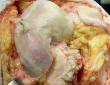
Figure 3 – Preoperative view of excised tibial plateau.
Source: Photo from the Hospital São Bento Cardioclínica archive – BH / MG
ANALYSIS ON OSTEOARTHRITIS LESIONS OF THE KNEE
Rev Bras Ortop. 2011;46(2):155-9
For the radiological classification of knee osteoar-thritis, we used the Ahlbäck classification modified by Keyes et al(7) (Table 1).
very severe injury:
Grade 4a – penetration of the subchondral bone, but not the overall diameter of the defect
Grade 4b – penetration throughout the entire diameter of defect
To locate the chondral injury and determine wheth-er it is linked to the integrity or lack of integrity of the anterior cruciate ligament (ACL), the medial tibial plateau was divided transversely into four zones: A, B, C and D from front to back(8) (Figure 4).
For the histological study, osteocartilaginous tissue samples were collected during total knee arthroplasty surgery, in the areas most affected. The samples were fixed in 10% formaldehyde and sent to the same laboratory.
grade Radiograph in AP view withone-foot support view with 20° of flexionRadiograph in lateral
1 Moderate destruction of cartilage (narrowing of the joint space)
2
Total destruction of cartilage (Obliteration or near obliteration of joint space)
3 Less than 5 mm wear of the tibial plateau
Posterior part of the tibial plateau intact
4 5 to 15 mm wear of tibial plateau
Extensive wear of posterior tibial plateau margin
5
Bone wear greater than 15 mm (severe subluxation of the tibia relative to femur)
> 10 mm anterior subluxation of the tibia Table 1 – Ahlbäck classification modified by Keyes and Goodfellow.
Source: Translated from: Ahlbäck S, Rydberg J. X-ray classification and examination techniques in gonarthrosis. Läkartidningen. 1980;77(22):2091-3; Keyes GW, Carr AJ, Miller RK, Goodfellow JW. The radiographic classification of medial gonarthrosis. Correlation with operation methods in 200 knees. Acta Orthop Scand. 1992;63(5):497-501.
For macroscopic analysis of injuries of the knee joint (Figure 2) and excised tibial plateau (Figure 3) ICRS (International Cartilage Repair Society) clas-sification of chondral surface and injuries was used, as described(14).
iCrS classification of the chondral surface and injuries:
Normal:
Grade 0
Nearly normal:
Grade 1a – superficial injuries/softening Grade 1b – 1a and/or surface cracks or fissures
Abnormal:
Grade 2a – length < 50% thickness
Severe injury:
Grade 3a – extension> 50% Grade 3b – to the calcified layer
Grade 3c – to the surface of the subchondral bone (without entering)
Figure 4 - Drawing of the division of the medial tibial plateau.
Figure 5 – Comparison between modified Ahlbäck radiological classification and ICRS classification of the chondral surface.
SOURCE: Data obtained from the study, Hospital São Bento Cardioclínica – BH / MG
Rev Bras Ortop. 2011;46(2):155-9
RESULTS
Regarding the Ahlbäck radiological classification modified by Keyes et al(7), of the 40 knees studied, three
(7.5%) were classified as grade 1, two (5%) as grade 2, 17 (42.5%) as grade 3, 16 (40%) as grade 4 and two (5%) as grade 5.
In the macroscopic examination of the knee follow-ing the criteria of the ICRS (International Cartilage Repair Society), 25 (62.5%) patients had very severe injuries and 15 (37.5%) had severe injuries.
Of the 40 patients, 32 (80%) had intact ACL and the injury was located in the anterocentral region of the knee medial tibial plateau. In the eight patients (20%) with ruptured ACL, the injury extended into the poste-rior tibial plateau.
The results of microscopic analysis of the material were similar, with the following description: histologi-cal sections of the hyaline cartilage showing a reduced number of chondrocytes, also with hypotrophic appear-ance, with reactive changes, diminished volume nuclei, some even pycnotic. The outer surface of the cartilage also shows small vacuolated areas, sometimes covered with vascular and conjunctiva neoformation. The trabec-ulae bone show moderate osteoclastic and osteoblastic activity, and conjunctive-vascular neoformation.
In the comparative study between the modified Ahl-bäck radiological classification and the macroscopic analysis, ICRS (International Cartilage Repair Society) criteria, demonstrated by plotting the composite column (Figure 5), we observed disagreement between the ra-diological classification and severity of chondral injury in Ahlbäck’s grades 1, 2 and 3. In these grades, chondral injury was severe or very severe. However, when the modified Ahlbäck radiological classification was grade 4 or 5, there was greater agreement with the ICRS clas-sification for chondral injury.
DISCUSSIOn
The treatment of knee osteoarthritis should be based on clinical examination, especially in relation to pain, deformity and disability in patients. The ra-diographic examination is an imaging method used to classify the grade of the injury, and associated with the clinical examination, it is of use in the sur-gical conduct.
Ahlbäck(5), based on a radiological study of 370
knees with primary knee osteoarthrosis, demonstrat-ed that the degenerative process was limitdemonstrat-ed to only one compartment of the knee and the medial joint space was 10 times more affected than the lateral. He defined five grades of joint degeneration, from narrowing of the joint space through to subluxation of the joint(6).
The Ahlbäck classification is probably the most commonly used system for classifying knee osteoarthritis, but some difficulties with the classifica-tion, such as its reproducibility and reliability, have been recognized by several authors (9,10-12).
In clinical practice, we observe a discrepancy be-tween modified Ahlbäck radiological classification and the intraoperative macroscopic findings, which prompted this research.
This comparative study between the modified Ahlbäck radiological classification and macroscopic analysis, ICRS criteria, showed that for the most se-vere injuries the macroscopic and radiological findings were similar and were confirmed by the histological
POSTERIOR REGION
CENTRAL REGION
ANTERIOR REGION
No. of patients
159
Rev Bras Ortop. 2011;46(2):155-9
study. In the Ahlbäck classification grades 1, 2 and 3 – and after a careful clinical examination the patient was indicated for surgery – the macroscopic exami-nation showed severe or very severe chondral injury. This discrepancy can be explained by the difficulty in determining the width of the joint space in the Ahl-bäck classification. When the joint space is not totally obliterated, with the femoral and tibial ends close to the joint line, is difficult to determine whether there is bone destruction, sometimes resulting in an incorrect choice between grades 1 and 3(10).
It is recognized that the radiographic findings may bear little relation to the symptoms. The width of the joint space, osteophytes, and subchondral changes may occur independently of the clinical syndrome called osteoarthritis. Radiograph is probably the best tool for measuring the progression of osteoarthritis(1).
With regard to the topographic location of the inju-ry in patients with intact ACL, the injuinju-ry was located in the anterocentral region of the medial tibial plateau. In patients with ruptured ACL, the injury extended into the posterior tibial plateau region, a finding that was also reported by other authors(8). White et al(8)
demonstrated that in osteoarthrosis with intact ACL, tibial erosion by no means reached the posterior tibi-al plateau. The combination of anterocentrtibi-al erosion and intact ACL provide a logical explanation for the clinical symptoms. The anterocentral position of the erosion of the joint surface explains why the varus deformity is present in the extension and not in the flexion. The intact cruciate ligaments, working with the preserved joint surface of the lateral compartment,
require the medial femoral condyle to roll back in flexion, out of the previous depression and into the intact cartilage of posterior tibial plateau.
We have observed that in osteoarthritis with intact ACL, the injury begins in the central tibial plateau. When ACL rupture occurs first and the patient devel-ops osteoarthritis, the injury begins in the posterior tibial plateau.
The histological study of osteocartilaginous tissue corroborated the ICRS classification of chondral injuries.
COnCLUSIOn
1) Grades 4 and 5 knee osteoarthritis, in the modi-fied Ahlbäck radiological classification, corroborated the macroscopic analysis, ICRS criteria, very severe chondral injury.
2) Grades 1, 2 and 3 knee osteoarthritis, in the modified Ahlbäck radiological classification, have shown disagreement with the macroscopic analysis, ICRS criteria.
3) The location of the injury in the tibial plateau showed correlation with the integrity or non-integri-ty of ACL. In patients with osteoarthritis and intact ACL, the injury was located in the anterior-central re-gion of the medial tibial plateau, and in patients with ruptured ACL, the lesion extended into the posterior tibial plateau. However, due to the number of cases it was not possible to make a statistical correlation. 4) The osteocartilaginous tissue pathology confirmed the ICRS classification of chondral injuries.
REFEREnCES
1. Altman R, Asch E, Bole G, Borenstein D, Brandt K, Christy W, et al. Develop-ment of criteria for the classification and reporting of osteoarthritis. Classification of osteoarthritis of the knee. Diagnostic and Therapeutic Criteria Committee of the American Rheumatism Association. Arthritis Rheum. 1986;29(8):1039-49.
2. Centers for Disease Control and Prevention (CDC). Arthritis prevalence and activity limitations--United States, 1990. MMWR Morb Mortal Wkly Rep. 1994;43(24):433-8.
3. Ravaud P, Chastang C, Auleley GR, Giraudeau B, Royant V, Amor B, et al. Assessment of joint space width in patients with osteoarthritis of the knee: a comparison of 4 measuring instruments. J Rheumatol. 1996;23(10):1749-55. 4. Guermazi A, Hunter DJ, Roemer FW. Plain radiography and magnetic resonance
imaging diagnostics in osteoarthritis: Validated staging and scoring J Bone Joint Surg Am. 2009;91(Suppl 1):54-62.
5. Ahlbäck S. Osteoarthrosis of the knee. A radiographic investigation. Acta Radiol Diagn (Stockh). 1968:Suppl 277:7-72.
6. Ahlbäck S, Rydberg J. X-ray classification and examination technics in gonar-throsis. Läkartidningen. 1980;77(22):2091-3.
7. Keyes GW, Carr AJ, Miller RK, Goodfellow JW. The radiographic classification of medial gonarthrosis. Correlation with operation methods in 200 knees. Acta Orthop Scand. 1992;63(5):497-501.
8. White SH, Ludkowski PF, Goodfellow JW. Anteromedimedial osteoarthritis of the knee. J Bone Joint Surg Br. 1991;73(4):582-6.
9. Galli M, Santis V, Tafuro L. Reliability of the Ahlbäck classification of knee osteoarthritis. Osteoarthritis Cartilage. 2003;11(8):580-4.
10. Weidow J, Cederlund CG, Ranstam J, Kärrholm J. Ahlbäck grading of osteoar-thritis of the knee. Poor reproducibility and validity based on visual inspection of the joint. Acta Orthop. 2006; 77(2):262-6.
11. Villardi AM, Mandarino M, Veiga LT. Avaliação da reprodutibilidade da clas-sificação de Ahlbäck modificada para osteoartrose do joelho. Rev Bras Ortop. 2006;41(5):157-61.
12. Albuquerque RP, Giordano V, Sturm L, Azevedo Júnior V, Leão A, Amaral NP. Análise da reprodutibilidade de três classificações para a osteoartrose do joelho. Rev Bras Ortop. 2008;43(8):329-35.
13. Jacobsen K. Gonylaxometry: Stress radiographic measurement of passive stability in the knee joints of normal subjects and patients with ligament injuries. Accuracy and range of application. Acta Orthop Scand. 1981;52(Suppl 194):1-263.
14. Clinical Münchenwiler Evaluation Group. ICRS Cartilage Injury Evaluation Package. Available at: http://www.cartilage.org/_files/ content management/ ICRS_evaluation.pdf. Accessed in December 2009.