Abstract
Objectives: To determine the prevalence of lower respiratory tract infection (LRTI) due to Chlamydia trachomatis in newborn infants and to describe the clinical, laboratory, and radiological characteristics of the disease.
Methods: A cross-sectional study carried out over a 12-month period. All infants up to 6 months of age admitted consecutively at the Centro Pediátrico Professor Hosannah de Oliveira of the Universidade Federal da Bahia in Salvador, Brazil, and diagnosed with LRTI according to clinical and/or radiological criteria were included in the study. C. trachomatis infection was diagnosed by the enzyme-linked immunosorbent assay (ELISA) for the detection of IgM-class antibodies. The prevalence of LRTI by C. trachomatis was determined and the prevalence ratios for the infection and clinical or laboratory variables were calculated.
Results: One hundred and ifty-one infants were submitted to serology for C. trachomatis and 15 (9.9%) tested positive. Chlamydial infection was found only in infants under 5 months of age, mainly in those aged under 2 months. Three of the infants with C. trachomatis infection were born by cesarean section. Conjunctivitis and eosinophilia had occurred in 33.3% of the cases. Chest X rays were abnormal in 92.0% of cases. There was an association between C. trachomatis infection and the duration of hospitalization exceeding 15 days (p = 0.0398) and oxygen therapy (p = 0.0484).
Conclusions: There was a high prevalence of C. trachomatis respiratory infection in the population studied. The infection was associated with a more severe form of the disease, emphasizing the importance of testing pregnant women for this infection to avoid infection in the newborn infant.
J Pediatr (Rio J). 2012;88(5):423-9: Chlamydia trachomatis, respiratory tract infection, pneumonia, bronchiolitis, infant.
O
riginala
rticle Copyright © by Sociedade Brasileira de Pediatria423 Introduction
According to the World Health Organization (WHO), 92 million cases of Chlamydia trachomatis infection occur in adults annually worldwide, 9.5 million in Latin America and the Caribbean.1 It is the most prevalent sexually transmitted disease in the United States (2.8 million cases/year).2 In Brazil, data on the prevalence of infection by this agent in
adults are based on isolated studies conducted in speciic
populations, with reported prevalence rates ranging from 2.1 to 25.7%3-5 depending on the study population and the diagnostic methods used.
In adults, C. trachomatis causes genital infections
that are usually asymptomatic; however, it is a signiicant
Chlamydia trachomatis: a major agent of
respiratory infections in infants from low-income families
Edna Lucia Souza,1 Renata Silva Girão,2 Juçara Magalhães Simões,3Carolina Ferraz Reis,2 Naiara Araújo Galvão,4 Sandra Cristina S. Andrade,2 Denise Mattedi F. Werneck,2 César A. Araújo-Neto,2 Leda Solano F. Souza1
1. PhD, Medicina e Saúde, Universidade Federal da Bahia (UFBA), Salvador, BA, Brazil. 2. Physician, UFBA, Salvador, BA, Brazil.
3. Biochemical Pharmacist, UFBA, Salvador, BA, Brazil. 4. Medical student, UFBA, Salvador, BA, Brazil.
No conflicts of interest declared concerning the publication of this article.
Financial support: This study was partially supported by Fundação de Amparo à Pesquisa do Estado da Bahia (FAPESB).
Suggested citation: Souza EL, Girão RS, Simões JM, Reis CF, Galvão NA, Andrade SC, et al. Chlamydia trachomatis: a major agent of respiratory infections in infants from low-income families. J Pediatr (Rio J). 2012;88(5):423-9.
Manuscript submitted Mar 15 2012, accepted for publication Jun 6 2012.
cause of inclusion conjunctivitis and respiratory infections in newborns6 as well as lower respiratory tract infection
(LRTI) in the irst 6 months of life.7-16 Around 10-20% of children of infected mothers develop pneumonia,6,17 with
signiicant medical and social repercussions that would be
preventable with good prenatal care.
Few studies have been conducted in Brazil on the role of C. trachomatis in the etiology of LRTI in newborn infants.11,18 The objective of this study was to determine the prevalence of LRTI due to C. trachomatis in infants admitted to a university teaching hospital in Salvador, Brazil, and to describe the clinical, laboratory, and radiological characteristics of the disease in these patients.
Methods
This cross-sectional, observational study was conducted between April 2006 and March 2007 at the Centro Pediátrico Professor Hosannah de Oliveira (CPPHO), a pediatric center of the Universidade Federal da Bahia (UFBA), Salvador, Brazil.
Study population
All infants up to 6 months of age admitted consecutively to the hospital and diagnosed with LRTI or pneumonia or bronchiolitis, according to clinical and/or radiological criteria, were included in the study. Infants with severe comorbidities unrelated to LRTI were excluded.
Diagnostic procedures and data collection
The following tests were performed: full blood count, erythrocyte sedimentation rate, C-reactive protein, serological studies, detection of respiratory viral infections by
indirect immunoluorescence for respiratory syncytial virus, adenovirus, inluenza A and B viruses, and parainluenza
1, 2 and 3 viruses, and polymerase chain reaction (PCR) for bocavirus and rhinovirus. C. trachomatis infection was diagnosed by enzyme-linked immunosorbent assay (ELISA) for the detection of IgM-class antibodies, using commercial kits (NovaTec®, Dietzenbach, Germany), strictly in accordance with the manufacturer’s instructions. The test was considered positive when the optical density of the evaluated serum was above the cut-off value determined for each plate. Blood cultures were performed whenever clinically indicated.
The infants were submitted to posteroanterior and lateral chest X rays. Two pediatric radiologists, blinded to the patient’s clinical history, independently evaluated the radiographies. When the two radiology reports failed to agree, the X rays were further evaluated by a third pediatric
radiologist. The inal radiology report was issued based on
agreement between two of the three evaluators.
The following variables were evaluated: gender, age, gestational age at birth, breastfeeding, type of delivery, mother’s age, mother’s education level, prenatal care, family income, maternal vaginal discharge, history of conjunctivitis, history of fever, duration of symptomatology at the time of admission to hospital, symptoms at admission, fever during hospitalization, diagnosis of pneumonia, diagnosis of bronchiolitis, radiological signs, use of oxygen, co-infection by cytomegalovirus, leukocyte count, eosinophil count, and duration of hospitalization.
Pneumonia was deined as the presence of coughing and/or dificulty in breathing, associated with radiological
changes.19 Bronchiolitis was deined as the occurrence of
tachypnea (≥ 50 breaths per minute) and/or dyspnea and/
or diffuse wheezing, associated or not with radiological
changes of hyperinlation and/or atelectasis.
Statistical analysis
The prevalence of LRTI by C. trachomatis was determined, and the prevalence ratios for the infection were calculated in accordance with the variables evaluated, which were selected to permit comparison with associations previously described in the literature and/or to evaluate the severity of the infection. Findings were considered
statistically signiicant when the probability of a type I
error was < 5% (p < 0.05).
Ethics approval
This study was approved by the Internal Review Board of the Maternidade Climério de Oliveira, UFBA, under protocol number 23/2004.
Results
Characteristics of the study population
Overall, 168 infants of 0-6 months of age were admitted to the CPPHO with LRTI, and 157 were included in the study; the remaining 11 being excluded based on the
exclusion criteria. Ninety-ive participants (60.5%) were
male and their mean age was 87.0±52.2 days (range 10-210 days; median 71 days). Mean age of the mothers was 23.8±6.1 years (range 14-41 years; median 23 years). Median family income was minimum wage. The principal complaints that led parents to seek the healthcare service were: coughing (94.3%), nasal obstruction (84.7%), and
breathing dificulties (75.2%).
Prevalence of C. trachomatis infection
One hundred and ifty-one infants (96.2%) were
Figure 1 - Summary study results
Six infants with C. trachomatis infection were co-infected with a respiratory virus: three cases of respiratory syncytial
virus and one case each of inluenza B, parainluenza virus
3 and rhinovirus.
Twelve infants with C. trachomatis infection were tested
for cytomegalovirus, with ive (41.7%) testing positive.
Demographic, clinical and laboratory characteristics of C. trachomatis respiratory infection cases
Of the 15 cases of C. trachomatis infection that were detected, eight were in boys (53.3%). Mean age of the infants was 63.5±28.9 days (range 35-117 days; median 52 days). Mean maternal age was 22.2±4.6 years (range 18-37 years). Fourteen mothers (93.3%) had attended prenatal care and six had had a vaginal discharge during pregnancy,
ive in the inal trimester. Twelve infants were born vaginally
and three by cesarean section. Six mothers had premature rupture of membranes. Of the three C. trachomatis-positive infants born by caesarean section, two had mothers who developed premature rupture of membranes. Five infants
had a history of conjunctivitis, three within the irst 8 days
of life. Of those with C. trachomatis infection, only one infant was premature and had a low birth weight. Thirteen infants were being breastfed, nine exclusively.
All infants had a cough, nasal obstruction and breathing
dificulties. There was a history of fever in ive cases (33.3%),
and 12 patients (80.0%) had a runny nose. The mean interval between the onset of the respiratory symptoms and admission to hospital was 12.9±10.8 days (range 2-37 days; median 9 days). Blood cultures were performed on 11 infants; however, only one patient tested positive for a nosocomial Klebsiella pneumoniae infection.
Ten infants with C. trachomatis infection were diagnosed with pneumonia at admission to hospital, while the
remaining ive were diagnosed with bronchiolitis. Wheezing
was detected in 11 babies (73.3%), four of whom had a viral co-infection. Crepitations were detected in 13 infants (86.7%). Mean leukocyte count in this group was 14,033±6,662 cells/mm3, and mean eosinophil count was 484±864 cells/mm3.
Table 1 - Radiological indings (n and %) in lower respiratory chlamydial infection in infants
Radiological indings n %
Atelectasis 1 8.4
Pleural effusion 1 8.4
Hyperinlation 7 58.4
Interstitial iniltrate 8 66.7
Pneumonia 1 8.4
None 1 8.4
to treatment with erythromycin, one was discharged in good clinical condition, and the other developed respiratory failure and was transferred to the ICU.
Radiological indings
All 15 infants were submitted to chest X rays. Twelve cases were evaluated by two radiologists, and one case was considered normal. The remaining three cases were not evaluated by the study radiologists; however, pediatricians described consolidation in two cases, while the other case
was considered normal. Hyperinlation and interstitial iniltrate were the principal indings (Table 1). One child
had a radiological diagnosis of pleural effusion associated
with hyperinlation and interstitial iniltrate. This inding
suggests a bacterial co-infection; however, blood culture was negative in this case.
Association between C. trachomatis infection and the variables studied
An association was found between C. trachomatis
infection and the following variables: symptomatology > 7 days, symptomatology > 15 days, no fever, oxygen therapy, co-infection by cytomegalovirus, and duration of hospitalization > 15 days (Table 2). To conduct a more thorough investigation into the possible effect of a viral co-infection on the associations found, prevalence ratios were calculated after excluding the infants with C. trachomatis and viral co-infections. In this subgroup, only the associations between C. trachomatis infection and oxygen therapy (p = 0.0484) or duration of hospitalization > 15
days (p = 0.0398) remained statistically signiicant.
Discussion
The prevalence of C. trachomatis respiratory infection of 9.9% found in this study is in agreement with data published in the literature reporting rates that range from 7.0% to
around 30.0% in the irst 6 months of life.7-16,20
Ejzenberg et al.11 reported a prevalence of 10.3% of positivity for C. trachomatis in infants < 6 months of
age admitted to a hospital in São Paulo, Brazil with LRTI. Other investigators have described higher prevalence rates of this infection in this age group.7-10 Differences in prevalence rates between studies may be due to the population evaluated, which may have included cases of mild or severe respiratory disease. Infected children may be asymtomatic.12 Symptomatic cases can vary in severity: only around 30% of infected infants develop pneumonia,21 and only 20% of infected infants require hospitalization.22 This study determined the prevalence of C. trachomatis infection in hospitalized infants with a more severe form of the disease.
A study conducted in Salvador, Brazil, in children under 5 years of age, admitted to hospital because of pneumonia, found a prevalence of C. trachomatis infection of 4.0%.23 The lower prevalence found in that study may be due to the age group evaluated, which included older children in whom the occurrence of C. trachomatis
infection is uncommon. Likewise, Pientong et al.24 also investigated this bacteria in nasopharyngeal secretions by PCR and restriction fragment length polymorphism in Thai children of 1 month to 2 years of age and hospitalized with acute bronchiolitis. The prevalence of C. trachomatis
infection was 2.4%, and occurred mainly in infants under 6 months of age. The selected population (children under 2 years of age diagnosed with bronchiolitis) may have contributed to the low prevalence observed.
Nasopharyngeal secretion cultures remain the gold standard for the diagnosis of C. trachomatis respiratory infection.15,17,21 Nevertheless, technical dificulties prevent their routine use in clinical practice. More recently, nucleic
acid ampliication tests have begun to take the place
of culture17 due to the high sensitivity and speciicity of these techniques.25 Notwithstanding, none of these techniques has the approval of the U. S. Food and Drug Administration for use in nasopharyngeal specimens from infants.17 There are several antigen-based detection
methods such as direct luorescent monoclonal antibody
staining and enzyme immunoassays for the detection of
C. trachomatis. However, the sensitivity of these tests with nasopharyngeal specimens ranges from 33 to 90%.21 In this study, direct detection of C. trachomatis IgM was used for diagnosis – sensitivity with this test being estimated at 75 to 89%15,16 and speciicity at 88%.16
Some factors associated with a greater risk of C. trachomatis respiratory infection, such as having an adolescent mother and poor socioeconomic level, were investigated in this study; however, no association was found between these variables and the infection. Nonetheless, some limitations in the evaluation of socioeconomic level must be considered, since the study population consisted almost entirely of infants born into families with limited
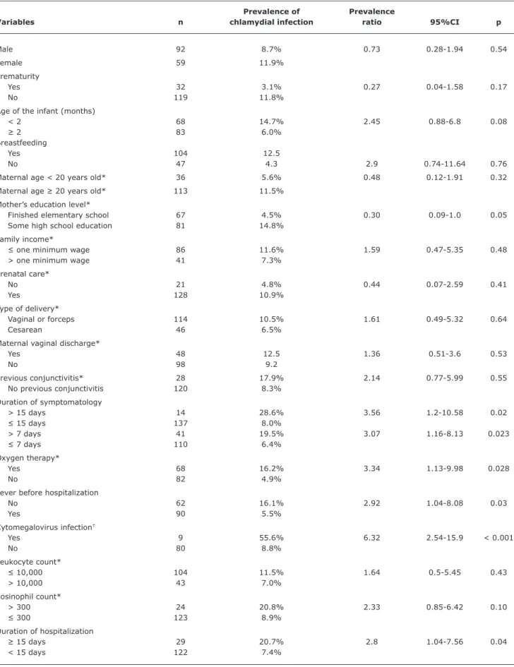
Table 2 - Prevalence ratio of Chlamydia trachomatis respiratory infection together with 95% conidence intervals and demographic, clinical and laboratory variables
Prevalence of Prevalence
Variables n chlamydial infection ratio 95%CI p
Male 92 8.7% 0.73 0.28-1.94 0.54
Female 59 11.9%
Prematurity
Yes 32 3.1% 0.27 0.04-1.58 0.17
No 119 11.8%
Age of the infant (months)
< 2 68 14.7% 2.45 0.88-6.8 0.08
≥ 2 83 6.0%
Breastfeeding
Yes 104 12.5
No 47 4.3 2.9 0.74-11.64 0.76
Maternal age < 20 years old* 36 5.6% 0.48 0.12-1.91 0.32
Maternal age ≥ 20 years old* 113 11.5%
Mother’s education level*
Finished elementary school 67 4.5% 0.30 0.09-1.0 0.05
Some high school education 81 14.8%
Family income*
≤ one minimum wage 86 11.6% 1.59 0.47-5.35 0.48
> one minimum wage 41 7.3%
Prenatal care*
No 21 4.8% 0.44 0.07-2.59 0.41
Yes 128 10.9%
Type of delivery*
Vaginal or forceps 114 10.5% 1.61 0.49-5.32 0.64
Cesarean 46 6.5%
Maternal vaginal discharge*
Yes 48 12.5 1.36 0.51-3.6 0.53
No 98 9.2
Previous conjunctivitis* 28 17.9% 2.14 0.77-5.99 0.55
No previous conjunctivitis 120 8.3%
Duration of symptomatology
> 15 days 14 28.6% 3.56 1.2-10.58 0.02
≤ 15 days 137 8.0%
> 7 days 41 19.5% 3.07 1.16-8.13 0.023
≤ 7 days 110 6.4%
Oxygen therapy*
Yes 68 16.2% 3.34 1.13-9.98 0.028
No 82 4.9%
Fever before hospitalization
No 62 16.1% 2.92 1.04-8.08 0.03
Yes 90 5.5%
Cytomegalovirus infection†
Yes 9 55.6% 6.32 2.54-15.9 < 0.001
No 80 8.8%
Leukocyte count*
≤ 10,000 104 11.5% 1.64 0.5-5.45 0.43
> 10,000 43 7.0%
Eosinophil count*
> 300 24 20.8% 2.33 0.85-6.42 0.10
≤ 300 123 8.9%
Duration of hospitalization
≥ 15 days 29 20.7% 2.8 1.04-7.56 0.04
< 15 days 122 7.4%
References
1. World Health Organization (WHO). Global prevalence and incidence of selected curable sexually transmitted infections. 2001. http://www.who.int/docstore/hiv/GRSTI/003.htm. Access: 02/08/2010.
The results of the present study call particular attention to the fact that C. trachomatis infection was found only in infants < 5 months of age, principally in those < 2 months of age, a group representing 75.0% of cases. The greater occurrence of this infection in infants of 2-3 months of age
is in agreement with the indings of other studies.12,13,20,26,27 Therefore, when there is a clinical suspicion of this infection, the fact that the infant is < 2-3 months of age should increase the etiological suspicion of C. trachomatis
respiratory infection.
In contrast to some reports in the literature that the occurrence of C. trachomatis respiratory infection is rare in infants delivered by cesarean section,21 20.0% of the infants in the present study with this diagnosis were delivered by this route. This event has already been described, and the authors suggest that the infection may be transmitted following membrane rupture or in the uterus and not exclusively during the baby’s passage through the birth canal.16,28,29
The reported association between chlamydial respiratory infection and conjunctivitis in 50.0% of cases26 was lower in the present study in which only 33.3% of the infants
had conjunctivitis, a inding that is in agreement with other
reports in the literature.13 Approximately 70% of infants with this respiratory bacterial infection failed to develop fever, thus
conirming the classic description of the condition, afebrile
pneumonia. The presence of eosinophilia > 300 cells/mm3 is
a common inding in C. trachomatis respiratory infections.21 Nevertheless, in the present study, this abnormality was
found in only ive infants (33.3%). These results are in
agreement with the study conducted by Carballal et al.27 in Argentinean children.
The results of the present study show an association between C. trachomatis infection and symptomatology for > 15 days prior to hospitalization, duration of hospitalization > 15 days, the use of oxygen therapy, and co-infection by cytomegalovirus. The slow progression of
C. trachomatis infection has already been reported in the literature.6,26 In the evaluation performed on the subgroup of infants with this bacterial infection and no co-infection, only the associations with oxygen therapy and a longer
period of hospitalization remained statistically signiicant.
Rours et al.20 conducted a retrospective evaluation of
C. trachomatis infection using PCR in a sample of Dutch infants and also reported the occurrence of more severe forms of the disease in positive cases; however, this may be because the sample population in that study consisted almost entirely of infants with underlying diseases, unlike the sample in the present study.
Chest X rays were abnormal in 92.0% of the infants enrolled in the present study, which is well above the rate found in infants with a viral infection. A greater number
of radiological indings together with a longer duration of
hospitalization and the use of oxygen therapy suggest that
C. trachomatis affects the lower respiratory tract more severely than respiratory viruses.
Some investigators have reported a strong association between C. trachomatis and other microorganisms,8 cytomegalovirus being the most common co-infection.8,9,26
The present results conirm this association, which has also
been reported in a previous study.30 Nevertheless, following exclusion of the group of infants co-infected with respiratory viruses, the association between C. trachomatis infection and
cytomegalovirus no longer remained statistically signiicant.
The low prevalence of C. trachomatis infection in infants with no other viral co-infections and the small number of patients investigated for cytomegalovirus may have hampered this analysis. Cytomegalovirus infection is associated with precarious socioeconomic conditions and other factors – such as adolescent motherhood and promiscuity,8 conditions that are also associated with C. trachomatis infection, possibly contributing to the elevated occurrence of co-infection between these microorganisms.
Some limitations of the present study include the selection of the study population. Therefore, the data obtained do not refer to infants with milder respiratory infections who did not require hospitalization and who represent the majority of cases. Furthermore, the use of a single diagnostic method may have underestimated the prevalence of C. trachomatis infection. On the other hand, concurrent data collection and the inclusion of a large number of infants who were submitted to clinical, laboratory, and radiological evaluation including interpretation of the X rays by two or three specialist radiologists allowed the
clinical and laboratory proile of the study population to be deined.
In conclusion, the prevalence of C. trachomatis infection in young infants admitted to a hospital for respiratory infections in Salvador, Brazil, was high, and this infection was associated with a more severe form of the disease. This emphasizes the importance of testing pregnant women for this infection in order to avoid transmission of the infection to the newborn infant. In addition, it is important to consider this etiologic agent when evaluating infants with a respiratory infection.
Acknowledgement
2. U.S. Preventive Services Task Force. Screening for chlamydial infection: U.S. Preventive Services Task Force recommendation statement. Ann Intern Med. 2007;147:128-34.
3. Simões JA, Giraldo PC, Faúndes A. Prevalence of cervicovaginal infections during gestation and accuracy of clinical diagnosis. Infect Dis Obstet Gynecol. 1998;6:129-33.
4. Araújo RS, Guimarães EM, Alves MF, Sakurai E, Domingos LT, Fioravante FC, et al. Prevalence and risk factors for Chlamydia trachomatis infection in adolescent females and young women in central Brazil. Eur J Clin Microbiol Infect Dis. 2006;25:397-400. 5. Ramos BR, Polettini J, Marcolino LD, Vieira EP, Marques MA, Tristão
AR, et al. Prevalence and risk factors of Chlamydia trachomatis cervicitis in pregnant women at the genital tract infection in obstetrics unit care at Botucatu Medical School, São Paulo State University-UNESP, Brazil. J Low Genit Tract Dis. 2011;15:20-4. 6. Schachter J, Grossman M, Sweet RL, Holt J, Jordan C, Bishop
E. Prospective study of perinatal transmission of Chlamydia trachomatis. JAMA. 1986;255:3374-7.
7. Harrison HR, English MG, Lee CK, Alexander ER. Chlamydia trachomatis infant pneumonitis: comparison with matched controls and other infant pneumonitis. N Engl J Med. 1978;298:702-8. 8. Stagno S, Brasield DM, Brown MB, Cassell GH, Pifer LL, Whitley
RJ, et al. Infant pneumonitis associated with cytomegalovirus, Chlamydia, Pneumocystis, and Ureaplasma: a prospective study.
Pediatrics. 1981;68:322-9.
9. Brasield DM, Stagno S, Whitley RJ, Cloud G, Cassell G, Tiller RE. Infant pneumonitis associated with cytomegalovirus, Chlamydia, Pneumocystis, and Ureaplasma: follow-up. Pediatrics. 1987;79:76-83.
10. Farrow JM, Mahony JB. Chlamydial pneumonia in Costa Rica: results of a case-control study. Bull World Health Organ. 1988;66:365-8.
11. Ejzenberg B, Melles H, Melles C, Dias R, Baldacci ER, Okay Y. Aerobic bacteria, Chlamydia trachomatis, Pneumocystis carinii and Cytomegalovirus as agents of severe pneumonia in small infants. Rev Inst Med Trop Sao Paulo. 1996;38:9-14.
12. Videla C, Celadilla MI, Mirsiglian A, Aguilar MC, Ricarte C, Carballal G. Chlamydia trachomatis in low tract acute respiratory infections in infants under 6 months of age. Infectol Microbiol Clin. 1996;8:83-91.
13. Portillo C, Lovera D, Arbo A. Chlamydia trachomatis as the agent of pneumonia in Paraguay. Rev Soc Bol Ped. 1997;36:S43-8. 14. Muhe L, Tilahun M, Lulseged S, Kebede S, Enaro D, Ringertz S,
et al. Etiology of pneumonia, sepsis and meningitis in infants younger than three months of age in Ethiopia. Pediatr Infect Dis J. 1999;18:S56-61.
15. Chen CJ, Wu KG, Tang RB, Yuan HC, Soong WJ, Hwang BT.
Characteristics of Chlamydia trachomatis infection in hospitalized infants with lower respiratory tract infection. J Microbiol Immunol Infect. 2007;40:255-9.
16. Mishra KN, Bhardwaj P, Mishra A, Kaushik A. Acute Chlamydia trachomatis respiratory infection in infants. J Glob Infect Dis. 2011;3:216-20.
17. Hammerschlag MR. Chlamydial and gonococcal infections in infants and children. Clin Infect Dis. 2011;53:S99-102.
18. Melles HH, Colombo S, Ejzenberg B. Pneumonia infantil por Chlamydia trachomatis: diagnóstico sorológico especíico. Rev Inst Adolfo Lutz. 1988;48:57-62.
19. Cherian T, Mulholland EK, Carlin JB, Ostensen H, Amin R, de Campo M, et al. Standardized interpretation of paediatric chest radiographs for the diagnosis of pneumonia in epidemiological studies. Bull World Health Organ. 2005;83:353-9.
20. Rours GI, Hammerschlag MR, Van Doornum GJ, Hop WC, de Groot R, Willemse HF, et al. Chlamydia trachomatis respiratory infection in Dutch infants. Arch Dis Child. 2009;94:705-7.
21. Hammerschlag MR. Chlamydia trachomatis and Chlamydia pneumoniae infections in children and adolescents. Pediatr Rev. 2004;25:43-51.
22. Rosenman MB, Mahon BE, Downs SM, Kleiman MB. Oral erythromycin prophylaxis vs watchful waiting in caring for newborns exposed to Chlamydia trachomatis. Arch Pediatr Adolesc Med. 2003;157:565-71.
23. Nascimento-Carvalho CM, Ribeiro CT, Cardoso MR, Barral A, Araújo-Neto CA, Oliveira JR, et al. The role of respiratory viral infections among children hospitalized for community-acquired pneumonia in a developing country. Pediatr Infect Dis J. 2008;27:939-41. 24. Pientong C, Ekalaksananan T, Teeratakulpisarn J, Tanuwattanachai
S, Kongyingyoes B, Limwattananon C. Atypical bacterial pathogen infection in children with acute bronchiolitis in northeast Thailand.
J Microbiol Immunol Infect. 2011;44:95-100.
25. Mylonas I. Female genital Chlamydia trachomatis infection: where are we heading? Arch Gynecol Obstet. 2012;285:1271-85. 26. Beem MO, Saxon EM. Respiratory-tract colonization and a
distinctive pneumonia syndrome in infants infected with Chlamydia trachomatis. N Engl J Med. 1977;296:306-10.
27. Carballal G, Mahony JB, Videla C, Cerqueiro C, Chernesky M.
Chlamydial antibodies in children with lower respiratory disease.
Pediatr Infect Dis J. 1992;11:68-71.
28. Gencay M, Koskiniemi M, Fellman V, Ammala P, Vaheri A, Puolakkainen M. Chlamydia trachomatis infection in mothers with preterm delivery and in their newborn infants. APMIS. 2001;109:636-40.
29. Bekler C, Kultursay N, Ozacar T, Sayiner A, Yalaz M, Akisu M.
Chlamydial infections in term and preterm neonates. Jpn J Infect Dis. 2012;65:1-6.
30. Souza EL, Souza LS, Mendes CM, Bastos CM, Sá AC, Simões JM, et al. Infección respiratória por Chlamydia trachomatis en lactentes internados en el Hospital Universitario de Salvador, Brasil. Paper presented at: XIV Congreso de la Asociación Sudamericana de Cirugía Torácica, V Congreso de la Asociación Latinoamericana de Tórax (ALAT) y LXV Congreso de la Sociedad Mexicana de Neumología y Cirugía de Tórax; 2006 Jul 3-7, Cancun, Mexico.
Correspondence: Edna Lucia Souza
Avenida Santa Luzia, 379/902, Horto Florestal CEP 40295-050 - Salvador, BA - Brazil