Case Report
Extrinsic Compression of Left Main Coronary Artery from Aneurysmal
Dilation of Pulmonary Trunk in an Adolescent. Involution after
Surgery Occlusion of Sinus Venosus Atrial Septal Defect and
Pulmonary Trunk Plasty for Reduction
Fernando T. V. Amaral, Lafaiete Alves Jr., João A. Granzotti, Paulo H. Manso, Moysés O. Lima Filho, Mauro C. Jurca,
Alfredo J. Rodrigues, Walter V. A. Vicente
Hospital das Clínicas da Faculdade de Medicina de Ribeirão Preto - Ribeirão Preto, SP - Brazil
Mailing Addres: Fernando T. V. Amaral •
Rua João Pandovan, 195
14024-030 - Ribeirão Preto, SP - Brazil E-mail: [email protected]
Manuscript received March 03, 2005; revised manuscript received May 18, 2006; accepted on March 18, 2006.
Introduction
Extrinsic compression of left coronary artery (LCA) by pulmonary artery trunk (PAT) is rare, and usually associated to IAC and/or pulmonary hypertension1-5. Even more rarerely it is associated to persistence of arterial canal6, tetralogy of Fallot (TOF)7, and atrioventricular septal defect8. Most studies that have been published report on adult patients that may present precordial pain and myocardial ischemia, requiring revascularization simultaneously to the correction of defect that causes PAT dilation1,4,6. We report the case of an adolescent with sinus venosus atrial septal defect (ASD), with proximal compression of LCA, with uneventful evolution after the occlusion of defect and PAT reduction plasty.
Case Report
A 16-year-old adolescent reporting complaints of stress dyspnea and atypical, occasional precordial pain was referred to the Clinics Hospital, at Ribeirão Preto Medical School, with an initial diagnosis of primary pulmonary hypertension, and with no structural defect shown on We report the case of an adolescent referred with initial diagnosis of pulmonary hypertension. Non-invasive investigation disclosed a sinus venous atrial septal defect with pulmonary hypertension. The hemodynamic study confirmed diagnosis, and also showed extrinsic compression of left main coronary artery by pulmonary trunk. Surgical closure of the defect in addition to pulmonary trunk plasty were undertaken. Two years after the surgery the patient is well, with clinical signs of mild pulmonary hypertension, and showing no evidence – also on echocardiogram – of left coronary artery trunk obstruction.
Key words
Pulmonary hypertension, coronary artery anomalies, atrial septal defect / surgery.
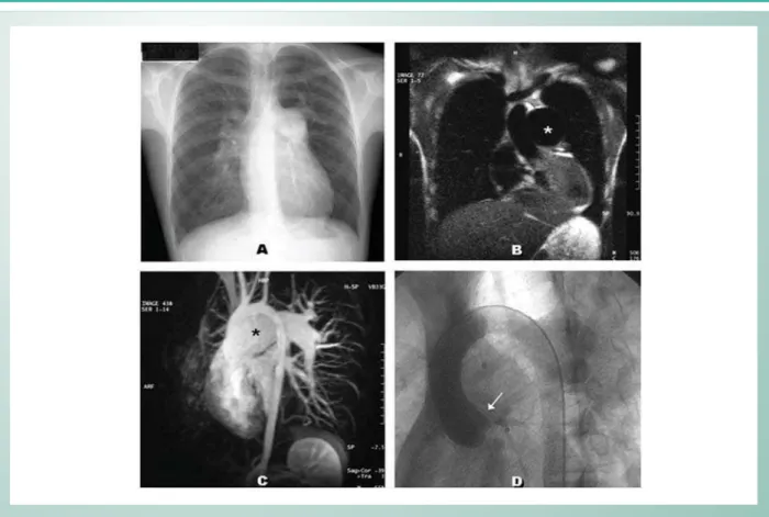
echocardiogram. The pacient was in good condition at physical exam, pulse was normal, and ++/6+ systolic murmur at pulmonary focus, with no thrill. Heart sound 2 was steadily unfolded, with a hyperphonetic pulmonary component. ECG showed signs of significant right ventricular overburden. Thoracic X-Ray showed pulmonary hyperflow, right branch of pulmonary artery dilation, and quite dilated PAT (Fig. 1A). Thoracic CAT scan (Fig. 1B), as well as magnetic angioresonance (Fig. 1C) pointed out the significant dilation of pulmonary branches and trunk, and relative aortic caliber decrease. Transesophageal echocardiogram showed a 10 mm sinus venosus IAC, with anomalous venous drainage of right upper pulmonary vein in right atrium. The hemodynamic study confirmed non-invasive findings and showed significant compression of LCA trunk by PAT (Fig. 1D). Aortic pressure was 126/93 mmHg, while pulmonary artery pressure was 110/70 mmHg , decreasing to 73/52 mmHg with O2 inhalation. Systemic vascular resistance (SVR) was 13 UWood, pulmonary vascular resistance (PVR) was 6.8 UWood. Pulmonary/ systemic flow ratio was 2.9. The patient was submitted to surgery on January 28, 2004. After pericardium opening, PAT diameter called the attention since it was three times larger than aortic diameter. The ASD was occluded with bovine pericardial strip, the anomalous pulmonary vein was directed into the left atrium, with reduction plasty of PAT. Immediate evolution was favorable and the patient was discharged on captopril one week after hospital admission. Six months later the patient was in good condition, and the hemodynamic study showed slight residual ASD, with pulmonary/systemic flow ratio at 1.05, PVR: 7 UWood, SVR: 13.7 UWood, pulmonary artery pressure: 75/52 mmHg, and aortic pressure: 110/83 mmHg. Extrinsic compression of LCA was also observed to have been reduced (Fig. 2A). Twenty-one months after the surgery the patient was asymptomatic, on 50 mg/day of captopril. Cardiac auscultation revealed a pulmonary click with S2 mildly hyperphonetic. Significant regression of right ventricular overburden was shown on the ECG; discreet reduction of PAT was shown on the X-Ray (Fig. 2B). Transthoracic echocardiogram showed pulmonary artery systolic pressure at 55 mm/Hg, intact interatrial septum, right ventricle hypertrophy, no compression of LCA (Fig. 2C), and PAT still dilated (Fig. 2D). Stress echocardiogram with dobutamine was negative for myocardial ischemia.
Case Report
Amaral et al
EXTRINSIC COMPRESSION OF LEFT MAIN CORONARY ARTERY FROM ANEURYSMAL DILATION OF PULMONARY TRUNK IN AN ADOLESCENT. INVOLUTION AFTER SURGERY OCCLUSION OF SINUS VENOSUS ATRIAL SEPTAL DEFACT (ASD) AND PULMONARY TRUNK PLASTY FOR REDUCTION
Arq Bras Cardiol 2007; 88(2) : e39-e41
Discussion
Described for the first time almost half a century ago9, extrinsic compression of LCA by PAT is not common. Comprehensive studies are rare, although information has been available in literature on the occurrence of 2 patients (1.8%) out of 48 IAC cases studied by Kothari et al10. Another relevant study was conducted in 1989 by Mitsudo1, and has stood as a landmark for topic discussion. While studying 38 patients in the 15-62-year-old range, the author found 7 cases (18%) of extrinsic compression of LCA. The figures reached 44% for cases with pulmonary hypertension associated.
Most cases described reveal a frequent association with IAC1,2,4,5,11; however, other conditions – such as persistence of arterial canal6, tetralogy of Fallot (TOF)7, and atrioventricular septal defect8 – may be involved. Primary pulmonary hypertension patients with significant PAT dilation may also report the same conditions3,12. Precordial pain may act as suspicion of such serious complication in patients with this type of cardiopathy and with significant PAT dilation on thoracic X-Ray. The ECG may reveal ischemic changes2, 3,6, and coronary obstruction may be visualized on echocardiogram and on nuclear magnetic resonance. However, diagnostic confirmation and the assessment of compression severity is usually more reliable through coronariography.
The case presented includes some characteristics which we understand justify this report. Although the topic has
been discussed based on fairly extensive studies in this country4, our patient seems to be the first case to be published in our journals. Additionally, the occurrence of such complication in an adolescent – as described – seems to be quite rare. To our knowledge, there is only one report in that age range11.
Another interesting aspect – also made evident in our case – is the conduct adopted. Some authors believe in the need of revascularization for cases with significant obstruction1. Others suggest that surgical reduction of PAT only may eliminate compression of the LCA, particularly if pulmonary hypertension is considered reversible2. Our patient had significant signs of severe pulmonary hypertension, with good response to O2. Almost 2 years after surgery, considerable involution of the obstruction could be seen on the echocardiogram.
To conclude, we could say that extrinsic compression of LCA by PAT is quite rare. As suggested, it may, therefore, be underdiagnosed4. Due to the possibility of obstruction involution, as demonstrated, many IAC cases that have been operated on without visualization of the coronaries may not have been properly documented. Cardioapathy patients that develop PAT increase (with or without pulmonary hypertension), must be carefully investigated, particularly if associated to precordial pain complaint. Due to the rare degree of the condition - which hinders any randomized therapy study – the authors find it hard to define whether routine revascularization for cases with significant obstruction
Fig. 1 -(pre-operative) - Aneurysmal dilation of PAT demonstrated by thorax X-Ray (A), CAT scan (B), and magnetic angioresonance (C); (D) Hemodynamic study: aortography on left anterior oblique incidence, with evidence of significant obstruction at coronary trunk (arrow).
Case Report
Amaral et al EXTRINSIC COMPRESSION OF LEFT MAIN CORONARY ARTERY FROM ANEURYSMAL DILATION OF PULMONARY TRUNK IN AN ADOLESCENT. INVOLUTION AFTER SURGERY OCCLUSION OF SINUS VENOSUS ATRIAL SEPTAL DEFACT (ASD) AND PULMONARY TRUNK PLASTY FOR REDUCTION
Arq Bras Cardiol 2007; 88(2) : e39-e41
References
1. Mitsudo K, Fujino T, Matsunaga K, Doi O, Nishihara Y, Awa J. The coronary angiographic findings in the patients with atrial septal defect and pulmonary hypertension (ASD + PH): compression of left main coronary artery by pulmonary trunk. Kokyu To Junkan. 1989; 37: 649-55.
2. Fujiwara K, Naito Y, Higashine S, Takagaki Y, Goto Y, Okamoto M. Left main coronary trunk compression by dilated main pulmonary artery in atrial septal defect. J Thorac Cardiovasc Surg. 1992; 104: 449-52.
3. Patrat JF, Jondeau G, Dubourg O, Lacombe P, Rigaud M, Bourdarias JP. Left main coronary artery compression during primary pulmonary hypertension. Chest. 1997; 112: 842-3.
4. Kajita LJ, Martinez EE, Ambrose JA, Lemos PA, Esteves A, Nogueira da Gama M. Extrinsic compression of the left main coronary artery by a dilated pulmonary artery: clinical, angiographic and hemodynamic determinants. Cathet Cardiovasc Intervent. 2001; 52: 49-54.
5. Gullu H, Kosar F, Battaloglu B. Left main coronary artery compression by dilated pulmonary trunk in a patient with atrial septal defect. Acta Cardiol. 2003; 58: 355-7.
6. Bijl M, Bronzwaer JGF, van Rossum AC, Verheugt FWA. Angina pectoris due to left main coronary artery compression in Eisenmenger ductus arteriosus.
Am Heart J. 1993; 125: 1767-71.
7. Sengupta PP, Saxena A, Rajani M. Left main coronary artery compression by aneurismal pulmonary artery in a patient with tetralogy of Fallot with absent pulmonary valve. Cathet Cardiovasc Intervent. 1999; 46: 438-40.
8. Manojkumar R, Grover A. Left main coronary artery compression by dilated main pulmonary artery in endocardial cushion defect. Indian Heart J. 2002; 54: 74-6.
9. Corday E, Gold H, Kaplan L. Coronary artery compression: an explanation for the cause of coronary insufficiency in pulmonary hypertension. Trans Am Coll Cardiol. 1957; 7: 93-103.
10. Kothari SS, Chatterjee SS, Sharma S, Rajani M, Wasir HS. Left main coronary artery compression by dilated main pulmonary artery in atrial septal defect. Indian Heart J. 1994; 46: 165-7.
11. Kito K, Fujiwara Y, Kimura T, Shimada Y. Left main coronary trunk compression by dilated main pulmonary artery in a patient with atrial septal defect. Masui. 2001; 50: 184-7.
12. Eksinar S, Gedevanishvili A, Koroglu M, Afzal A, Oto A, Conti V, et al. Extrinsic compression of the left main coronary artery in pulmonary hypertension. JBR-BTR. 2005; 88: 190-2.
Fig. 2 -(post-operative) - A) Six months after surgery, hemodynamic study shows decrease of coronary obstruction (arrow); B) Two years after surgery, evaluation shows discreet decrease of PAT on thoracic X-Ray (B) and echocardiogram showing no evidence of left coronary artery (C), although PAT still dilated (D).
is required or not. However, basead on the case reported here, and on the small amount of information available in the literature, it does seem that for young patients with no injury
in other arteries and with reversible pulmonary hypertension, the most approppriate conduct is correction of cardiac defect associated to PAT reduction plasty.