Research Institute of San Diego, San Diego, California, United States of America,4Government Central Laboratories, Ministry of Health, Jerusalem, Israel,5Department of Pathology and Laboratory Medicine, University of California Irvine, Irvine, California, United States of America
Abstract
Human infection with non-typhoidal Salmonella serovars (NTS) infrequently causes invasive systemic disease and
bacteremia. To understand better the nature of invasive NTS (iNTS), we studied the gene content and the pathogenicity of bacteremic strains from twelve serovars (Typhimurium, Enteritidis, Choleraesuis, Dublin, Virchow, Newport, Bredeney,
Heidelberg, Montevideo, Schwarzengrund, 9,12:l,v:- and Hadar). Comparative genomic hybridization using aSalmonella
enterica microarray revealed a core of 3233 genes present in all of the iNTS strains, which include the Salmonella
pathogenicity islands 1–5, 9, 13, 14; five fimbrial operons (bcf, csg, stb, sth,sti); three colonization factors(misL, bapA, sinH);
and the invasion gene,pagN. In the iNTS variable genome, we identified 16 novel genomic islets; various NTS virulence
factors; and six typhoid-associated virulence genes (tcfA,cdtB,hlyE,taiA, STY1413, STY1360), displaying a wider distribution
among NTS than was previously known. Characterization of the bacteremic strains in C3H/HeN mice showed clear differences in disease manifestation. Previously unreported characterization of serovars Schwarzengrund, 9,12:l,v:-, Bredeney and Virchow in the mouse model showed low ability to elicit systemic disease, but a profound and elongated shedding of serovars Schwarzengrund and 9,12:l,v:- (as well as Enteritidis and Heidelberg) due to chronic infection of the mouse. Phenotypic comparison in macrophages and epithelial cell lines demonstrated a remarkable intra-serovar variation, but also
showed thatS. Typhimurium bacteremic strains tend to present lower intracellular growth than gastroenteritis isolates.
Collectively, our data demonstrated a common core of virulence genes, which might be required for invasive salmonellosis, but also an impressive degree of genetic and phenotypic heterogeneity, highlighting that bacteremia is a complex phenotype, which cannot be attributed merely to an enhanced invasion or intracellular growth of a particular strain.
Citation:Suez J, Porwollik S, Dagan A, Marzel A, Schorr YI, et al. (2013) Virulence Gene Profiling and Pathogenicity Characterization of Non-TyphoidalSalmonella Accounted for Invasive Disease in Humans. PLoS ONE 8(3): e58449. doi:10.1371/journal.pone.0058449
Editor:Jose Alejandro Chabalgoity, Facultad de Medicina, Uruguay
ReceivedAugust 15, 2012;AcceptedFebruary 5, 2013;PublishedMarch 7, 2013
Copyright:ß2013 Suez et al. This is an open-access article distributed under the terms of the Creative Commons Attribution License, which permits unrestricted use, distribution, and reproduction in any medium, provided the original author and source are credited.
Funding:Work in the Infectious Diseases Research Laboratory was supported by grant number 249241 from the European Community’s Seventh Framework Program (PF7/2007-2013) and a GIF Research grant number 1096.39.11/2010 to O.G. Work in the McClelland lab was supported by grants from the United States National Institute of Allergy and Infectious Diseases, the United States Department of Agriculture, and the United States - Israel Binational Agricultural Research and Development Fund. The funders had no role in study design, data collection and analysis, decision to publish, or preparation of the manuscript.
Competing Interests:The authors have declared that no competing interests exist. * E-mail: [email protected]
Introduction
Salmonella enterica is a Gram-negative, facultative intracellular human and animal pathogen posing a major public health concern worldwide [1]. The single species S. enterica includes more than 2,600 serovars, which are taxonomically classified into six subspecies, sharing high sequence similarity [2]. Subspecies I serovars, adapted for mammals and avian hosts, are responsible for more than 99% of allSalmonella infections in humans. These serovars can be divided into two clinically relevant groups according to the disease they cause. Infections with the human-restricted serovarsS. Typhi andS. Paratyphi elicit an invasive, life-threatening systemic disease referred to as typhoid or enteric fever [3]. On the other hand, non-typhoidal serovars (NTS) will normally cause in humans, self-limited gastroenteritis, associated with intestinal inflammation and diarrhea [4]. Nevertheless, in developed countries up to 5% of NTS cases may be invasive,
extra-intestinal disease leading to bacteremia and focal systemic infections. Moreover, in Sub-Saharan Africa invasive non-typhoidal Salmonella (iNTS) have emerged as a major cause of bloodstream infection in adults and children, with an estimated annual incidence of 175–388 cases per 100 000 children and 2000–7500 cases per 100 000 HIV-infected adults [5].
Different factors including the genetic background and the immunological status of the host are now known to predispose to iNTS disease and were recently reviewed [6]. Nevertheless, since certain NTS serovars such asS. Choleraesuis andS. Dublin are inclined to cause bacteremia more frequently than others [7], it is clear that pathogen characteristics are greatly important as well. Indeed, the unique genetic features of a dominant African invasive genotype of S. Typhimurium ST313, which was shown to comprise a distinct prophage repertoire, unique drug resistant elements, and evidence for genome degradation [8], further
emphasize the contribution of pathogen genetics to the invasive outcome.
Genomic diversity across bacterial strains is largely shaped by gain of functions via horizontal gene transfer [9]. Foreign acquired DNA containing functionally related genes is designated Genomic Island or Islet (GI). GIs that encode virulence genes are referred to as Pathogenicity Islands (PIs) and their presence in the genome of bacterial pathogens distinguish them from closely related non-pathogenic strains or species (reviewed in [10]).
In the species S. enterica, 21 Salmonella Pathogenicity Islands (SPIs) have been identified thus far [11], in addition to the
Salmonella genomic island 1 (SGI-1) [12] and the high-pathoge-nicity island (HPI) [13]. SPIs are considered to be ‘quantum leaps’ in the evolution ofSalmonella[14], playing a fundamental role in pathogenesis [15] and host specificity [16]. While certain SPIs (such as SPI-1 and SPI-2) have been studied in depth, other SPIs have been identified more recently and much less is known about their distribution acrossSalmonellaserovars and the role they play in disease.
Characterization of the mechanisms underlying invasive man-ifestation by NTS is essential to a more general understanding of the biology and pathogenicity ofSalmonella. To better understand the genetic profile and the virulence of iNTS strains, we determined the virulence gene content in S. Typhimurium invasive and gastroenteritis strains and in invasive isolates from eleven additional serovars. Furthermore, the pathogenicity of invasive isolates representing the 12 serovars was characterized in the mouse model and virulence-associated phenotypes were compared between invasive and gastroenteritis isolates in tissue cultures modelsin-vitro.
Materials and Methods
Bacterial Strains and Growth Conditions
Bacterial strains utilized in this study are listed in Table S1 and include low-passage clinical strains isolated from blood (N = 66), stool (N = 68) or unknown source (N = 1) from 12 NTS serovars as well as Salmonellareference sequenced strains (N = 9). All clinical isolates were obtained from the Israeli National Salmonella
Reference Center after serotyping according to the White-Kauffmann-LeMinor scheme by agglutination with O- and H-antigen specific sera. All Salmonella reference strains were purchased from the Salmonella Genetic Stock Center (SGSC) at the University of Calgary. Bacterial cultures were routinely maintained in Luria-Bertani (LB; BD Difco) liquid medium at 37uC. LB or xylose lysine deoxycholate (XLD; BD Difco) agar plates were used when appropriate. The clinical isolates that were analyzed by CGH were selected based on patient’s age informa-tion and included strains isolated from 2,patients,60 year-old to minimize age-related bias.
Comparative Genomic Hybridization (CGH)
Genomic DNA fromS. Typhimurium LT2 and clinical strains was extracted from overnight cultures grown in LB using the GenElute kit (Sigma-Aldrich) according to manufacturer’s instruc-tions. DNA labeling and hybridization to the STv7E Salmonella
microarray (http://www.sdibr.org/Faculty/mcclelland/ mcclelland-lab/mcclelland-protocols) were performed as previous-ly described [17]. An Agilent microarray scanner G2505B was used for image acquisition and signal intensities were quantified with the Spotreader software (Niles Scientific). Data normaliza-tion, analysis, and determining the presence or absence of genes, were described elsewhere [17].
Phylogenetic Analysis
A maximum parsimony tree was constructed according to the presence-absence data obtained by the CGH of the clinical isolates and discrete data that were inferred from the genome sequences of reference taxa (in total 5375 genes646 taxa were analyzed). The tree was obtained using the Close-Neighbor-Interchange algo-rithm with search level 1, in which the initial trees were obtained with the random addition of positions (5 replicates). Branch lengths were calculated using the average pathway method to represent the number of changes. The bootstrap consensus tree inferred from 100 replicates, and the evolutionary analyses were conducted in MEGA5.
PCR and Southern Blot Hybridization
Primers used in this study are listed in Table S2. DNA primers were purchased from IDT and PCR was carried out using ReddyMix PCR (Thermo Scientific) or PfuUltra II Fusion HS DNA Polymerase (Stratagene). For Southern blot analysis, 1mg of genomic DNA was digested at 37uC for 16–18 h with PstI, subjected to electrophoresis in 1.0% agarose gels before being capillary transferred and cross-linked onto Hybond-N membranes (Amersham Biosciences). Genomic DNA fromE. coliDH5awas included as a negative control in all hybridizations. S. Typhi CT18, S. Typhimurium DT104, S. Typhimurium 14028 s or SARC13 were used as positive controls. Southern blots were processed using the DIG DNA Labeling and Detection Kit (Roche Applied Sciences), followed by an anti-DIG detection according to the manufacturer’s protocol.
RT-PCR
RNA was extracted fromSalmonella cultures that were subcul-tured into fresh LB broth and grown aerobically to late-exponential-phase using the Qiagen RNAprotect Bacteria Re-agent and the RNeasy mini kit (Qiagen) according to the manufacturer’s instructions, including an on-column DNase digest. Purified RNA was secondarily treated with an RNase-free DNase I followed by ethanol-precipitation and 150 ng of DNase I-treated RNA was subjected to a first strand cDNA synthesis, using the iScript cDNA synthesis kit (Bio-Rad Laboratories) according to the kit protocol. 2ml from the reverse transcription reaction were used as a template for a PCR amplification using the primers listed in Table S2.
Tissue Culture Conditions and Bacterial Infection The human epithelial HeLa and the murine macrophages-like RAW264.7 cell-lines were purchased from ATCC and cultured in a high glucose (4.5 g/L) Dulbecco’s Modified Eagle Medium (DMEM) supplemented with 10% heat-inactivated fetal bovine serum (FBS), 1 mM pyruvate and 2 mM L-Glutamine. All cell-lines were cultured at 37uC in a humidified atmosphere with 5% CO2. Epithelial cells and RAW264.7 macrophages were seeded at 56104 and 2.56105 cells/ml, respectively in a 24-well tissue culture dish 18 to 24 h prior to bacterial infection and experiments were carried out using the gentamicin protection assay as was previously described [18]. HeLa cells were infected at multiplicity of infection (MOI) of,1:100 withSalmonellastrains that had been subcultured from an overnight culture and grown for 3 h to late logarithmic phase under aerobic conditions. RAW264.7 cells were infected at MOI of ,1:1 using overnight stationary grown cultures. At the desired time points post infection (p.i.), cells were washed three times with Phosphate Buffered Saline (PBS) and harvested by addition of lysis buffer (0.1% SDS, 1% Triton X-100 in PBS). Appropriate dilutions were plated on LB-agar plates for
bacterial enumeration by CFU count. Salmonella invasion was determined by the number of intracellular Salmonella at 2 h p.i. divided by the number of infecting bacteria. Survival and intracellular multiplication (in macrophages and in non-phago-cytic cells, respectively) were determined by the number of recovered intracellular Salmonella at 24 h p.i. divided by the number of intracellular Salmonella at 2 h p.i. An unpaired t test with two tails was used to determine the significance of the differences between the compared data and p,0.05 was considered to be statistically significant. All isolates analyzed in TC were examined and found to be gentamicin sensitive and lysis buffer resistant.
Virulence Experiments in Mice
Six-weeks-old female C3H/HeN and BALB/c mice were purchased from Harlan laboratories and housed at the Sheba Medical Center animal facility under specific pathogen free conditions. All the mice were kept in sterilized cages and given food and waterad libitum. The mice were monitored daily, and any that showed extreme distress or became moribund were sacrificed. Experiments in this study were approved and carried out according to the national animal care guidelines and the institutional ethic committee of the Sheba Medical Center (approval No 601/10). Groups of 4–6 mice were infected intraperitoneally (i.p.) with ,56103CFU in 0.3 ml saline. Fecal pellets and organs harvested at end-points were homogenized in
cold saline, diluted and plated on selective XLD-agar plates (which distinguish the blackSalmonellacolonies) for CFU enumeration. An unpaired t test with two tails was used to determine the significance of the differences between the compared data and p,0.05 was considered to be statistically significant.
Results and Discussion
Core and Variable Genome of iNTS
Bacteremia and systemic infection challenge the pathogen with distinct environmental conditions and stresses, different from the ones in the gastrointestinal ambient [19]. It is therefore expected that a defined set of genes including specific virulence factors will be required and positively selected during this particular lifestyle. To better understand the genetic profile of iNTS and reveal their virulence factor composition we have analyzed the gene content of bacteremic and gastroenteritisS. Typhimurium isolates (five from each group) in addition to bacteremic strains from eleven other NTS serovars (Enteritidis, Choleraesuis, Dublin, Virchow, New-port, Bredeney, Heidelberg, Montevideo, Schwarzengrund, 9,12:l,v:- and Hadar). These serovars were chosen to provide a global view onto the iNTS phenomenon and were of the leading cause of NTS bacteremia in Israel, collectively responsible for over 85% of NTS invasive cases during 1995–2011. All of the invasive isolates analyzed in this study accounted for systemic disease in humans and were isolated from patients’ blood as listed in Table S1.
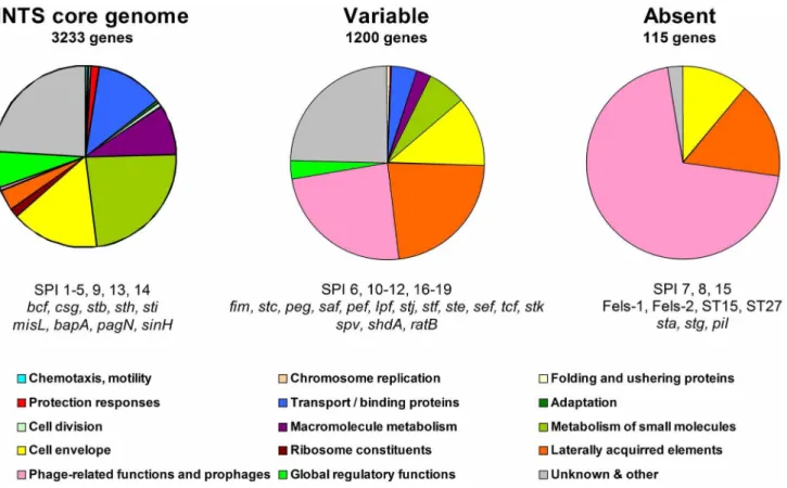
Figure 1. Core, variable and absent genes in iNTS strains.Microarray-based comparative genomic hybridization was used to determine the presence and absence of theSalmonellaORFs represented on theS. entericaSTv7E microarray. After excluding redundant probes (corresponding to the same ORF) and ambiguous predictions, we were able to assign 4548Salmonellagenes into three groups. A set of 3233 common genes was determined to be present in all of the bacteremic strains and defined as the core genome of iNTS (left panel). 1200 genes with varying distribution were missing from, at least, one serovar and were considered as the variable iNTS genome (center panel). 115 genes were absent from all the 16 blood strains analyzed in this study (right panel). Functional classification of the genes is shown and assigned using GenProtEC (http://genprotec.mbl. edu/).
doi:10.1371/journal.pone.0058449.g001
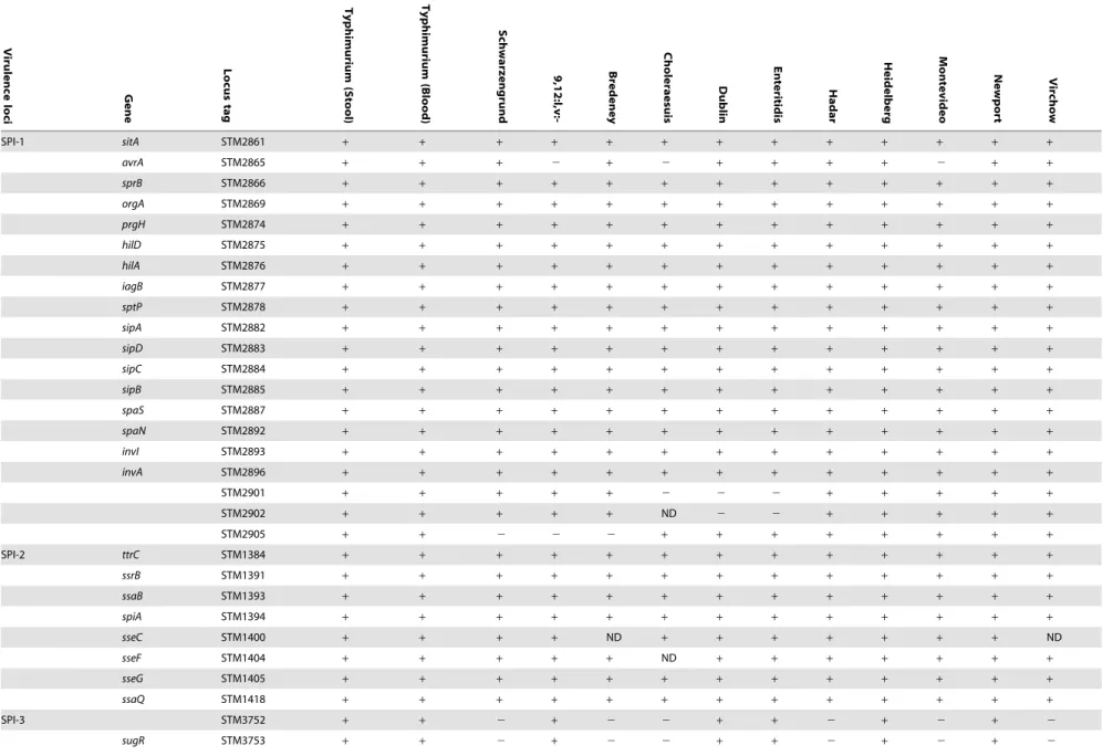
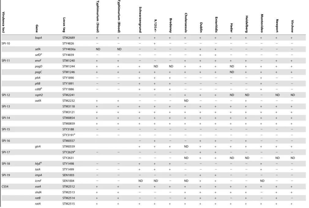
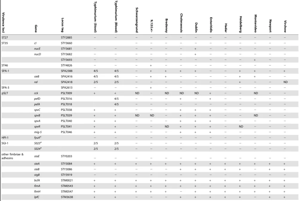
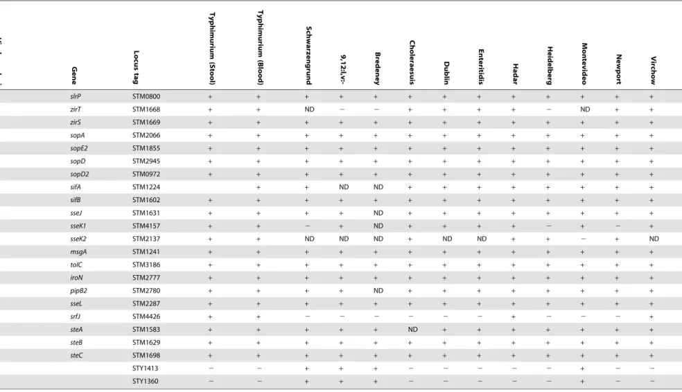
Table 1.Distribution of virulence genes across NTS bacteremic isolates.
Virulenc
e
loci Gene
Locus
tag
Typhimurium
(Stool)
Typhimurium
(Blood)
Schwa
rzengrund
9,12:l,
v:-Bredeney
Chole
raesuis Dub
lin
Enteritidis
Hadar
Heidelberg Mont
evideo
Newpor
t
Virchow
SPI-1 sitA STM2861 + + + + + + + + + + + + +
avrA STM2865 + + + 2 + 2 + + + + 2 + +
sprB STM2866 + + + + + + + + + + + + +
orgA STM2869 + + + + + + + + + + + + +
prgH STM2874 + + + + + + + + + + + + +
hilD STM2875 + + + + + + + + + + + + +
hilA STM2876 + + + + + + + + + + + + +
iagB STM2877 + + + + + + + + + + + + +
sptP STM2878 + + + + + + + + + + + + +
sipA STM2882 + + + + + + + + + + + + +
sipD STM2883 + + + + + + + + + + + + +
sipC STM2884 + + + + + + + + + + + + +
sipB STM2885 + + + + + + + + + + + + +
spaS STM2887 + + + + + + + + + + + + +
spaN STM2892 + + + + + + + + + + + + +
invI STM2893 + + + + + + + + + + + + +
invA STM2896 + + + + + + + + + + + + +
STM2901 + + + + + 2 2 2 + + + + +
STM2902 + + + + + ND 2 2 + + + + +
STM2905 + + 2 2 2 + + + + + + + +
SPI-2 ttrC STM1384 + + + + + + + + + + + + +
ssrB STM1391 + + + + + + + + + + + + +
ssaB STM1393 + + + + + + + + + + + + +
spiA STM1394 + + + + + + + + + + + + +
sseC STM1400 + + + + ND + + + + + + + ND
sseF STM1404 + + + + + ND + + + + + + +
sseG STM1405 + + + + + + + + + + + + +
ssaQ STM1418 + + + + + + + + + + + + +
SPI-3 STM3752 + + 2 + 2 2 + + 2 + 2 + 2
sugR STM3753 + + 2 + 2 2 + + 2 + 2 + 2
Virulence
and
Genetic
Characterizatio
n
of
iNTS
ONE
|
www.ploson
e.org
4
March
2013
|
Volume
8
|
Issue
3
|
i
STM3754 + + 2 + 2 2 + + 2 + 2 2 2
rhuM STM3755 + + 2 + 2 2 + + + + 2 2 2
misL STM3757 + + + + + + + + + + + + +
marT STM3759.S + + + + + + + + + + + + +
mgtC STM3764 + + + + + + + + + + + + +
STM3780 + + + + + + 2 2 + + + 2 +
SPI-4 siiD STM4260 + + + + + + + + + + + + +
siiE STM4261 + + + + + + + + + + + + +
siiF STM4262 + + + + + + + + + + + + +
SPI-5 pipA STM1087 + + + ND + + + + + + + + +
pipB STM1088 + + + + + + + + + + + + +
sopB STM1091 + + + + + + + + + + + + +
pipD STM1094 + + + + + + + + + + + + +
SPI-6 sciL STM0277 + + + + + 2 2 2 + + + + 2
sciR STM0284 + + + + + + + 2 + + + + 2
sciS STM0285 + + + + + + + 2 + + + + 2
safA STM0299 + + ND + 2 + 2 ND + 2 2 2 2
safB STM0300 + + + + + + + + + 2 + + +
tcfAb STY0345
2 2 + + + + 2 2 2 + + 2 +
tinR STY0349 2 2 + + + + 2 2 2 + + 2 +
pagN STM0306 + + + + + + + + + + + + +
SPI-7 pilQ STY4545 2 2 2 2 2 ND 2 2 2 2 2 2 2
pilV STY4550 2 2 2 2 2 2 2 2 2 2 2 2 2
tviE STY4656 2 2 2 2 2 2 ND 2 2 2 2 2 ND
vexE STY4651 2 2 ND 2 ND ND 2 2 2 2 2 2 2
vexC STY4653 2 2 2 2 ND ND 2 2 2 ND 2 2 2
vexAa STY4655
2 2 2 2 2 2 2 2 2 2 2 2 2
SPI-8 STY3283 2 2 2 2 2 ND 2 2 2 2 2 2 2
STY3281a
2 2 2 2 2 2 2 2 2 2 2 2 2
SPI-9 STY2878 + + + + + + + + + + + + +
Virulence
and
Genetic
Characterizatio
n
of
iNTS
5
March
2013
|
Volume
8
|
Issue
3
|
Table 1.Cont.
Virulence
loc
i
Gene
Locu
s
tag
Typhimurium
(Stool)
Typhimurium
(Blood)
Schw
arzengrund
9,12:
l,v:-Bredeney
Ch
oleraesuis
Dublin
Enteritidis
Hadar
Heidelberg Mont
evideo Newport Virchow
bapA STM2689 + + + + + + + + + + + + +
SPI-10 STY4826 2 2 2 + 2 2 2 2 2 2 2 2 2
sefA STY4836a ND ND 2 2 2 2 + + 2 2 2 2 2
sefDa STY4839
2 2 2 2 2 2 + + 2 2 2 2 2
SPI-11 envF STM1240 + + 2 2 2 + + + + + 2 + +
pagD STM1244 + + + ND ND + + + ND + + + +
pagC STM1246 + + + + + + + + ND + + + +
pltA STY1890 2 2 + + + 2 2 2 2 2 + 2 2
pltB STY1891 2 2 + + + 2 2 2 2 2 + 2 2
cdtBb STY1886
2 2 + + + 2 2 2 2 2 + 2 2
SPI-12 sspH2 STM2241 2 2 2 + + + ND ND 2 ND ND
oafA STM2232 + + 2 2 2 ND 2 2 2 + 2 2 2
SPI-13 STM3118 + + + + + + + + + + + + +
STM3121 + + + + + + + + + + + + +
SPI-14 STM0854 + + + + + + + + + + + + +
STM0859 + + + + + + + + + + + + +
SPI-15 STY3188 2 2 2 2 2 2 2 2 2 2 2 2 2
STY3191a
2 2 2 2 2 2 2 2 2 2 2 2 2
SPI-16 STM0557 2 + 2 2 + + 2 + 2 2 2
gtrA STM0559 + + + ND + + + + + + +
SPI-17 STY2629a
2 2 2 2 2 2 + + 2 2 2 2 2
STY2631 2 2 2 ND + + ND ND 2 ND ND
SPI-18 hlyEb STY1498
2 2 + + + 2 2 2 2 2 + 2 2
taiA STY1499 2 2 + + + 2 2 2 2 2 + 2 2
SPI-19 impA SEN1003 2 2 2 2 2 2 + + 2 2 2 2 2
icmF SEN1004 2 2 ND ND 2 ND + + 2 2 ND 2 2
CS54 xseA STM2512 + + + + + + + + + + + + +
shdA STM2513 + + 2 2 2 + + + + + 2 + +
ratB STM2514 + + 2 2 2 + + + 2 + 2 + 2
ratA STM2515 + + + + + + + + + + + + +
Virulence
and
Genetic
Characterizatio
n
of
iNTS
ONE
|
www.ploson
e.org
6
March
2013
|
Volume
8
|
Issue
3
|
i
sinI STM2516 + + + ND ND + + + + ND + + +
sinH STM2517 + + + + + + + + + + + + +
Gifsy-1 gipA STM2600 4/5 2 2 2 + 2 2 + 2 2 2 +
gogB STM2584 4/5 2 2 2 + 2 2 2 2 2 2 2
Gifsy-2 sodC1 STM1044 4/5 2 2 2 + + + 2 + 2 + 2
gvrA STM1034 4/5 2 2 2 ND + 2 2 + 2 + 2
sseI STM1051 4/5 2 2 2 + + + ND ND 2 ND ND
gtgE STM1055 4/5 2 2 2 + + + 2 ND 2 2 2
Gifsy-3 sspH1b
2 2 2 2 2 2 2 2 2 2 2 2 2
Fels-1 nanH STM0928 2 2 2 2 2 2 2 2 2 2 2 2 2
sodCIII STM0924 2 2 2 2 2 2 2 2 2 2 2 2 2
Fels-2 STM2695 2 2 2 2 2 2 + 2 2 2 2 2 2
STM2723 2 2 2 2 2 2 2 2 2 2 2 2 2
STM2739 2 2 2 2 2 2 + 2 2 2 2 2 2
Def4 STM4196 + + 2 2 2 + + + 2 + 2 2 +
STM4198
STM4205
STM4213
ST64b Sb46a 4/5
2 2 2 + + + 2 + 2 2 2
ssek3b 3/5 1/5
2 2 2 2 + + 2 2 2 2 2
SopEF STY4619 2 2 2 2 2 2 2 2 2 2 2 2 2
sopEFa,b STY4609 1/5
2 2 2 + + + + 2 + +
S.Typhi phage ST10 STY1046 3/5 3/5 ND ND
STY1048 1/5 +
STY1054 1/5 +
STY1071 2/5 +
S.Typhi phage ST18 STY2019 2/5 +
STY2028 2/5 +
STY2036 1/5 +
ST15 gam STY1601 2 2 2 2 2 2 2 2 2 2 2 2 2
Virulence
and
Genetic
Characterizatio
n
of
iNTS
7
March
2013
|
Volume
8
|
Issue
3
|
Table 1.Cont.
Virulence
loc
i
Gene
Locu
s
tag
Typhimurium
(Stool)
Typhimurium
(Blood)
Schw
arzengrund
9,12:
l,v:-Bredeney
Ch
oleraesuis
Dublin
Enteritidis
Hadar
Heidelberg Mont
evideo Newport Virchow
ST27 STY2885 2 2 2 2 2 2 2 2 2 2 2 2 2
ST35 cI STY3660 2 2 2 2 2 2 2 2 2 2 2
nucE STY3681 2 2 2 2 2 2 + 2 2 2 2 2 2
nucD STY3682 2 2 2 2 2 2 + 2 2 2 2 2 2
STY3693 2 2 2 2 2 2 2 2 2 2 + 2 2
ST46 STY4826 2 2 2 + 2 2 2 2 2 2 2 2 2
SPA-1 SPA2388 4/5 4/5 2 + + + + 2 2 + + 2 +
sieB SPA2416 4/5 4/5 2 + + 2 2 2 2 + + 2 2
ral SPA2418 2/5 2/5 2 2 2 2 2 2 2 + 2 2 ND
SPA-3 SPA2613 2 2 2 + 2 2 2 2 2 2 2 2 2
pSLT rck PSLT009 + + ND 2 ND ND ND + 2 2 ND 2 2
pefD PSLT016 4/5 2 2 2 + 2 + 2 2 2 2 2
pefA PSLT018 4/5 2 2 2 2 2 2 2 2 2 2 2
spvC PSLT038 + + 2 2 2 + + + 2 2 2 2 2
spvB PSLT039 + + ND ND 2 + + + 2 2 ND 2 2
spvA PSLT040 + + 2 2 2 + + + 2 2 2 2 2
spvR PSLT041 + + 2 2 ND + + + 2 ND 2 2 2
mig-5 PSLT046 + + 2 2 2 + + + 2 2 2 2 2
HPI-1 fyuAa
2 2 2 2 2 2 2 2 2 2 2 2 2
SGI-1 S023a 2/5 2/5
2 2 2 2 2 2 2 2 2 2 2
S024a 2/5 2/5
2 2 2 2 2 2 2 2 2 2 2
other fimbriae &
adhesins staE STY0203 2 2 2 2 2 2 2 2 2 2 2 2 2
steA STY3084 + + + + + + + + + + + + +
steB STY3086 2 2 2 2 2 + + + + + 2 + +
stgB STY3919 2 2 2 2 2 2 2 2 2 2 2 2 2
bcfA STM0021 + + + + + + + + + + + + +
fimA STM0543 + + + + + + + + + + + + +
fimH STM0547 + + + + + 2 + + + + + + +
lpfC STM3638 + + 2 2 2 + + + + + 2 + +
Virulence
and
Genetic
Characterizatio
n
of
iNTS
ONE
|
www.ploson
e.org
8
March
2013
|
Volume
8
|
Issue
3
|
i
csgA STM1144 + + + + + + + + + + + + +
stbC STM0338 + + + + + + + + + + + + +
stcB STM2151 + + 2 2 2 + 2 2 + + 2 + +
pegD SEN2144A 2 2 + + + 2 + + 2 2 2 2 2
stfD STM0197 + + 2 2 2 + + + + + 2 + +
sthD STM4592 + + + + + + + + + + + + +
stiC STM0175 + + + + + + + + + + + + +
stjC STM4573 + + 2 + 2 2 2 2 + + + + +
stkE SPA0181 2 2 2 2 2 2 2 2 + + 2 2 +
stdD STM3026 + + 2 2 2 2 + + 2 + 2 2 2
stdC STM3027 + + + + + + + + + + + + +
regulators zur STM4241 + + + + + + + + + + + + +
hfq STM4361 + + + + + + + ND + + + ND ND
igeR STM0410 + + 2 2 2 + + + + + 2 + +
relA STM2956 + + + + + + + + + + + + +
phoQ STM1230 + + + + + + + + + + + + +
phoP STM1231 + + + + + + + + + + + + +
fur STM0693 + + + + + + + + + + + + +
hns STM1751 + + + + + + + + + + + + +
hnr STM1753 + + + + + + + + + + + + +
rpoE STM2640 + + + + + + + + + + + + +
rpoS STM2924 + + + + + + + + + + + + +
oxyR STM4125 + + + + + + + + + + + + +
slyA STM1444 + + + + + + + + + + + + +
ompR STM3502 + + + + + + + + + + + + +
rtcR STM3522 + + 2 2 2 + + + 2 + 2 2 2
sirA STM1947 + + + + + + + + + + + + +
Effectors outside of SPIS and other virulence factors
virK STM2781
+ + + + + + + + + + + + +
mig-14 STM2782 + + + + + + + + + + + + +
Virulence
and
Genetic
Characterizatio
n
of
iNTS
9
March
2013
|
Volume
8
|
Issue
3
|
Table 1.Cont.
Virulenc
e
loci Gene
Locus
tag
Typhimurium
(Stool)
Typ
himurium
(Blood)
Schwar
zengrund 9,12:l,
v:-Bredeney
Choler
aesuis Dub
lin
Enterit
idis
Hadar
Heidelberg Montevid
eo
Newport Virc
how
slrP STM0800 + + + + + + + + + + + + +
zirT STM1668 + + ND 2 2 + + + + 2 ND + +
zirS STM1669 + + + + + + + + + + + + +
sopA STM2066 + + + + + + + + + + + + +
sopE2 STM1855 + + + + + + + + + + + + +
sopD STM2945 + + + + + + + + + + + + +
sopD2 STM0972 + + + + + + + + + + + + +
sifA STM1224 + + ND ND + + + + + + + +
sifB STM1602 + + + + + + + + + + + + +
sseJ STM1631 + + + + ND + + + + + + + +
sseK1 STM4157 + + 2 + ND + + + + 2 + 2 +
sseK2 STM2137 + + ND ND ND + ND ND + + 2 + ND
msgA STM1241 + + + + + + + + + + + + +
tolC STM3186 + + + + + + + + + + + + +
iroN STM2777 + + + + + + + + + + + + +
pipB2 STM2780 + + + + ND + + + + + + + +
sseL STM2287 + + + + + + + + + + + + +
srfJ STM4426 + + 2 2 2 2 2 2 + 2 2 2 +
steA STM1583 + + + + + ND + + + + + + +
steB STM1629 + + + + + + + + + + + + +
steC STM1698 + + + + + + + + + + + + +
STY1413 2 2 + + + 2 2 2 2 2 + 2 2
STY1360 2 2 + + + 2 2 2 2 2 + 2 2
The presence-absence of genes associated withSalmonellavirulence were determined in fiveS. Typhimurium stool isolates (115043, 88359, 93561, 98001, 130100) and five blood isolates (103259, 111682, 116449, 93130, 98666) in addition to 11 invasive strains from serovars Schwarzengrund (124983), 9,12:l,v:- (94293), Bredeney (96115), Choleraesuis (90958), Dublin (74007), Enteritidis (122205), Hadar (121851), Heidelberg (78646), Montevideo (111072), Newport (91532) and Virchow (103033). Where not stated otherwise, presence was determined by CGH using theSalmonellaSTv7E microarray.a, presence was determined or confirmed by southern blot;b, presence was determined or confirmed by PCR. A plus sign indicates the presence of the gene; white blocks indicate an absence; a variable presence among the blood or stool isolates ofS. Typhimurium is shown by the number of positive isolates/ total number of isolates (N = 5); ND, no data (CGH was not conclusive).
doi:10.1371/journal.pone.0058449.t001
Virulence
and
Genetic
Characterizatio
n
of
iNTS
ONE
|
www.ploson
e.org
10
March
2013
|
Volume
8
|
Issue
3
|
CGH approach using the S. enterica STv7E microarray was applied to determine on a single-gene resolution the distribution of 5648 Salmonella ORFs represented on the array. The STv7E microarray contains nearly all (.96%) genes encoded in S. Typhimurium LT2,S. Typhimurium SL1344,S. Typhi TY2,S. Typhi CT18,S. Paratyphi A SARB42,S. Enteritidis PT4 and the LT2 pSLT virulence plasmid. To verify some of the microarray results and to explore the presence of certain genetics elements, not represented on the array (such as the Gifsy-3 and the ST64B bacteriophages, HPI-1 and SGI-1) we also used Southern blot hybridizations and PCR of selected targets. Overall, from a total of 5656SalmonellaORFs examined by all methods (CGH, PCR and southern blot), 3233 ORFs (57%) were shared by all 16 bacteremic isolates from 12 serovars and therefore considered as the ‘‘core’’ genome of the iNTS strains (Fig. 1). This pool of common genes includes besides many ancestral housekeeping ORFs, horizontally transferred genes encoded within SPIs 1–5, 9, 13 and 14; five fimbrial clusters (bcf, csg, stb, sthandsti); the SPI-6 encoded invasin,
pagN; and the CS54 invasin,sinH. Although these genes can be found also in reference or gastroenteritis isolates (Table S3), their common presence in all of the invasive isolates (from 12 serovars) suggests that they might be universally required by NTS serovars for invasive manifestation in the human host.
By contrast, 1200 genes were found to be absent (or had no close homologue) in at least one invasive isolate, demonstrating a large degree of genetic diversity among these bacteremic strains (that displayed the same invasive phenotype). The accessory
genome included many mobile elements-encoded genes such as the virulence plasmid and prophages, but also multiple metabolic operons, colonization factors and known virulence genes (Fig. 1). In addition, we identified 16 previously uncharacterized genomic islets composed of 2–8 ORFs presenting inconsistent distribution among the NTS serovars (Text S1 and Table S4). Sporadic and patchy distribution indicates either ancestral acquisition following by loss in multiple subsequent lineages, or independent gaining by lateral transfer at different stages duringSalmonellaevolution. Both scenarios elucidate the dynamics and plasticity of the NTS genome.
115 ORFs were found to be entirely absent from all of the iNTS strains and are mainlyS. Typhi,S. Paratyphi, Fels-1, and Fels-2 genes (Fig. 1).
The identified core genome is very different from a previously reported core genome ofSalmonella enterica subspecies I [20] (see Table S3) and may reflect both technical (e.g. usage of a
pan-Salmonella vs.S. Typhi CT18 arrays) and biological (e.g. choice of serovars and isolates) differences. Highlighted list, presenting the distribution of 200 virulence-associated genes is shown in Table 1 and the complete data set, including comparison to sequenced reference genomes can be viewed in Table S3.
Additionally, intra-serovar comparison between invasive (N = 5) and gastroenteritis (N = 5)S. Typhimurium isolates revealed 127 ORFs that were found to be conserved in the invasive strains, but variable among the examined stool isolates (Table S5). These genes included the pef (plasmid-encoded fimbriae) operon; the Figure 2. Presence and expression of typhoid-associated virulence factors among NTS serovars.(A) PCR analysis was used to confirm CGH results and to determine the distribution of three typhoid-associated genes in 35 clinical isolates from the 12 NTS serovars. PCR amplicons of 353-bp, 294-bp, and 335-bp indicate the presence ofcdtB,hlyEandtcfA, respectively. Tested isolates are numbered from 1–35 according to Table S1, with the isolates that were characterized in mice and subjected to CGH analysis in bold.S.Typhi CT18 (CT18) andS.Typhimurium SL1344 (SL1344) were used as positive and negative controls, respectively. (B) Reverse transcription-PCR was applied to examine expression ofcdtB,hlyE, taiAandtcfA genes in serovars Schwarzengrund, Montevideo, 9,12:l,v:- and Bredeney. Bacterial RNA was extracted from Salmonellacultures grown to late logarithmic phase in LB, followed by treatment with DNase I and reverse transcription. cDNA was used as template for PCR amplification using the primers listed in Table S2.SalmonellaRNA without a reverse transcriptase treatment (-RT) and purified gDNA were used as negative and positive controls, respectively.
doi:10.1371/journal.pone.0058449.g002
periplasmic [Cu,Zn]-superoxide dismutase, sodC; an attachment/ invasion protein (STM1043) and the region spanning STM2740-STM2771 containing two sugar phosphotransferase systems (STM2750-2752 and STM3256-3260). To get a further insight into their distribution we examined their presence in a larger collection of 15 invasive and 15 enteritisS. Typhimurium isolates (including the isolates analyzed by CGH). PCR analysis showed that
pefA, SodC and gogB were variably present in both invasive and enteritis isolates, whilesseI, STM2759 (as a probe for the STM2740-STM2771 region), and STM3260 (gatC) were found in all of the invasive isolates (15/15) and in 14/15 of the enteritis strains (Table S6). Although these results confirmed the variable presence among
S. Typhimurium isolates, they did not indicate a clear origin-related distribution for any of the aforementioned genes.
Distribution of SPIs and type Three Secretion System (T3SS) Effectors among iNTS
Many of the virulence factors ofS.entericaare encoded by genes organized on SPIs that have been acquired by horizontal transfer
SPIs identified in the iNTS strains could be divided into three categories: (i) intact (or mostly intact) SPIs with universal presence in all of the iNTS isolates (SPIs 1–5, 9, 13 and 14); (ii) SPIs that are completely absent from this iNTS collection (SPIs 7, 8 and 15); and (iii) SPIs with variable or mosaic presence across the serovars (SPIs 6, 10–12 and 16–19).
Interestingly, SPI-18 which was considered to be S. Typhi-specific [11], was found to be present in several NTS serovars (see below). Also, SPI-17 that was described before inS. Typhi [22],S. Enteritidis,S. Gallinarum [23] andS. Typhimurium LT2 [24] was now identified inS. Dublin as well. TheYersiniahigh pathogenicity island (HPI) that was recently found in S. Senftenberg poultry isolates [13] was not present in any of the tested iNTS isolates; however, the SGI-1 [25] was identified in two invasive (116449 and 98666) and two gastroenteritis (93561 and 115043) S. Typhimurium isolates.
With the exception ofavrAthat was found to be variable across isolates, other SPI-1, SPI-2, and SPI-5 encoded effector genes were conserved in their presence. The SPI-1 and SPI-2 T3SSs-substrate gene,sspH1, was absent from all examined isolates and at least ten
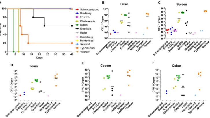
Figure 4. Pathogenicity of iNTS strains in mice. Twelve groups of 4–5 female C3H\HeN mice were challenged i.p. with,56103CFU of bacteremic strains from serovars Schwarzengrund (isolate number 124983), 9,12:l,v:- (94293), Bredeney (96115), Choleraesuis (90958), Dublin (74007), Enteritidis (122205), Hadar (121851), Heidelberg (78646), Montevideo (111072), Newport (91532), Typhimurium (103259), and Virchow (103033). Survival of the mice during 40 days post-infection is shown (A). At end-points (as shown in A), harvested organs were homogenized and serial dilutions were plated onto XLD-agar plates for CFU count. Bacterial load in each mouse is represented as CFU/organ by individual dots in the liver (B), spleen (C), ileum (D), cecum (E), and colon (F). Geometric mean for each serovar in the different sites is shown by a horizontal line.
doi:10.1371/journal.pone.0058449.g004
other T3SS effector genes (all encoded outside of SPI-1, SPI-2 and SPI-5) were variably distributed, includingsspH2, sseK1, sseK2, srfJ, and the prophages encoded effectors: gogB, sseI, gipA, gtgE, sseK3
and sopE. Many of these effectors including SopE were shown to play a role inSalmonellavirulence (reviewed in [26]); however their absence from multiple iNTS isolates suggests that they are unnecessary for invasive manifestation in the human host.
SopE translocation into cultured cells leads to actin cytoskeletal rearrangements and membrane ruffling [27] and many of theS. Typhimurium epidemic strains identified so far were reported to carry sopE [28]. To learn more about sopE distribution among bacteremic strains we screened by PCR a larger collection of 35 clinical isolates from blood and stool sources representing the above 12 serovars (Table S1). These analyses indicated the absence of sopE from 4/5 of the blood isolates of serovars Typhimurium, Schwarzengrund (3 isolates) and from at least one isolate of serovar Enteritidis (isolate 10325; data not shown), supporting the notion that SopE is nonessential for invasive salmonellosis in humans.
Distribution of Fimbriae and Colonization Factors
S.entericasubspecies I serovars harbors a wide array of putative fimbriae and pili central to bacterial adherence [29]. Salmonella
serovars were shown to harbor a unique combination of fimbrial genes, believed to play a role in host tropism and colonization
[30]. Among 20 fimbrial operons examined, a core set of 5 operons (bcf, csg, stb, sth and sti) seemed to be conserved in all bacteremic isolates, whereas three operons (stg, sta and pil), considered as ‘‘typhoid’’ fimbriae were missing from all serovars. The peg fimbrial cluster (SEN2144A-SEN2145B) that was first identified inS. Enteritidis PT4 [23] and was recently shown to be required for systemic infection of this serovar in mice [31], was revealed, for the first time, also in serovars Bredeney, 9,12:l,v:-, Dublin and Schwarzengrund (Table 1).
Besides fimbriae, additional colonization factors including the SPI-3 encoded adhesin,misL; the SPI-4 encoded adhesin,siiE; the SPI-6 encoded invasin,pagN; the SPI-9 encoded biofilm-associated protein,bapA; and the CS54 invasin,sinHwere all present through the entire iNTS collection, implying that they may play a role in an invasive lifestyle of NTS, possibly by contributing to tissue tropism and/or to niche-specificity.
Presence and Expression of Typhoid-associated Virulence Factors in NTS Serovars
CGH analysis identified a curious presence of several virulence factors that were originally characterized in S. Typhi and are believed to play a role in enteric fever manifestation. One of these factors is the cytolethal distending toxin CdtB, shown to be expressed by intracellular S. Typhi and cause cell-cycle arrest, severe host cell distension and enlargement of the nucleus [32]. In Figure 5. Persistent infection of iNTS strains in the mouse model.Following challenge with,56103CFU of the bacteremic strains, fecal pellets were collected from the C3H\HeN mice at the indicated time points during 40 days (or until the animal was sacrificed). Pellets were weighed, homogenized in PBS and plated onto XLD-agar plates to determine the number of CFU/g stool. Shedding ofS.Choleraesuis (D) andS.Dublin (E) is shown only until 3 days p.i., as the mice had to be euthanized. Dots represent independent CFU counts in pellets of individual mice.
doi:10.1371/journal.pone.0058449.g005
S. Typhi,cdtB(STY1886) is encoded within SPI-11 together with
pltA(STY1890) andpltB(STY1891) that were shown to form an intracellular tripartite toxin that is transported to the cell surface via a vesicular mechanism [33]. CGH revealed the presence of
cdtB, pltA and pltB in isolates of four NTS serovars including Montevideo, Schwarzengrund, Bredeney and 9,12:l,v:- (Table 1). To broaden and verify the above CGH results we expanded the number of tested strains and examined by PCR 35 clinical isolates (Table S1). This analysis confirmed the array results and indicated the presence of cdtB in 13/13 clinical strains from serovars Schwarzengrund, 9,12:l,v:-, Bredeney and Montevideo (Fig. 2A).
Another S. Typhi toxin is the pore-forming hemolysin HlyE (ClyA) that accumulates in the periplasm of S. Typhi [34]. hlyE
(STY1498) is encoded within SPI-18 together with the Typhi-associated invasin A (taiA, STY1499), and is required for efficient invasion of human epithelial cells. Functional transfer ofS.Typhi
hlyE to S. Typhimurium was shown to improve colonization of deep organs (spleen, liver) in mice [35,36]. TaiA is a secreted, 27 kDa invasin that increases Salmonella uptake by human macro-phages and is controlled by the virulence-related regulator PhoP [35]. Like in the case ofcdtB, CGH indicated the presence of
SPI-18 in the invasive isolates of Schwarzengrund, Montevideo, 9,12:l,v:- and Bredeney (Table 1 and Fig. 2A).
These results are in line with a recent report by den Bekkeret al. [37], showing the presence ofcdtBandhlyE-taiAinS. Schwarzen-grund and Montevideo isolates, but also reveal their presence, for the first time, in serovars 9,12:l,v:- and Bredeney.
S. Typhi carries a fimbriae cluster known as the Typhi-colonization factor (tcf) operon (STY0345- STY0348) encoded in SPI-6 and was suggested to play a role in host specificity of S. Typhi to humans [38]. In addition to Typhi and Paratyphi A, the
tcfoperon was found present in serovars Choleraesuis, Schwarzen-grund and Heidelberg [29,39] and recently also in Virchow and Montevideo [37]. Our results confirmed the presence of tcf in blood isolates of these serovars and further identified its previously unknown presence in 9,12;l,v;- and Bredeney (Table 1 and Fig. 2A).
After finding the presence ofcdtB, hlyE and tcfAin clinical isolates of serovars Schwarzengrund, 9,12:l,v:-, Bredeney and Montevi-deo, we have further examined a larger collection of 40 invasive and 43 gastroenteritis isolates from these four serovars (including the relevant 13 isolates shown in Fig. 2). The results presented in Table S7 showed that whiletcfAwas found in 3/4 invasive isolates Figure 6. Invasion of bacteremic and enteritis strains into human epithelial cells.Clinical isolates from 12 NTS serovars from blood (invasive) and stool (gastrointestinal) sources were grown to late logarithmic phase in LB medium and used to infect HeLa cells at a MOI of,100:1. The invasion of each strain (source and isolate number are indicated below each bar) is shown in relation to the invasion of the stool isolate in each serovar. InS. Typhimurium, invasion is presented relative to median value of the stool isolates (isolate 93561). Indicated values present the mean and the standard error of the mean (SEM; represented by the error bars) of at least 4 independent infections. ND, no data, as the source of isolate 4311– 10781 from serovar Dublin is not known.
doi:10.1371/journal.pone.0058449.g006
ofS. Schwarzengrund (as well as in the two reference sequenced genomes CVM19633 and SL480) it was uncommonly found in only 1/12 gastroenteritis isolates; indicating a variable and possibly source-related distribution oftcfAinS. Schwarzengrund. Yet,tcfAwas found in all of the isolates from serovars 9,12:l,v:- (15 invasive and 13 gastroenteritis), Bredeney (13 invasive and 12 gastroenteritis) and Montevideo (8 invasive and 6 gastroenteritis). The distribution ofcdtBandhlyEwas more unified and found in all of the isolates examined (Table S7), indicating that their presence is typical to, at least, clinical isolates, from serovars 9,12:l,v:-, Bredeney and Montevideo and is not associated with a specific source (i.e. invasive vs. gastroenteritis).
The presence of, so called, ‘‘typhoid-virulence genes’’ in a subset of NTS serovars was interesting and prompted us to test if they are also expressed. RNA purified from the invasive isolates of serovars Schwarzengrund, Montevideo, 9,12:l,v:- and Bredeney was used for a reverse-transcription PCR analysis. The results presented in Fig. 2B demonstrated that tcfA, hlyE, taiA and cdtB are readily expressed in these four serovarsin-vitro, under conditions known to induceSalmonellainvasion [40].
Moreover, homologs of two other yet uncharacterized typhoid genes, STY1413 and STY1360 were also identified in serovars Schwarzengrund, Montevideo, 9,12:l,v:- and Bredeney (Table 1). These two genes are characterized by a distinct G+C content and
present amino acid sequence similarity to the EspN2-2 (EHU09761; E-value 6e-72) and EspS (YP_003363979; E-value 5e-14), T3SS effectors of E. coli and Citrobacter rodentium, respectively, implying that they might function as secreted effector proteins in theseSalmonellaserovars.
Collectively, these results revealed an unknown presence of several typhoid virulence genes in serovars Schwarzengrund, Montevideo, 9,12:l,v:- and Bredeney, establishing that the distribution of these factors outside of typhoidal serovars is wider than previously known. Furthermore, we demonstrated, for the first time, native expression ofcdtB, andtaiAin NTS serovars, and presented the first evidence for variable presence of tcfA in S. Schwarzengrund and for its native expression in serovars 9,12:l,v:-and Bredeney; suggesting that their role is not limited to enteric fever caused by Typhi and Paratyphi. Nevertheless, the absence of these factors from other iNTS isolates indicates that their function is still not universally required by all invasive Salmonellae and cannot explain invasive manifestation by other strains.
Phylogenetic Clustering of the iNTS Strains
Data obtained from the CGH (5374 positions) were used to determine the phylogenetic relationship of the various clinical isolates and corresponding reference sequenced strains (46 taxa in total). In particular, we were interested to see how invasive isolates Figure 7. Intracellular growth of invasive and enteritis strains in epithelial cells.Clinical isolates (N = 41) from blood and stool sources were grown to late logarithmic phase in LB medium and used to infect epithelial HeLa cells at a MOI of,100:1. Intracellular replication (ratio between recovered CFU at 24 h/CFU at 2 h p.i.) is shown in relation to the stool isolate of each serovar. InS. Typhimurium, replication is presented relative to median value of the stool isolates (isolate 88359). Indicated values present the mean and the SEM of at least 4 independent infections.
doi:10.1371/journal.pone.0058449.g007
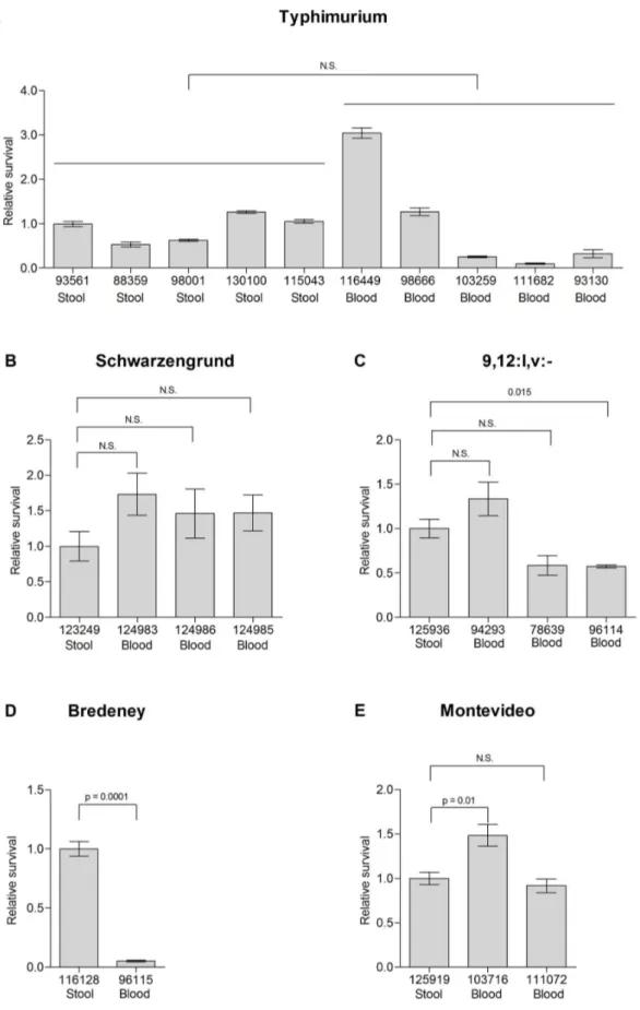
Figure 8. Survival of invasive and enteritis strains in macrophages. Salmonellaisolates from blood and stool sources from serovars Typhimurium (A), Schwarzengrund (B), 9,12:l,v:-(C), Montevideo (D) and Bredeney (E) were grown to stationary phase in LB medium and used to infect RAW2464.7 cells at a MOI of,1. Survival is shown in relation to the stool isolate of each serovar. InS. Typhimurium, survival is presented relative to median value of the stool isolates (isolate 93561). Indicated values present the mean and the SEM of at least 4 independent infections.
doi:10.1371/journal.pone.0058449.g008
will be clustered in relation to reference genomes and among themselves. A maximum parsimony tree clustered most of the bacteremic isolates together with their corresponding reference genomes and with the clinical gastroenteritis isolates (in the case of
S. Typhimurium, Fig. 3) and did not group together invasive isolates from different serovars. These results suggested that genetic variation was kept among different bacteremic strains, although displaying the same invasive phenotype (and not a genetic convergence towards a uniform gene content).
Pathogenicity of iNTS Strainsin-vivo
Next, we were interested in comparing the virulence of the bacteremic strains in a murine host and asked whether these isolates will present a comparable virulence phenotype. The pathogenicity of 12 bacteremic isolates (that were subjected to CGH analysis) was characterized in C3H/HeN (Nramp1+/+) inbreed mice, by comparing animals survival, colonization in systemic and intestinal sites, and bacterial shedding. The results of an i.p. infection indicated profound differences between the serovars in all of the three examined parameters. Clearly, only isolates, from serovars Typhimurium, Dublin and Choleraesuis, and to a lesser extent, Enteritidis were able to establish an acute systemic infection in this mouse strain (Fig. 4), indicating that a former systemic colonization in human patients did not restrict their host-tropism and the known potential of these serovars to cause systemic infection in mice [41–43] was maintained. Serovars Dublin and Choleraesuis killed all the mice in 4 and 7 days, respectively; serovars Typhimurium and Enteritidis killed 80% and 40% of the mice, respectively; while serovars Schwarzen-grund, 9,12:l,v:-, Bredeney, Hadar, Heidelberg, Montevideo, Newport and Virchow did not elicit any animal mortality (Fig. 4A). Not surprisingly, infection with the four serovars, which caused mortality (Dublin, Choleraesuis, Typhimurium and En-teritidis), was characterized by relatively high bacterial load in systemic sites (Fig. 4B–C) and in the intestinal tract (Fig. 4D–F).
Another parameter that was studied is bacterial shedding, which was monitored during 40 days p.i. (or until the animal had to be sacrificed). Notably, shedding was not coincident with the severity of the infection or signs of disease, as serovars Heidelberg, Schwarzengrund and 9,12:l,v:-, which showed only low to mild systemic infection, and Enteritidis caused the most profound and prolonged shedding, during a persistent infection in the mouse (Fig. 5). On the other hand, serovars Montevideo and Virchow could not infect the mice at all and did not provoke any bacterial shedding. To the best of our knowledge, this is the first report describing the pathogenicity of serovars Schwarzengrund, 9,12:l,v:-, Bredeney, and Virchow in the mouse model.
The high level of virulence demonstrated by serovars Typhi-murium, Enteritidis, Dublin and Choleraesuis is well consistent with previous studies characterizing their pathogenicity in the mouse model [41–43]. Also, the poor ability of serovars Hadar, Heidelberg, Montevideo and Newport to infect mice is in agreement with other reports that showed low CFU counts in systemic sites following infection with these serovars [41–47]. These observations suggest that althoughSalmonellamay enhance its virulence after passage through its host [48], a former systemic infection in humans did not confer adaptive changes facilitating infection in mice.
Taken together, these experiments established that different serovars known to infect mice [41] retained this ability even after systemic manifestation in humans and that invasive infection of NTS in humans does not lead to phenotypic convergence or to virulence uniformity in the murine host. Interestingly, in addition to an inter-serovar variation in the induction of systemic disease,
previously attributed to the presence of the virulence plasmid [45], we demonstrated clear differences in the ability of these clinical strains to promote long-term shedding in mice during persistent infection, with particularly high shedding of serovars Schwarzen-grund, 9,12:l,v:- and Enteritidis. For that reason, infection of C3H/HeN mice with these isolates may be used as an informative model to study host-pathogen interactions during chronic Salmo-nellainfection.
Are iNTS Strains more Virulent than Gastrointestinal Isolates?
One possible explanation for Salmonella dissemination beyond the intestinal epithelium during salmonellosis is an enhanced virulence capability of the iNTS strains compared to non-invasive isolates. To test this notion, we analyzed epithelial cell invasion, intracellular replication and macrophage survival following infection with clinical isolates from blood (invasive) and stool (gastroenteritis) sources. Invasion experiments using the human epithelial HeLa cell-line showed substantial degree of intra-serovar variation and at least in serovars Typhimurium (Fig. 6A), Schwarzengrund (6B), 9,12:l,v:- (6C), Bredeney (6D), Hadar (6H), Heidelberg (6I) and Virchow (6L) stool isolates were equally (or more) invasive than at least one of the corresponding blood isolates. Only stool isolates from serovars Choleraesuis (6E), Enteritidis (6G), Montevideo (6J) and Newport (6K) were significantly less invasive than the compared blood isolates.
Interestingly, when the intracellular replication ofS. Typhimur-ium isolates was examined, we found that gastroenteritis isolates tended to present higher intracellular growth than invasive isolates (p = 0.007; Fig. 7A). Higher intracellular replication of gastroen-teritis strains compared to the invasive isolates was also observed in serovars Bredeney (7D), Choleraesuis (7E), Hadar (7H) and Heidelberg (7I); while comparable replication was found in serovars Schwarzengrund (Fig. 7B), 9,12:l,v:- (7C), Enteritidis (7G) and Virchow (7L). Only blood isolates from serovars Montevideo (7J) and Newport (7K; 2/12 serovars studied) replicated better than the tested stool isolates. It is therefore tempting to speculate that invasive manifestation might restrain the ability of the pathogen to replicate within epithelial cells, perhaps due to adaptive changes that occurred during the systemic infection.
Intracellular survival of clinical isolates fromS. Typhimurium and the four serovars found to contain typhoid virulence genes (Fig. 2B) was also tested in macrophage-like RAW 264.7 cells. Again, stool isolates from serovars Typhimurium, Schwarzen-grund, 9,12:l,v:-, and Montevideo presented similar macrophage survival to some of the bacteremic strains (Fig. 8 A-D) and the stool isolate of serovar Bredeney survived even better than the blood strain (Fig. 8E). Comparable results were also attained in the mouse model, as blood and stool isolates of serovar Schwarzen-grund (124983 and 123249, respectively) reached, 7 days p.i., to similar bacterial loads in the gastrointestinal and systemic sites, following i.p. infection of BALB/c mice (data not shown).
Accumulatively, these experiments indicated a substantial degree of intra-serovar variation in virulence-associated pheno-types, and established that iNTS strains do not generally present superior invasion or macrophage survival in vitro and are not hyper-virulent in mice, suggesting that invasive manifestation in humans is not likely due to an elevated virulence potentialper seof the infected strain.
Conclusions
To better understand the nature of invasive NTS strains we have characterized the pathogenicity and studied the gene
bacteremic isolates (such as the lack of SspH1 and SspH2 from serovars Schwarzengrund, 9,12:l,v:- Bredeney, and Montevideo, or the absence of SopE from blood isolates of Typhimurium, Enteritidis and Schwarzengrund), suggesting they are dispensable for invasive infection. Additionally, we revealed an unknown presence of typhoid virulence genes (tcfA, cdtB, hlyE, taiA, STY1413, and STY1360) and demonstrated, for the first time, native expression of, cdtB, and taiA in several NTS serovars, establishing that the distribution, and likely the function, of these factors outside of typhoidal serovars is broader than assumed.
With the possible exception oftcfAthat was found to be more frequently associated with invasive isolates ofS. Schwarzengrund than with gastroenteritis isolates, we did not identify a clear source-related distribution of virulence genes. This observation may indicate that potential genetic differences between invasive and gastroenteritis isolates may be more subtle and possibly result from variances that were not analyzed in this study such as genes expression, point mutations or DNA polymorphism.
In the second part of the study we have characterized the pathogenicity of bacteremic strains in an experimental murine infection model and demonstrated a clear variation in disease and in bacterial shedding. We showed that human invasive isolates from serovars Typhimurium, Dublin and Choleraesuis were able to establish an acute systemic infection in C3H/HeN mice, whereas isolates of Schwarzengrund, 9,12:l,v:-, Heidelberg and Enteritidis elicited a persistent infection accompanied by prolonged pathogen shedding. Intra-serovar comparison in epithelial and macrophages cell lines of 41 clinical isolates from the 12 studied serovars exhibited a prominent variation in the virulence-associated phenotypes of isolates from the same serovars and established that blood isolates are not generally more invasive than stool isolates. An intriguing trend of superior intraepithelial cell replication of gastroenteritis isolates compared to invasive isolates was observed in serovars Typhimurium, Bredeney, Choleraesuis, Hadar and Heidelberg, suggesting possible higher intracellular fitness of the former. Collectively, these findings highlight that bacteremia is a complex phenotype, which cannot be explained merely by increased invasion or intracellular growth of a certain strain.
Supporting Information
Table S1 Bacterial strains used in this study. Laboratory strains
and clinical isolates used in the study are listed. Isolate or strain designation, the source and patient age (whom the strain was isolated from) are given. Clinical strains, which were included in the analysis shown in Fig. 2 are numbered 1–35. The specific isolates that were used for the CGH analysis and for mice infection experiments are indicated by the plus (+) sigh. SGSC,Salmonella
Genetic Stock Center; NA, data is not available. (DOC)
Table S2 Primers used in this study.
(DOC)
Table S3 Prediction of presence-absence of genes in NTS isolates
by CGH. The STv7bESalmonellamicroarray was used for CGH to determine the presence-absence of 5374 ORFs represented on the
is indicated by the number ‘‘0’’ and colored in red. Low signal or ambiguous data (1) are shown in white. The presence-absence of genes in the reference strains was determined using an in-house script that BLAST the DNA sequences of the array probes against the following reference genomes: E. coli K12 (accession number NC_000913.2),S. bongoriNCTC12419 (NC_015761.1),S. arizonae
RSK2980 (NC_010067.1), S. Typhi CT18 (NC_003198.1), S.
Typhi Ty2 (NC_004631.1),S. Paratyphi B SPB7 (NC_010102.1),S. Paratyphi C RKS4594 (NC_012125.1), S. Paratyphi A 9150 (NC_006511.1), S. Paratyphi A AKU12601 (NC_011147.1), S. Schwarzengrund CVM19633 (NC_011094.1),S. Choleraesuis SC-B67 (AE017220.1), S. Dublin CT_02021853 (CP001144.1), S. Enteritidis P125109 (NC_011294.1), S. Gallinarum 287/91 (NC_011274.1),S. Hadar SL485 (ABFG00000000),S. Heidelberg SL476 (CP001120.1), S. Montevideo SARB30 (AESU00000000) and SARB31 (AESR00000000),S. Newport SL254 (CP001113.1) and SL317 (ABEW00000000), S. Typhimurium LT2 (AE006468.1), SL1344 (FQ312003.1), 14028s (CP001363.1), ST4/74 (NC_016857.1) and ST313 (NC_016854.1) and S. Virchow SL491 (ABFH00000000). Data is also compared to a previous report analyzing the core genome ofS. entericasubs. 1 [20]. (XLS)
Table S4 Novel genomic islets identified in this study. Discrete
regions with variable presence among the 12 NTS serovars are listed. Plus (+) indicates the presence of the islets, minus sign (-) indicates its absence and plus-minus sign (6) indicates partial or mosaic presence.
(DOC)
Table S5 Distribution of conserved genes found in five invasive S. Typhimurium isolates. S. Typhimurium genes, which were found by CGH to be conserved in five invasive isolates (116449, 98666, 103259, 111682 and 93130), but with variable presence in the five gastroenteritis isolates (93561, 88359, 98001, 130100 and 115043) analyzed by CGH are listed. Locus tag, gene symbol and the function of 127 identified genes is indicated. Prediction of present genes among the invasive isolates is indicated by the number ‘‘2’’ and blue color, while prediction of absent genes is indicated by the number ‘‘0’’ and colored in red. Low signal or ambiguous data (1) are shown in white.
(XLS)
Table S6 Distribution of pefA, sodC, sseI, STM2759, gatC and
gogBamong invasive and enteritis isolates ofS.Typhimurium. The presence of pefA, sodC, sseI, STM 2759, gatC and gogB was examined by PCR in 15 blood and 15 stool isolates of S. Typhimurium. The primers used for this analysis are listed in Table S2. A ‘‘+’’ sign indicates gene presence and ‘‘–‘‘ sign indicates its absence.
(DOCX)
Table S7 Distribution ofcdtB, hlyE, andtcfAamong invasive and
enteritis isolates of serovars Schwarzengrund, 9,12:l,v:-, Bredeney and Montevideo. The presence of cdtB, hlyE, and tcfA was examined by PCR in clinical isolates of serovars Schwarzengrund, 9,12:l,v:-, Bredeney and Montevideo from blood (invasive) and stool (gastroenteritis) sources. The primers used for this analysis
are listed in Table S2. A ‘‘+’’ sign indicates gene presence and ‘‘–‘‘ sign indicates its absence.
(DOC)
Text S1 Presence of thespvoperon and the virulence plasmid;
Prophage and bacteriophage remnants; Identification of novel genomics islets.
(DOCX)
Acknowledgments
We thank Dr. Lea Valinsky and Dr. Israel Nisan from the Government Central Laboratories for sharing clinical isolates, and to Dr. Shirley
Horn-Saban from the microarray unit of the Weizmann Institute of Science for valuable help with the arrays scanning.
Author Contributions
Conceived and designed the experiments: JS AD AM MM GR OG. Performed the experiments: JS AD AM YS OG. Analyzed the data: JS SP AD PD GR OG. Contributed reagents/materials/analysis tools: SP PD MM VA. Wrote the paper: JS MM OG.
References
1. Galanis E, Lo Fo Wong DM, Patrick ME, Binsztein N, et al. (2006) Web-based surveillance and globalSalmonelladistribution, 2000–2002. Emerg Infect Dis 12: 381–388.
2. Edwards RA, Olsen GJ, Maloy SR (2002) Comparative genomics of closely related salmonellae. Trends Microbiol 10: 94–99.
3. Parry CM, Hien TT, Dougan G, White NJ, Farrar JJ (2002) Typhoid fever. N Engl J Med 347: 1770–1782.
4. Zhang S, Kingsley RA, Santos RL, Andrews-Polymenis H, Raffatellu M, et al. (2003) Molecular pathogenesis of Salmonella enterica serotype typhimurium-induced diarrhea. Infect Immun 71: 1–12.
5. Feasey NA, Dougan G, Kingsley RA, Heyderman RS, Gordon MA (2012) Invasive non-typhoidalsalmonelladisease: an emerging and neglected tropical disease in Africa. Lancet.
6. Gordon MA (2011) Invasive nontyphoidal Salmonella disease: epidemiology, pathogenesis and diagnosis. Curr Opin Infect Dis 24: 484–489.
7. Jones TF, Ingram LA, Cieslak PR, Vugia DJ, Tobin-D’Angelo M, et al. (2008) Salmonellosis outcomes differ substantially by serotype. J Infect Dis 198: 109– 114.
8. Kingsley RA, Msefula CL, Thomson NR, Kariuki S, Holt KE, et al. (2009) Epidemic multiple drug resistantSalmonella Typhimuriumcausing invasive disease in sub-Saharan Africa have a distinct genotype. Genome Res 19: 2279–2287. 9. Lawrence JG (2005) Common themes in the genome strategies of pathogens.
Curr Opin Genet Dev 15: 584–588.
10. Gal-Mor O, Finlay BB (2006) Pathogenicity islands: a molecular toolbox for bacterial virulence. Cell Microbiol 8: 1707–1719.
11. Sabbagh SC, Forest CG, Lepage C, Leclerc JM, Daigle F (2010) So similar, yet so different: uncovering distinctive features in the genomes ofSalmonella enterica
serovars Typhimurium and Typhi. FEMS Microbiol Lett 305: 1–13. 12. Akiba M, Nakamura K, Shinoda D, Yoshii N, Ito H, et al. (2006) Detection and
characterization of variantSalmonellagenomic island 1s fromSalmonellaDerby isolates. Jpn J Infect Dis 59: 341–345.
13. Petermann SR, Sherwood JS, Logue CM (2008) TheYersiniahigh pathogenicity island is present inSalmonella entericaSubspecies I isolated from turkeys. Microb Pathog 45: 110–114.
14. Groisman EA, Ochman H (1996) Pathogenicity islands: bacterial evolution in quantum leaps. Cell 87: 791–794.
15. Hensel M (2004) Evolution of pathogenicity islands ofSalmonella enterica. Int J Med Microbiol 294: 95–102.
16. Baumler AJ, Tsolis RM, Ficht TA, Adams LG (1998) Evolution of host adaptation inSalmonella enterica. Infect Immun 66: 4579–4587.
17. Porwollik S, Wong RM, McClelland M (2002) Evolutionary genomics of
Salmonella: gene acquisitions revealed by microarray analysis. Proc Natl Acad Sci U S A 99: 8956–8961.
18. Gal-Mor O, Valdez Y, Finlay BB (2006) The temperature-sensing protein TlpA is repressed by PhoP and dispensable for virulence ofSalmonella entericaserovar Typhimurium in mice. Microbes Infect 8: 2154–2162.
19. Guiney DG (1997) Regulation of bacterial virulence gene expression by the host environment. J Clin Invest 99: 565–569.
20. Anjum MF, Marooney C, Fookes M, Baker S, Dougan G, et al. (2005) Identification of core and variable components of theSalmonella entericasubspecies I genome by microarray. Infect Immun 73: 7894–7905.
21. Ochman H, Lawrence JG, Groisman EA (2000) Lateral gene transfer and the nature of bacterial innovation. Nature 405: 299–304.
22. Parkhill J, Dougan G, James KD, Thomson NR, Pickard D, et al. (2001) Complete genome sequence of a multiple drug resistantSalmonella entericaserovar Typhi CT18. Nature 413: 848–852.
23. Thomson NR, Clayton DJ, Windhorst D, Vernikos G, Davidson S, et al. (2008) Comparative genome analysis of Salmonella Enteritidis PT4 and Salmonella Gallinarum 287/91 provides insights into evolutionary and host adaptation pathways. Genome Res 18: 1624–1637.
24. Vernikos GS, Parkhill J (2006) Interpolated variable order motifs for identification of horizontally acquired DNA: revisiting theSalmonella pathoge-nicity islands. Bioinformatics 22: 2196–2203.
25. Boyd DA, Peters GA, Ng L, Mulvey MR (2000) Partial characterization of a genomic island associated with the multidrug resistance region ofSalmonella entericaTyphymurium DT104. FEMS Microbiol Lett 189: 285–291. 26. McGhie EJ, Brawn LC, Hume PJ, Humphreys D, Koronakis V (2009)Salmonella
takes control: effector-driven manipulation of the host. Curr Opin Microbiol 12: 117–124.
27. Galan JE, Zhou D (2000) Striking a balance: modulation of the actin cytoskeleton bySalmonella. Proc Natl Acad Sci U S A 97: 8754–8761. 28. Mirold S, Rabsch W, Rohde M, Stender S, Tschape H, et al. (1999) Isolation of
a temperate bacteriophage encoding the type III effector protein SopE from an epidemicSalmonella typhimuriumstrain. Proc Natl Acad Sci U S A 96: 9845–9850. 29. Townsend SM, Kramer NE, Edwards R, Baker S, Hamlin N, et al. (2001)
Salmonella entericaserovar Typhi possesses a unique repertoire of fimbrial gene sequences. Infect Immun 69: 2894–2901.
30. Baumler AJ, Tsolis RM, Heffron F (1997) Fimbrial adhesins of Salmonella typhimurium. Role in bacterial interactions with epithelial cells. Adv Exp Med Biol 412: 149–158.
31. Silva CA, Blondel CJ, Quezada CP, Porwollik S, Andrews-Polymenis HL, et al. (2012) Infection of Mice by Salmonella enterica Serovar Enteritidis Involves Additional Genes That Are Absent in the Genome of Serovar Typhimurium. Infect Immun 80: 839–849.
32. Haghjoo E, Galan JE (2004)Salmonella typhi encodes a functional cytolethal distending toxin that is delivered into host cells by a bacterial-internalization pathway. Proc Natl Acad Sci U S A 101: 4614–4619.
33. Spano S, Ugalde JE, Galan JE (2008) Delivery of aSalmonella Typhiexotoxin from a host intracellular compartment. Cell Host Microbe 3: 30–38. 34. Oscarsson J, Westermark M, Lofdahl S, Olsen B, Palmgren H, et al. (2002)
Characterization of a pore-forming cytotoxin expressed bySalmonella enterica
serovars typhi and paratyphi A. Infect Immun 70: 5759–5769.
35. Faucher SP, Forest C, Beland M, Daigle F (2009) A novel PhoP-regulated locus encoding the cytolysin ClyA and the secreted invasin TaiA ofSalmonella enterica
serovar Typhi is involved in virulence. Microbiology 155: 477–488. 36. Fuentes JA, Villagra N, Castillo-Ruiz M, Mora GC (2008) TheSalmonella Typhi
hlyEgene plays a role in invasion of cultured epithelial cells and its functional transfer toS.Typhimuriumpromotes deep organ infection in mice. Res Microbiol 159: 279–287.
37. den Bakker HC, Moreno Switt AI, Govoni G, Cummings CA, Ranieri ML, et al. (2011) Genome sequencing reveals diversification of virulence factor content and possible host adaptation in distinct subpopulations ofSalmonella enterica. BMC Genomics 12: 425.
38. Folkesson A, Advani A, Sukupolvi S, Pfeifer JD, Normark S, et al. (1999) Multiple insertions of fimbrial operons correlate with the evolution ofSalmonella
serovars responsible for human disease. Mol Microbiol 33: 612–622. 39. Bronowski C, Winstanley C (2009) Identification and distribution of accessory
genome DNA sequences from an invasive African isolate ofSalmonella Heidelberg. FEMS Microbiol Lett 298: 29–36.
40. Ibarra JA, Knodler LA, Sturdevant DE, Virtaneva K, Carmody AB, et al. (2010) Induction ofSalmonellapathogenicity island 1 under different growth conditions can affect Salmonella-host cell interactions in vitro. Microbiology 156: 1120– 1133.
41. Helmuth R, Stephan R, Bunge C, Hoog B, Steinbeck A, et al. (1985) Epidemiology of virulence-associated plasmids and outer membrane protein patterns within seven commonSalmonellaserotypes. Infect Immun 48: 175–182. 42. Suar M, Jantsch J, Hapfelmeier S, Kremer M, Stallmach T, et al. (2006) Virulence of broad- and narrow-host-range Salmonella entericaserovars in the streptomycin-pretreated mouse model. Infect Immun 74: 632–644.
43. Swearingen MC, Porwollik S, Desai PT, McClelland M, Ahmer BM (2012) Virulence of 32Salmonellastrains in mice. PLoS One 7: e36043.
44. Collins FM, Mackaness GB, Blanden RV (1966) Infection-immunity in experimental salmonellosis. J Exp Med 124: 601–619.
45. Gulig PA, Danbara H, Guiney DG, Lax AJ, Norel F, et al. (1993) Molecular analysis ofspvvirulence genes of theSalmonellavirulence plasmids. Mol Microbiol 7: 825–830.
46. Karasova D, Havlickova H, Sisak F, Rychlik I (2009) Deletion ofsodCIand
spvBCinSalmonella entericaserovar Enteritidis reduced its virulence to the natural