Quantification of Cell-Free
mSHOX2
Plasma
DNA for Therapy Monitoring in Advanced
Stage Non-Small Cell (NSCLC) and Small-Cell
Lung Cancer (SCLC) Patients
Bernd Schmidt1, Julia Beyer1, Dimo Dietrich2, Ines Bork1, Volker Liebenberg3, Michael Fleischhacker1*
1Clinic for Internal Medicine I, Department of Pneumology, UKH, Halle/Saale, Germany,2University Hospital Bonn, Institute of Pathology, Bonn, Germany,3Metanomics Health GmbH, Berlin, Germany
Abstract
Purpose
Most patients suffering from advanced lung cancer die within a few months. To exploit new therapy regimens we need better methods for the assessment of a therapy response.
Material and Methods
In a pilot study we prospectively enrolled 36 patients with advanced NSCLC and SCLC (34 stage IV, 2 stage IIIB) of whom 34 received standard platinum-based chemo/radiothera-py and two were treated with a tyrosine kinase inhibitor. We measured the levels of extracel-lular methylatedSHOX2DNA (mSHOX2) in plasma before and during therapy until
re-staging. ThemSHOX2analysis was blinded with respect to the clinical data making it an
observational study.
Results
According to the re-staging of 31 first-line patients, 19 patients were classified as non-responders while 12 patients were in the responder group. We observed a tight correlation between radiological data and the change of plasmamSHOX2level as the equivalent for a therapy response. A ROC analysis showed a high discriminatory power for both patient groups already one week after therapy start (AUC 0.844). Additionally, a Kaplan-Meier and Cox Proportional Hazards analyses revealed a strong relationship between survival and plasmamSHOX2value p0.001 (hazard ratio 11.08) providing some evidence for
mSHOX2also being a predictive marker.
Conclusion
The longitudinal measurement of extracellular plasmamSHOX2DNA yields information about the response to cytotoxic treatment and allows an early assessment of treatment OPEN ACCESS
Citation:Schmidt B, Beyer J, Dietrich D, Bork I, Liebenberg V, Fleischhacker M (2015) Quantification of Cell-FreemSHOX2Plasma DNA for Therapy Monitoring in Advanced Stage Non-Small Cell (NSCLC) and Small-Cell Lung Cancer (SCLC) Patients. PLoS ONE 10(2): e0118195. doi:10.1371/ journal.pone.0118195
Academic Editor:Srikumar P Chellappan, H. Lee Moffitt Cancer Center & Research Institute, UNITED STATES
Received:September 16, 2014
Accepted:January 6, 2015
Published:February 12, 2015
Copyright:© 2015 Schmidt et al. This is an open access article distributed under the terms of the Creative Commons Attribution License, which permits unrestricted use, distribution, and reproduction in any medium, provided the original author and source are credited.
Data Availability Statement:All relevant data are within the paper.
response for lung cancer patients. If confirmed in a larger study this would be a valuable tool for selecting and guiding a cytotoxic treatment.
Introduction
Lung cancer is still a health problem and in 2012 there were more than 409,000 new lung can-cer cases in Europe [1]. The five-year survival rates for lung cancer at all stages is 16% and only slightly better than it was 30 years ago [2]. In recent years several new therapy regimens were introduced including a variety of different multimodal treatments for patients with locally ad-vanced, late stage and metastatic disease [3]. Advances in the systemic therapies not only lead to an improved survival but also to a reduction of cancer-related symptoms and a higher quali-ty of life [4]. Nevertheless, the therapeutic window is still small, and it is important to have a method for an early response evaluation to choose the optimal therapy. The method of choice for the assessment of treatment response is a re-staging after two to four cycles of systemic therapy (i.e. after 6 to 12 weeks) using an imaging technique like CT, MRI, or PET. Apart from the high costs, these techniques are not very sensitive [5][6]. An alternative would be the use of biomarkers likeCYFRA-21,SCCA,CEAandCA-125for NSCLC patients andProGRPandNSE
for SCLC patients to correlate them with therapy response [7]. Unfortunately there is no uni-versal marker that useful for all different lung cancer histologies and there is not enough evi-dence for any of them to be routinely used in the clinic.
Mandel and Metais were the first to describe their observation of the presence of extracellu-lar nucleic acids in humans [8]. Tumor-associated genetic alterations can be found in cell-free nucleic acids isolated from all different body fluids [9,10][11]. According to our current knowl-edge all tumor-associated alterations found in tumor cells can also be detected in extracellular nucleic acids, including epigenetic alterations associated with the development of malignant tu-mors. DNA methylation and cytosine methylation are a hallmark of mammalian chromatin, play a role in the regulation of development and are important in basic biological processes like embryogenesis and cell differentiation [12] [13]. As such, DNA methylation regulates gene transcription and epigenetic alterations in oncogenes and tumor suppressor genes and are of key importance to cancer development [14]. Recently, the methylation of theSHOX2gene (mSHOX2) has been described as a novel and powerful marker for an early detection of pa-tients with lung cancer based on the analysis of bronchial aspirates and plasma [15] [16] [17], the evaluation of paramalignant and malignant pleural effusions [18], the examination of nee-dle aspirates for lung cancer staging [19] and as a predictor for outcome in NSCLC patients [20]. This study was performed to evaluate i) whether the quantitative analysis ofmSHOX2
plasma DNA correlates with treatment response in lung cancer patients and ii) to determine the best time for performing the analysis of this biomarker.
Material and Methods
Patients
We prospectively enrolled 36 patients which were consecutively referred to our outpatient clinic for diagnosis and treatment of lung cancer. We included patients with a late stage/advanced his-tologically proven lung tumor (independent of the typ of lung cancer) who were eligible for a chemo/radio-chemotherapy and had signed a written consent to participate in this study. When the clinical data wase combined with themSHOX2measurements we realized that five patients had received a treatment before enrollment in our study. All other 31 patients received a
first-preparation of the manuscript. The specific roles of these authors are articulated in the‘author contributions’section.
Competing Interests:Volker Liebenberg has been employed by Metanomics Health GmbH from 2010 until July 2012. Metanomics Health GmbH is a 100% subsidiary of BASF, chemical company and has not been active in research or development of DNA related lung cancer biomarkers before or during the time of Volker Liebenberg’s employment. Volker Liebenberg and Dimo Dietrich have been employees and are stockholders of Epigenomics AG, a company that aims to commercialize the DNA methylation marker SHOX2. Volker Liebenberg and Dimo Dietrich are coinventors and own patents on methylation biomarkers and related technologies. Patents:“A Method for Amplification of Nucleic Acids”
line therapy. The details of the clinical data of all patients are summarized in Tables1–3. The
specimens for the histopathological diagnosis were obtained by bronchoscopy and/or computed tomography (CT). All but one patient received a standard platinum-based combination chemo-therapy and if necessary an additional radiochemo-therapy according to existing guidelines. [21]. As part of the diagnostic workup all lung cancer patients are screened for EGFR mutations. Patients UKH10 and UKH 031 demonstrated an activating EGFR mutation and were treated with Erloti-nib. After three therapy cycles the patients were re-staged by physicians of the local tumor board based on repeat-CT. The response evaluation and the assignment of the patient as re-sponders and non-rere-sponders, respectively were carried out according to RECIST v1.1 criteria. The study has been approved by the Institutional Review Board (IRB) at the University Hospital of Halle/Saale. Informed consent (written) was obtained from all donors.
Preparation of plasma samples
We obtained 2 × 8.5 mL EDTA blood from all patients at the time of diagnosis (pre-therapy = baseline) and every time the patients were checked for their blood counts or when they received a chemotherapy treatment (usually at intervals of 7 to 10 days). The patients were followed until the end of three therapy cycles, i.e. the time of re-staging. The plasma was prepared by spinning the blood samples (within 1 to 2 hrs after blood drawing) for 15 min at 500x g. After careful transfer of the plasma supernatant into a new tube the sample was spun for a second time for 15 min at 2500x g. All samples were stored in 3–4 mL aliquots at -80°C until use.
Real-time quantification of
mSHOX2
plasma DNA
Free-circulating DNA from 3.5 mL plasma samples was isolated and bisulfite converted using the Epi proColon Plasma Quick Kit (Epigenomics AG, Berlin, Germany). DNA isolation and bi-sulfite conversion was carried out following the instruction with minor modifications. The DNA was finally eluted from the beads with 68μL elution buffer. Together with the patient
sam-ples we measured a calibrator sample (i.e. 5 ng artificially methylated bisulfite converted DNA). The sensitive and quantitative qPCR analysis ofmSHOX2was carried out as previously de-scribed [16])[18]. Each sample was measured in six PCR replicates and a relative methylation value (= PMR, percent methylation reference) formSHOX2was calculated using the adapted
ΔΔCT method [16]. ThemSHOX2DNA quantification was performed after all prospectively collected plasma samples were complete, i.e. making this analysis an observational study.
Statistics
Differences of methylation levels (PMR) in blood plasma of reponders and non-responders at base line and follow-up time points 1–8 were tested using unpaired two-sample Wilcox tests
(Mann Whitney) given that the PMR data was not normally distributed. The p-values were Bonferroni corrected. Other descriptive data characteristics used were median and median ab-solute deviation (MAD). Responder Operator Characteristics (ROC) curves were used to visu-alize the capability of theSHOX2marker to discriminate between responders and non-responders at different time points. Overall survival was calculated using the Kaplan-Meier and univariate Cox Proportional Hazards regression models. The analyses were carried out with the SPSS 21 software package (IBM, Armonk, NY) and R, respectively [22].
Results
first-line therapy (Tables1and2) while five patients had been treated before enrollment into the study (Table 3). All but two patients demonstrated a wild-typeEGFRgene and received a stan-dard platinum-based chemotherapy, while the two patients with an activatingEGFRmutation were treated with TKI. Seven of these 31 patients demonstrated a baseline PMR value1%
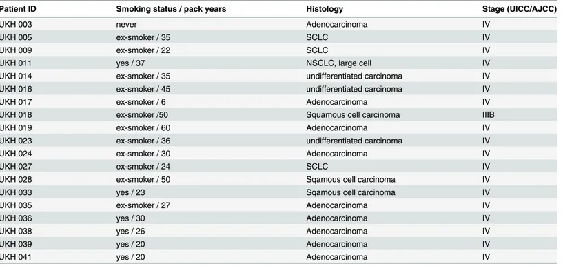
Table 1. Clinical data of patients not responding to the therapy.
Patient ID Smoking status / pack years Histology Stage (UICC/AJCC)
UKH 003 never Adenocarcinoma IV
UKH 005 ex-smoker / 35 SCLC IV
UKH 009 ex-smoker / 22 SCLC IV
UKH 011 yes / 37 NSCLC, large cell IV
UKH 014 ex-smoker / 35 undifferentiated carcinoma IV
UKH 016 ex-smoker / 45 undifferentiated carcinoma IV
UKH 017 ex-smoker / 6 Adenocarcinoma IV
UKH 018 ex-smoker /50 Squamous cell carcinoma IIIB
UKH 019 ex-smoker / 60 Adenocarcinoma IV
UKH 023 ex-smoker / 36 undifferentiated carcinoma IV
UKH 024 ex-smoker / 30 Adenocarcinoma IV
UKH 027 ex-smoker / 24 SCLC IV
UKH 028 ex-smoker / 50 Sqamous cell carcinoma IV
UKH 033 yes / 23 Sqamous cell carcinoma IV
UKH 035 ex-smoker / 27 Adenocarcinoma IV
UKH 036 yes / 30 Adenocarcinoma IV
UKH 038 yes / 26 Adenocarcinoma IV
UKH 039 yes / 20 Adenocarcinoma IV
UKH 041 yes / 20 Adenocarcinoma IV
Clinical data forfirst line patients who did not respond to the therapy. Fifteen of the patients were male and four were female. The median age of this patient group is 63 years.
doi:10.1371/journal.pone.0118195.t001
Table 2. Clinical data offirst-line patients responding to the therapy.
Pat ID Smoking status / pack years Histology Stage (UICC/AJCC)
UKH 007 ex-smoker / 35 SCLC IV
UKH 012 yes / 42 SCLC IV
UKH 015 ex-smoker / 17 Adenocarcinoma IV
UKH 022 ex-smoker / 35 undifferentiated carcinoma IV
UKH 025 ex-smoker / 25 SCLC IV
UKH 026 yes / 40 Adenocarcinoma IIIB
UKH 029 ex-smoker / 29 Adenocarcinoma IV
UKH 030 yes / 33 Adenocarcinoma IV
UKH 031 never Adenocarcinoma IV
UKH 034 ex-smoker / 15 Adenocarcinoma IV
UKH 040 never Adenocarcinoma IV
UKH 042 ex-smoker / 33 Sqamous cell carcinoma IV
Ten of the patients were male and two were female. The median age of this patient group is 61.5 years. Patient UKH 031 demonstrated an EGFR mutation and was treated with TKI.
which is assumed to be the level of technical/biological variance. The clinical data from the 31 pa-tients are summarized in Tables1and2. All patients who clinically responded to the therapy demonstrated a decrease of theirmSHOX2plasma DNA (Fig. 1). In this group of responders a decrease ofmSHOX2DNA was seen in most of the patients already at the time of first blood draw (i.e. day 7–10 after therapy start). The median PMR values of the patients responding to
the therapy were 4.06% at baseline and dropped to 0.62%, 0.12%, 0.05% at blood draws 1, 2 and 3 after therapy start. In contrast, 8/19 of the non-responding patients demonstrated a reduction ofmSHOX2after start of therapy but themSHOX2levels did not change as much as in the re-sponding patients (Fig. 1). In fact the medianmSHOX2values in this group of non-responders were 26.45% at baseline and dropped to 6.86%, 7.78%, 7.96% at blood draws 1, 2 and 3 after ther-apy start. None of the non-responding patients who demonstrated amSHOX2value of1% pre-therapy (ranging from 1.1% to 362%) showed a sustained reduction below 1% PMR. (Fig. 1). This observation holds true also for patient UKH 010 who demonstrated an activating EGFR mutation but did not respond to the TKI therapy (Table 3).
The PMR values are calculated as relative amounts of themSHOX2gene compared to the ß-actin reference gene (ACTB). TheSHOX2locus is frequently amplified in lung tumors
Fig 1. Trend curves for patients responding (black curves) and not responding (gray curves) to the therapy.The patients included in this figure are limited to the ones with a baseline mSHOX2 value of at least 1% PMR. The first blood draw (x = 0) is the point of diagnosis, i.e. before treatment and defines the baseline methylation of SHOX2. For the first eight blood draws Bonferroni corrected p-values from unpaired two sample Wilcox tests are given at the bottom.
doi:10.1371/journal.pone.0118195.g001
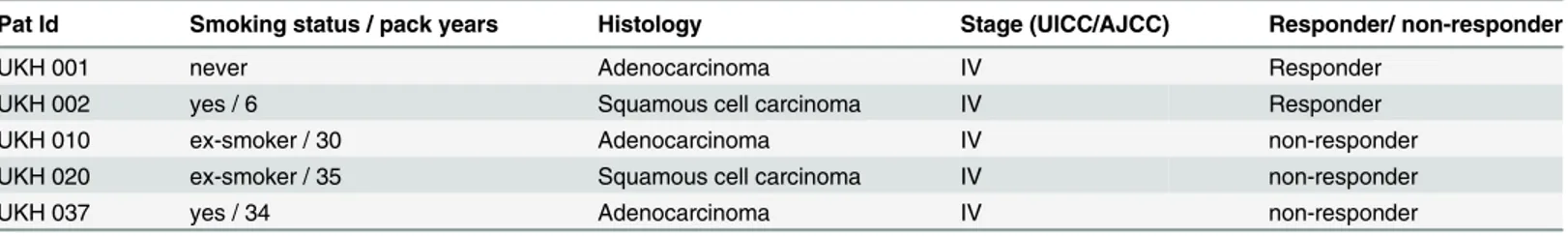
Table 3. Clinical data of second-line patients.
Pat Id Smoking status / pack years Histology Stage (UICC/AJCC) Responder/ non-responder
UKH 001 never Adenocarcinoma IV Responder
UKH 002 yes / 6 Squamous cell carcinoma IV Responder
UKH 010 ex-smoker / 30 Adenocarcinoma IV non-responder
UKH 020 ex-smoker / 35 Squamous cell carcinoma IV non-responder
UKH 037 yes / 34 Adenocarcinoma IV non-responder
In this group were four male and one female patients. The only female patient in this group (UKH 001) did respond to therapy. The median age of this patient group is 64 years. Patient UKH 010 demonstrated an EGFR mutation and was treated with TKI.
(Schneider et al BMC Cancer 2011) and this copy number variation leads tomSHOX2PMRs over 100% in patients with very high levels of free circulating tumor DNA.
The median PMR formSHOX2at baseline was 4.06% for the 12 responders and 26.45% for the 19 non-responders but this difference is not significant. Interestingly, the ROC curve analy-ses for the 24 patients with a PMR1% showed that the baseline valuesmSHOX2did not dis-criminate between responders and non-responders (area under the curve 0.593), while a discrimination of these two patient populations based on values obtained after start of the ther-apy demonstrated a high sensitivity and specificity already starting at blood draw one with an AUC of 0.844 which increased to 1.000 at blood draw 7 (Fig. 2). The classification of the pa-tients as responders and non-responders is based on CT scans as the gold standard which was performed by the local tumor board and was completely independent from the
mSHOX2measurements.
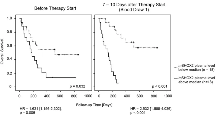
A Kaplan-Meier analysis including the 31 patients (i.e. 24 patients with a PMR1% plus 7 patients with a PMR1%) demonstrated a strong relationship between the survival time and a CT-based assignment of patients into responder and non-responder. The hazard ratio in this
Fig 2. ROC curves for the discrimination of responders from non-responder at different blood draws. Only patients with a baseline PMR1% were included. The first blood draw (time 0) is the point before treatment (= baseline methylation). Blood draws 1 to 8 were taken during the therapy at intervals of 7 to 10 days.
calculation is 18 and the P-value0.00003 (data not shown). Interestingly, there is a similarly strong relationship between the survival time and the PMR ofmSHOX2. When the PMR values from all 36 patients were used for a Kaplan-Meier analysis we could demonstrate that even the plasmamSHOX2baseline levels showed a trend toward significance (Fig. 3). At blood draw one this difference reached statistical significance which might be interpreted as evidence that this marker also has a predictive value.
Discussion
Due to more powerful drugs [23] and the introduction of a targeted therapy for molecularly selected patient subgroups like patients with anEGFRactivation mutation, impressive im-provements in the treatment of lung cancer patients were achieved [24]. Additionally, more effective maintenance regimens have shown beneficial effects for patients with advanced stage NSCLC. There is also a survival advantage for patients who are treated with second-line che-motherapy as compared to best supportive care and clinical trials testing new combinations in the second line setting for refractory disease were initiated [25][26]. In order to select the best treatment options, a rapid, specific and sensitive method for the assessment of a therapy re-sponse is of crucial importance. The standard procedure for advanced stage lung cancer pa-tients after induction therapy is a CT scan to evaluate the tumor response [21]. Apart from the costs, the sensitivity of this imaging technique is not very high and the inter-observer vari-ability in the measurement of the tumor size is prone to misinterpretation of tumor response [27].
In the last few years several biomarkers have been tested for their usefulness as an indica-tor for therapy moniindica-toring. Amongst them were eight immunohistochemical biomarkers, none of which could predict chemotherapy response and survival rate and there was only a
Fig 3. Cox Proportional Hazards and Kaplan-Meier survival analysis of all 36 patients.The median plasmamSHOX2value at baseline was 2.88% PMR, while the median one week after therapy start was 2.16% PMR.
weak correlation between marker level and treatment response [28]. Neuron specific enolase (NSE) was used for monitoring SCLC patients and found to be only useful in patients with an increased pre-treatment level [29]. The levels of lactate dehydrogenase and chromogranin A did not correlate with treatment response [30]. A longitudinal measurement of soluble inter-leukin 2 receptor demonstrated a reduction in serum concentration during a therapy but was not a sign for disease remission [31] and thymidine kinase was unable to discriminate be-tween the various response groups of lung cancer patients [32]. There are some additional biomarkers for therapy monitoring in lung cancer patients likeCYFRA 21–1and nucleosome
levels which might be better suited but none of them is routinely used in the clinic [33] [34] [35] [36]. In addition there is a growing number of genes which are shown to be hyper- or hypomethylated in lung cancer patients which might be useful as biomarkers [37]. The most frequently analyzed genes likep16,DAPK,APC,RASSF1A,MGMT,FHIT,RARß and GSTP1
were applied as a means of detecting lung cancer or as prognostic factor but none of them had been used for therapy monitoring. Interstingly, all of these methylation markers could not only be detected in tissue but were also found in extracellular nucleic acids isolated from bronchial lavage supernatants, sputum, plasma or serum [37]. The aim of this analysis was to answer the question whether a quantitative measurement ofmSHOX2plasma DNA using a real-time PCR is a useful tool to follow advanced stage lung cancer patients receiving chemo/ radio-chemotherapy. Therefore we enrolled all eligible patients who were consecutively ad-mitted to our outpatient department. We considered this approach a proof-of-principle study and for this reason we were more interested in the inclusion of as many patients as pos-sible rather than the establishment of a uniform patient cohort. As a consequence the histo-logic distribution of the patients included in this analysis does not match exactly the figures given in the literature [38].
Our results demonstrate that a quantitative determination of plasmamSHOX2DNA ap-pears to be useful for the monitoring of a treatment response for advanced stage lung cancer patients. The turn-over rate of cell-free DNA is rather high as the half-life of extracellular nu-cleic acids was determined to be less than six hours in an animal model [39]. This correlates well with our observation of a fast and strong decline of plasmamSHOX2DNA in patients re-sponding to the therapy which holds true for the monitoring of NSCLC and SCLC patients alike. An additional advantage of this method is its applicability for patients with a very low pre-therapeuticmSHOX2value. We demonstrated that amSHOX2measurement taken one week after the start of a therapy is able to divide between responders and non-responders with a very high specificity. If this result can be verified in a large study, physicians have the possibil-ity to switch therapies or spare patients from an invalid therapy. Additionally, we used the data from all 36 patients, i.e. including the patients with amSHOX2baseline value of zero and the second-line patients for a Kaplan-Meier survival curve analysis. Interestingly, we demonstrated that the patients with a baseline plasmamSHOX2level below the median had a slightly longer survival time. When this analysis was performed at blood draw one after therapy start this dif-ference between patients responding to the therapy and non-responders was statistically signif-icant with a p0.001. The question whether patients with a very lowmSHOX2baseline value behave differently from patients with a high(er)mSHOX2level and whether it might be possi-ble to define a pre-therapeutic cut-off value which has a predictive meaning has to be answered in a large study which will be conducted in a multicentric approach.
Acknowledgments
We greatly acknowledge the participation of the patients in this study and the help of Dana Reinicke and Ute Völker. The Epi proLung BL Reflex Assays were kindly provided by Epigenomics.
Author Contributions
Conceived and designed the experiments: BS MF. Performed the experiments: JB. Analyzed the data: BS JB DD IB VL MF. Contributed reagents/materials/analysis tools: DD IB. Wrote the paper: BS DD VL MF. Collection of patient samples: IB JB.
References
1. Ferlay J, Steliarova-Foucher E, Lortet-Tieulent J, Rosso S, Coebergh JW, et al. (2013) Cancer incidence and mortality patterns in Europe: estimates for 40 countries in 2012. Eur J Cancer 49: 1374–1403. doi: 10.1016/j.ejca.2012.12.027PMID:23485231
2. Siegel R, Naishadham D, Jemal A (2013) Cancer statistics, 2013. CA Cancer J Clin 63: 11–30. doi:10. 3322/caac.21166PMID:23335087
3. Reck M, Heigener DF, Mok T, Soria JC, Rabe KF (2013) Management of non-small-cell lung cancer: re-cent developments. Lancet 382: 709–719. doi:10.1016/S0140-6736(13)61502-0PMID:23972814 4. Scheff RJ, Schneider BJ (2013) Non-Small-Cell Lung Cancer: Treatment of Late Stage Disease:
Che-motherapeutics and New Frontiers. Semin Intervent Radiol 30: 191–198. doi:10.1055/s-0033-1342961 PMID:24436536
5. Kratochwil C, Haberkorn U, Giesel FL (2010) [PET/CT for diagnostics and therapy stratification of lung cancer]. Radiologe 50: 684–691. doi:10.1007/s00117-009-1960-6PMID:20652216
6. William WN Jr, Pataer A, Kalhor N, Correa AM, Rice DC, et al. (2013) Computed tomography RECIST assessment of histopathologic response and prediction of survival in patients with resectable non-small-cell lung cancer after neoadjuvant chemotherapy. J Thorac Oncol 8: 222–228. doi:10.1097/JTO. 0b013e3182774108PMID:23287849
7. Cho WC (2007) Potentially useful biomarkers for the diagnosis, treatment and prognosis of lung cancer. Biomed Pharmacother 61: 515–519. PMID:17913444
8. Mandel P, Metais P (n.d.) Les acides nucleiques du plasma sanguin chez l’homme. C.R.Acad.Sci.Paris 142, 241–243. 1948.
9. Fleischhacker M, Schmidt B (2007) Circulating nucleic acids (CNAs) and cancer—a survey. Biochim Biophys Acta 1775: 181–232. PMID:17137717
10. Jung K, Fleischhacker M, Rabien A (2010) Cell-free DNA in the blood as a solid tumor biomarker—a critical appraisal of the literature. Clin Chim Acta 411: 1611–1624. doi:10.1016/j.cca.2010.07.032 PMID:20688053
11. Alix-Panabieres C, Schwarzenbach H, Pantel K (2012) Circulating tumor cells and circulating tumor DNA. Annu Rev Med 63: 199–215. doi:10.1146/annurev-med-062310-094219PMID:22053740 12. Smith ZD, Meissner A (2013) DNA methylation: roles in mammalian development. Nat Rev Genet 14:
204–220. doi:10.1038/nrg3354PMID:23400093
13. Gibney ER, Nolan CM (2010) Epigenetics and gene expression. Heredity (Edinb) 105: 4–13.
14. Suva ML, Riggi N, Bernstein BE (2013) Epigenetic reprogramming in cancer. Science 339: 1567–1570. doi:10.1126/science.1230184PMID:23539597
15. Schmidt B, Liebenberg V, Dietrich D, Schlegel T, Kneip C, et al. (2010) SHOX2 DNA methylation is a biomarker for the diagnosis of lung cancer based on bronchial aspirates. BMC Cancer 10: 600. doi:10. 1186/1471-2407-10-600PMID:21047392
16. Kneip C, Schmidt B, Seegebarth A, Weickmann S, Fleischhacker M, et al. (2011) SHOX2 DNA methyl-ation is a biomarker for the diagnosis of lung cancer in plasma. J Thorac Oncol 6: 1632–1638. doi:10. 1097/JTO.0b013e318220ef9aPMID:21694641
17. Dietrich D, Kneip C, Raji O, Liloglou T, Seegebarth A, et al. (2012) Performance evaluation of the DNA methylation biomarker SHOX2 for the aid in diagnosis of lung cancer based on the analysis of bronchial aspirates. Int J Oncol 40: 825–832. doi:10.3892/ijo.2011.1264PMID:22108652
19. Darwiche K, Zarogoulidis P, Baehner K, Welter S, Tetzner R, et al. (2013) Assessment of SHOX2 meth-ylation in EBUS-TBNA specimen improves accuracy in lung cancer staging. Ann Oncol. doi:10.1093/ annonc/mdt547PMID:24436961
20. Dietrich D, Hasinger O, Liebenberg V, Field JK, Kristiansen G, et al. (2012) DNA methylation of the homeobox genes PITX2 and SHOX2 predicts outcome in non-small-cell lung cancer patients. Diagn Mol Pathol 21: 93–104. doi:10.1097/PDM.0b013e318240503bPMID:22555092
21. Goeckenjan G, Sitter H, Thomas M, Branscheid D, Flentje M, et al. (2010) [Prevention, diagnosis, ther-apy, and follow-up of lung cancer]. Pneumologie 64 Suppl 2: e1–164. doi:10.1055/s-0029-1243837 PMID:20217630
22. R- project for Statistical Computing website. Available:http://www.r-project.org/. Accessed 2014 Nov 17.
23. Soria JC, Mauguen A, Reck M, Sandler AB, Saijo N, et al. (2013) Systematic review and meta-analysis of randomised, phase II/III trials adding bevacizumab to platinum-based chemotherapy as first-line treatment in patients with advanced non-small-cell lung cancer. Ann Oncol 24: 20–30. doi:10.1093/ annonc/mds590PMID:23180113
24. Thatcher N, Heighway J (2010) Maintenance and consolidation therapy in patients with unresectable stage III/IV non-small cell lung cancer. Oncologist 15: 1034–1042. doi: 10.1634/theoncologist.2009-0292PMID:20930098
25. Fathi AT, Brahmer JR (2008) Chemotherapy for advanced stage non-small cell lung cancer. Semin Thorac Cardiovasc Surg 20: 210–216. doi:10.1053/j.semtcvs.2008.09.002PMID:19038730 26. Varughese S, Jahangir KS, Simpson CE, Boulmay BC (2012) A paradigm shift in the treatment of
ad-vanced non-small cell lung cancer. Am J Med Sci 344: 147–150. doi:10.1097/MAJ. 0b013e318246e1b8PMID:22317902
27. Erasmus JJ, Gladish GW, Broemeling L, Sabloff BS, Truong MT, et al. (2003) Interobserver and intraobserver variability in measurement of non-small-cell carcinoma lung lesions: implications for as-sessment of tumor response. J Clin Oncol 21: 2574–2582. PMID:12829678
28. Toffart AC, Timsit JF, Couraud S, Merle P, Moro-Sibilot D, et al. (2013) Immunohistochemistry evalua-tion of biomarker expression in non-small cell lung cancer (Pharmacogenoscan study). Lung Cancer. doi:10.1016/j.lungcan.2013.08.029PMID:24636699
29. Splinter TA, Carney DN, Teeling M, Peake MD, Kho GS, et al. (1989) Neuron-specific enolase can be used as the sole guide to treat small-cell lung cancer patients in common clinical practice. J Cancer Res Clin Oncol 115: 400–401. PMID:2547802
30. Johnson PW, Joel SP, Love S, Butcher M, Pandian MR, et al. (1993) Tumour markers for prediction of survival and monitoring of remission in small cell lung cancer. Br J Cancer 67: 760–766. PMID:8385978 31. Brunetti G, Bossi A, Baiardi P, Jedrychowska I, Pozzi U, et al. (1999) Soluble interleukin 2 receptor
(sIL2R) in monitoring advanced lung cancer during chemotherapy. Lung Cancer 23: 1–9. PMID: 10100141
32. Holdenrieder S, von PJ, Duell T, Feldmann K, Raith H, et al. (2010) Clinical relevance of thymidine ki-nase for the diagnosis, therapy monitoring and prognosis of non-operable lung cancer. Anticancer Res 30: 1855–1862. PMID:20592392
33. Holdenrieder S, Stieber P, von PJ, Raith H, Nagel D, et al. (2004) Circulating nucleosomes predict the response to chemotherapy in patients with advanced non-small cell lung cancer. Clin Cancer Res 10: 5981–5987. PMID:15447981
34. Holdenrieder S, von PJ, Dankelmann E, Duell T, Faderl B, et al. (2009) Nucleosomes and CYFRA 21–1 indicate tumor response after one cycle of chemotherapy in recurrent non-small cell lung cancer. Lung Cancer 63: 128–135. doi:10.1016/j.lungcan.2008.05.001PMID:18571761
35. Holdenrieder S, von PJ, Dankelmann E, Duell T, Faderl B, et al. (2008) Nucleosomes, ProGRP, NSE, CYFRA 21–1, and CEA in monitoring first-line chemotherapy of small cell lung cancer. Clin Cancer Res 14: 7813–7821. doi:10.1158/1078-0432.CCR-08-0678PMID:19047109
36. Holdenrieder S, Stieber P, von PJ, Raith H, Nagel D, et al. (2006) Early and specific prediction of the therapeutic efficacy in non-small cell lung cancer patients by nucleosomal DNA and cytokeratin-19 frag-ments. Ann N Y Acad Sci 1075: 244–257. PMID:17108218
37. Fleischhacker M, Dietrich D, Liebenberg V, Field JK, Schmidt B (2013) The role of DNA methylation as biomarkers in the clinical management of lung cancer. Expert Rev Respir Med 7: 363–383. doi:10. 1586/17476348.2013.814397PMID:23964627
38. Hammerschmidt S, Wirtz H (2009) Lung cancer: current diagnosis and treatment. Dtsch Arztebl Int 106: 809–818. doi:10.3238/arztebl.2009.0809PMID:20038979