(Hymenoptera: Braconidae) Infected with
Nosema
sp.
(Microsporidia: Nosematidae): Implications in Integrated
Pest Management Strategy
Nadia Kermani1, Zainal-Abidin Abu Hassan2, Amalina Suhaimi1, Ismail Abuzid1, Noor Farehan Ismail1, Mansour Attia1, Idris Abd Ghani1*
1School of Environmental and Natural Resource Sciences, University National Malaysia, Bangi, Malaysia,2Faculty of Medicine, University Technology MARA UITM, Shah Alam, Malaysia
Abstract
The diamondback moth (DBM)Plutella xylostella(L.) has traditionally been managed using synthetic insecticides. However, the increasing resistance of DBM to insecticides offers an impetus to practice integrated pest management (IPM) strategies by exploiting its natural enemies such as pathogens, parasitoids, and predators. Nevertheless, the interactions between pathogens and parasitoids and/or predators might affect the effectiveness of the parasitoids in regulating the host population. Thus, the parasitism rate of Nosema-infected DBM by Cotesia vestalis (Haliday) (Hym., Braconidae) can be negatively influenced by such interactions. In this study, we investigated the effects ofNosemainfection in DBM on the parasitism performance ofC. vestalis. The results of no-choice test showed thatC. vestalishad a higher parasitism rate on non-infected host larvae than onNosema-treated host larvae. TheC. vestalisindividuals that emerged fromNosema-infected DBM (F1) and their progeny (F2) had smaller pupae, a decreased rate of emergence, lowered fecundity, and a prolonged development period compared to those of the control group. DBM infection byNosemasp. also negatively affected the morphometrics ofC. vestalis. The eggs of femaleC. vestalisthat developed inNosema-infected DBM were larger than those of females that developed in non-infected DBM. These detrimental effects on the F1 and F2 generations ofC. vestalismight severely impact the effectiveness of combining pathogens and parasitoids as parts of an IPM strategy for DBM control.
Citation:Kermani N, Abu Hassan Z-A, Suhaimi A, Abuzid I, Ismail NF, et al. (2014) Parasitism Performance and Fitness ofCotesia vestalis(Hymenoptera: Braconidae) Infected withNosemasp. (Microsporidia: Nosematidae): Implications in Integrated Pest Management Strategy. PLOS ONE 9(6): e100671. doi:10.1371/ journal.pone.0100671
Editor:Nicolas Desneux, French National Institute for Agricultural Research (INRA), France
ReceivedSeptember 9, 2013;AcceptedMay 30, 2014;PublishedJune 26, 2014
Copyright:ß2014 Kermani et al. This is an open-access article distributed under the terms of the Creative Commons Attribution License, which permits unrestricted use, distribution, and reproduction in any medium, provided the original author and source are credited.
Funding:This research was funded by grant No. (02-01-02 -SF0601) and (05-01-02-SF1019) from the Ministry of Sciences, Technology and Innovation, Malaysia (MOSTI) and Research University Grant (OUP): OUP-2012-043, OUP-2013-043. The funders had no role in study design, data collection and analysis, decision to publish, or preparation of the manuscript.
Competing Interests:The authors have declared that no competing interests exist.
* Email: [email protected]
Introduction
The diamondback moth (DBM) Plutella xylostella (L.) (Lep., Plutellidae) is a cosmopolitan pest that causes serious damage to a wide variety of cruciferous and other crops. This pest has been routinely controlled using chemical insecticides; however, the excessive use of these products has caused several concerns related to the development of resistance [1–4], the presence of pesticide residues in the environment and human food [1], and the impact of pesticide applications on populations of non-target organisms [5,6]. Microsporidia have long been considered an attractive alternative to synthetic chemical insecticides for pest management because of their significant environmental and economic advan-tages over chemical insecticides. They are environmentally safe, acceptable, cause no pollution and are also considered important regulators of population dynamics [7]. Recently, many investiga-tions have been focusing on the use of microsporidia as biological control agents of some economically important insect pests [8]. For example,Paranosema locustae(Nosema) was developed as a
long-term agent for grasshopper control in the USA [9] and in different areas of Argentina [10,11].
such as the fat body, Malpighian tubules and reproductive tissues depending on the microsporidian and host species [23–25]. Infective spores from midgut infections are released by cell lysis and passed into the lumen of the gut to the environment through the feces and silk of the infected hosts. The spores can also be released when an infected host dies [26].
The microsporidium Vairimorpha imperfecta (Nosema bombycis Negali) [27] has been reported to cause a major problem in the rearing of DBM and its parasitoids in the laboratory [28]. The effects of biopesticides on beneficial and non-target insects are generally assessed by measuring their acute (mortality) and sublethal toxicities [6,29]. Sublethal physiological effects of biopesticides, such as elongation of developmental period reduc-tion in the number of eggs produced per female, and changes in survival patterns, are considered important information to ensure safeguarding of non-target organisms, including beneficial insects, such as natural enemies [5]. The prevalence of microsporidial diseases in the host population has a negative impact on parasitoid population dynamics in the field such as Muscidifurax raptor (Hymenoptera: Pteromalidae), Trichogramma nubilale (Hymenop-tera: Trichogrammatidae) and Macrocentrus grandii(Hymenoptera: Braconidae) [30–34]. However, the impact of microsporidial diseases on DBM larval parasitoids was not studied.
Cotesia vestalis(Haliday) [ =C. plutellae(Kurdjumov)] (Hymenop-tera: Braconidae) is a specialist primary solitary larval endopar-asitoid that attacks the first and second instars of DBM [35].Cotesia vestalis is found in many regions including Europe, Asia, North America, the Caribbean and Australia [36]. However, it is more prevalent in warm climates, especially in the lowland areas of the tropics [37–39]. It is one of the important biological control agents commonly used in combination with other methods in integrated DBM management programs [2]. This endoparasitoid not only attacks DBM at a high rate [2,40] but also successfully reduces its feeding damage by killing the most damaging larval stage of the pest, the fourth instar.
In this study we investigated the effects ofNosema-infected DBM on: (1) the parasitism rate byC.vestalisand its F2 progeny and (2) the body size, fecundity, and morphometric characteristics ofC. vestalisthat developed onNosema-infected hosts.
Materials and Methods
Plant material
Cabbage plants (Brassica oleraceae var. capitata) were regularly grown to the six- to seven-leaf growth stage in plastic pots containing a mixture of sandy loam soil and peat moss (4:1) in screen houses at the National University of Malaysia (UKM). No pesticides were applied throughout the entire study period.
Insect sources
Healthy DBM larvae of the University Putra Malaysia strain were obtained from the Malaysian Agricultural Research and Development Institute (MARDI). This strain originated from crucifer crops in Serdang, Malaysia. It has been reared for several generations on an artificial diet in the insectary at MARDI. The colony is regularly examined microscopically to confirm that it is free of microsporidian infection. The larvae were brought to the UKM parasitology laboratory and reared on potted cabbage (B. oleracea var. capitata) in screen cages (38 cm626 cm626 cm) as stock culture maintained at 2561uC, 12 h:12 h (light:dark) photoperiod, and 45% to 65% relative humidity. Cotton wool soaked in 10% honey solution was offered to adults as food.
Cocoons (pupae) of C. vestalis were obtained from MARDI, Cameron Highlands, Pahang, Malaysia. The parasitoids were
reared at the UKM parasitology laboratory by using DBM larvae as hosts and cotton wool soaked in 10% aqueous honey solution as a food source for the adults. For oviposition, 200 second instar DBM larvae were introduced on a potted cabbage in a wooden cage (38 cm626 cm626 cm) covered with a fine cloth mesh. The larvae were allowed to feed on cabbage leaves for 1 h in order to produce damage that would attract the parasitoids. A total of 15 mated female parasitoids were released into the cage and left to oviposit for 2 d. Cabbage plants were replaced in the cages as needed for larval development until pupation. Cocoons of C. vestaliswere collected and kept individually in glass vials until adult
emergence. After emergence, each female was caged
(15 cm615 cm68 cm) with a few males until all females were mated and used in the experiment. DBM andC. vestaliscultures were maintained in the laboratory at 2561uC, 45% to 65% RH, and 12 h:12 h (light:dark) photoperiod.
Microsporidia
Nosema sp. spore suspensions were harvested from naturally infected DBM collected regularly from cabbage fields in the area of Cameron Highlands, Pahang. No special permits were required for field collection and sample processing. Collection permission was obtained from the land owners. The field studies did not involve endangered or protected species. DBM were ground, purified, and then centrifuged as described in our previous study [41]. The spores were counted using a haemocytometer under a light microscope with 406magnification following the Cantwell formula [42]. Spore suspensions ranging from 16102to 16105 spores/mL were prepared by diluting the original spore suspen-sions with distilled water before storage at 4uC until further use.
Exposure of DBM larvae toNosema infection
One microliter ofNosemasp. spore suspension at concentrations (treatments) of 16102, 16103, 16104, and 16105spores/mL were spread evenly on the surface of leaf discs (5 mm diameter) of the rape plantBrassica junceaby using the bulb end of a Pasteur pipette. Sterile water was used as a control. Second instar DBM larvae were selected randomly from uninfected colonies and placed into wells of 24-cell plastic culture plates. Each well contained one larva and one leaf disc. The control and treated larvae were kept separately under laboratory conditions mentioned above. After 24 h, 150 second instar larvae were selected from each treatment (concentration) for use in the next bioassay.
Effects ofNosema-infected DBM larvae on parasitism by
C. vestalis
The second instar DBM larvae selected for use in bioassays were randomly placed in wooden cages (30 cm630 cm630 cm)
con-taining potted cabbage plants. Each cage contained 30 second instar larvae and there were five cages for each concentration of Nosemasp. A single mated female parasitoid was introduced into each cage for parasitism and then removed from the cages after 24 h. The DBM larvae were allowed to develop on the cabbage until pupation (cocoon formation). Cabbage plants were added as needed. Before pupation, the larvae emerge from the host and then spin a silky cocoon near the remains of the host. The parasitoid cocoons were collected using forceps, weighed using a digital electronic balance within 24 h and kept in clean ventilated plastic containers until adult emergence. The number of cocoons, non-parasitized larvae (forming DBM pupae), and emerged adult parasitoids for each concentration ofNosemasp. were recorded.
parasitize uninfected DBM larvae to determine the effect ofNosema sp. on the second generation (F2) of C. vestalis. One mated F1 femaleC.vestalisfrom each treatment was randomly selected and introduced into a cage containing a cabbage plant and 30 uninfected second instar DBM larvae. Each cage considers a replication and there were three replications for each treatment. FemaleC.vestalisadults were removed from the cages after 24 h, and new plants were added until cocoons formed or larvae pupated. Data were recorded as described for the first generation.
Effects ofNosema-infected DBM larvae onC. vestalis
juvenile development
Only a single spore concentration was used in these experi-ments. The second instar larvae of DBM were fed with cabbage discs contaminated with 103 spores/mL as previously described. Control larvae were fed cabbage sprayed with distilled water. After 24 h, 50 second instar larvae of DBM from each treatment were divided into 10 groups of five individuals, and then placed in ventilated plastic containers (28 cm619 cm611 cm) with cabbage leaves. One mated female parasitoid was introduced into each of the containers through a small hole in the container’s lid. After insertion of the parasitoid, a cotton pad soaked in honey solution was inserted through the hole as a food source for the parasitoid. Each arena contained a ratio of one female parasitoid to five DBM larvae to ensure that the majority of the hosts were parasitized. The containers were maintained under the laboratory conditions. On the basis of previous observations, we removed the parasitoids 4 h after their introduction in order to avoid superparasitism. Each larva from all treatments was individually placed in a separate 100 mL plastic container lined with moist filter paper and a fresh cabbage leaf as a food source for host larval development. The larvae were maintained under the same laboratory condi-tions. Leaves were replaced with fresh ones as necessary until parasitoid pupation. Cocoons were then removed, placed individ-ually in 300 mL clean plastic containers and subsequently monitored for adult emergence. Cocoons were inspected twice daily until adult emergence (F1). The adult stage was considered to have begun when the adult completely left the cocoon. The following parameters were recorded: number of days from oviposition until the larvae egress from their host (duration of egg to larva stage) and the number of days from the cocoon appearance until adult emergence (duration of pupa stage). Adult female parasitoids that emerged from experimental DBM were kept with males that developed in healthy hosts for 24 h for mating. Mated females were then provided with uninfected second instar DBM larvae to assess the effect of Nosema sp. on the development of second-generation (F2)C. vestalisby using the same experimental procedure as described for the F1 generation.
Effects ofNosema-infected DBM larvae onC. vestalis
fecundity, and egg and body size
TenC. vestalisfemales (1-day-old) from each of the F1 and F2 generations which had previously been treated with 103spores/mL were selected randomly and killed by freezing for 30 min. Each female was placed on a glass microscope slide with 10% saline solution (NaCl 0.85%). The abdomen was opened using a pair of
#0 insect micropins to expose the ovaries under a dissecting microscope (Olympus, Tokyo, Japan). A glass coverslip was placed over the ovaries, and the mature eggs were counted (fecundity) using a stereomicroscope equipped with a lens (SMZ1500; Nikon, Japan) and a camera (Digital Sight DS-5M; Nikon). Mature eggs are transparent and spindle-shaped with a narrow pedicel at one end whereas immature eggs are smaller, opaque and barely
discernible in the distal portions of the ovariole. A total of 25 eggs from each treatment were randomly selected and their length and width were measured.
Body size was assessed by measuring the hind tibia by using an ocular micrometer mounted on a dissecting microscope at 206
magnification (Leica Microsystems, Bannockburn, IL, USA). The length of the hind tibia has been used as an indicator of the body size of other parasitoids [43]. Ten F1 individuals (males and females) from the infected and uninfected groups were deep-frozen on the day of emergence and placed on microscope slides within a droplet of saline solution. The hind tibia length was measured. The body length from the top of the head to the tip of the abdomen, the wing length (distance measured between thoracic attachment points to distal tip of a detached wing), the antenna length, and ovipositor length were also measured.
Statistical Analysis
Data were tested for normality by using the Anderson- Darling test. Transformation of data was not needed because the variances were normal and homogeneous. Parasitism rates were calculated as [% parasitism = (number ofC. vestaliscocoons/total numbers of C. vestaliscocoons +P. xylostella pupae6100]. Regression analysis was used to evaluate any correlation between parasitism rates and Nosemasp. dose. Parasitism rates, percent emergence and cocoon weight were analyzed using one-way analysis of variance (ANOVA), and differences between treatment means were separated using Tukey’s test at a 5% level of significance. The statistical analyses were conducted using MiniTab software version 16. The difference between untreated and treated parasitoid means for developmental time, fecundity, and morphometrics was determined on the basis oft-test (P,0.05).
Results
Effects ofNosemasp. on parasitism, cocoon weight and adult emergence ofC. vestalis
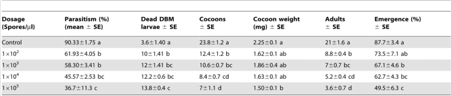
Percentage parasitism differed significantly across different treatments (F= 12.6; df= 4, 20; P,0.05, Table 1). Parasitism of controls was significantly higher than that of larvae treated with Nosemasp. Parasitism ofC. vestalis on DBM larvae infected with Nosema sp. decreased with increasing spore dose. As expected, there was a significant negative correlation between parasitism of Nosema-infected DBM larvae byC. vestalisand spore dose for both F1 and F2 generations (F1: r2=20.81, F(1,23)= 44.03,P,0.05; F2: r2=20.80, F(1,13)= 24.71,P,0.05).
Significantly fewerC. vestalispupae (F= 43.5;df= 4, 20;P,0.05) and adults (F= 60.7;df= 4, 20;P,0.05) developed from the host larvae fed each concentration of Nosema sp. spores than those developed from uninfected DBM larvae. Cotesia vestalis cocoons obtained from the infected had lower weight (F= 2.68;df= 4,120; P,0.05) than those obtained from the uninfected larvae.
Nosemasp. had a significantly negative effect on the percentage of adult emergence (F= 6.98; df= 4, 20;P,0.05) at all concen-trations tested (Table 1). The effect was more pronounced from host larvae fed 16105 spores/mL because less than 50% of the adults emerged from the cocoons compared with (87.7%) those that emerged developed from uninfected larvae.
noted between the Nosema-infected treatments and the control (Table 2).
Effects ofNosema-infected DBM larvae onC. vestalis
juvenile development
In general, the development time of different stages ofC. vestalis from theNosema-infected DBM larvae was significantly prolonged (t=24.31,P,0.05,df= 21 for the egg to larval period andt=2 2.46,P,0.05,df= 28 for the pupal period) compared with that of different stages of C. vestalis from the healthy DBM larvae (Figure 1A).
Similar to the effect ofNosemaon the development time of the F1 generation, the F2 generation ofC.vestalisoriginating from F1 females was longer in their larval (Figure 1,t=22.93;df= 26;P, 0.05) and pupal stages (t=22.17;df= 27,P,0.05) than those of their non-treated counterparts (Figure 1B).
Effects ofNosema-infected DBM larvae onC. vestalis
fecundity and egg and body size
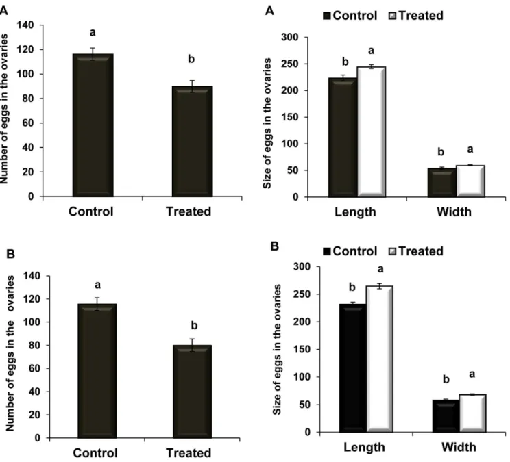
Adult femaleC. vestalishave two ovaries (Figure 2A); each ovary comprises several ovarioles in which the eggs develop. The eggs are spindle shaped and transparent, with a narrow peduncle at the front end (Figure 2B). As shown in Figure (3A), the number of ovarian eggs was lower (89.964.8) in females reared from infected DBM larvae than in those reared from healthy ones (116.265, t= 3.83, df= 17; P,0.05). As shown in Figure (3B), the mean number of eggs produced by F2 females was significantly lower (8065.4) than those produced by the controls (115.665.5,t= 4.65, df= 17;P,0.05).
The F1 parasitoids from treated hosts had significantly larger eggs (Figures 2C and 4A) (length:t=23.22;df= 42;P,0.05; and width:t=22.27;df= 36;P,0.05) than the controls. Similarly, the F2 parasitoids had larger eggs (Figures 2D and 4B) than the
controls (length:t=25.27;df= 46,P,0.05 and width:t=24.77; df= 45;P,0.05).
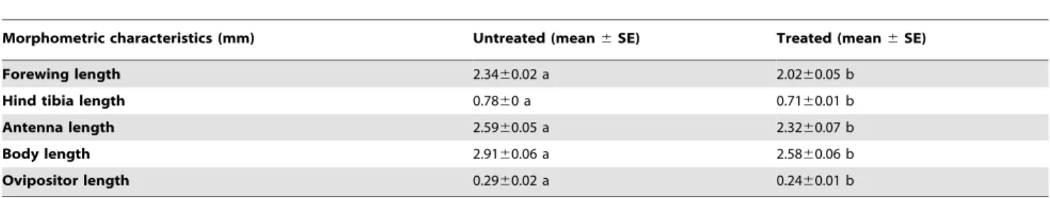
The parasitoids that developed in DBM larvae infected with 103 spores/mLNosemasp. had significantly lower forewing length (t= 6, df= 19;P,0.05), antennal length (t= 3.1, df= 25;P,0.05), body size as estimated by hind tibial length (t= 2.6, df= 25; P,0.05), body length (t= 3.5, df= 27; P,0.05), and ovipositor length (t= 2.6, df= 25; P,0.05) compared to those of the controls (Table 3).
Discussion
Parasitism rates of Nosema-infected DBM larvae by C. vestalis were significantly reduced. The infection also caused a significant decrease in the survival of this braconid parasitoid. As expected, the highest parasitism rates were obtained from the control treatment (90.3% in F1 and 87.9% in F2), followed by those of DBM larvae treated with 102spores/mL (61.9% in F1 and 70.6% in F2). In general, the adult emergence rates of C. vestalisfrom uninfected DBM larvae were significantly higher than those ofC. vestalisfrom infected DBM larvae. Parasitoids develop inside the host body, and microsporidial spores invade the host tissues such as the gut, fat body, and Malpighian tubules [44]. Thus,Nosemasp. is likely to invade the immature parasitoid body as well.Nosema spores can adversely affect the physiological processes of the developing parasitoids, resulting in the formation of small cocoons or inhibition of cocoon formation. Even, if cocoons are formed, the parasitoids might fail to emerge as adults. Similar results were reported for Glyptapanteles liparidis, Microplitis tristis, and Tachinae-phagus zealandicus[45–47]. Such results can be attributed to the lack of nutritional reserves in infected parasitoids that are required for chewing their way out of the cocoon [47]. The last study reported 60% emergence failure among infected parasitoids compared with 44% emergence failure among uninfected parasitoids. Increasing Table 1.Mean % parasitism, cocoons number and weight, number of adults and percentage of emergence of F1C. vestalis
developed within uninfected andNosema-infected DBM larvae.
Dosage (Spores/ml)
Parasitism (%) (mean6SE)
Dead DBM larvae6SE
Cocoons
6SE
Cocoon weight (mg)6SE
Adults
6SE
Emergence (%)
6SE
Control 90.3361.75 a 3.661.40 a 23.861.2 a 2.2560.1 a 2161.6 a 87.763.4 a
16102 61.9364.05 b 1061.41 b 12.461.2 b 1.6260.1 ab 8.860.4 b 73.567.1 ab
16103 58.3063.41 b 1261.41 bc 10.660.7 bc 1.8660.4 ab 760.7 bc 67.164.6 b
16104 45.5762.53 bc 12.260.6 bc 8.460.7 cd 1.6360.1 ab 5.260.4 cd 62.764.3 bc
16105 36.7611.3 c 13.860.4 c 761.1 d 1.5060.1 b 3.660.7 d 49.566.3 c
doi:10.1371/journal.pone.0100671.t001
Table 2.Mean % parasitism, cocoons number and weight, number of adults and percentage of emergence of F2C. vestalis
developed within uninfected DBM larvae.
Dosage (Spores/ml)
Parasitism (%) (mean6SE)
Dead DBM larvae6SE
Cocoons
6SE
Cocoons weight
(mg)6SE Adults6SE
Emergence (%)
6SE
Control 87.9866.07 a 5.360.8 a 21.761.5 a 1.8260.05 a 1960.5 a 87.964.5 a
16102 70.6764.17 ab 12.661.8 b 12.461.8 b 1.5860.06 ab 8.460.3 b 70.6610.3 ab
16103 67.7763.93 ab 15.661.7 bc 9.761.2 bc 1.3660.05 bc 6.361.5 bc 63.867.3 b
16104 44.3613.1 b 13.362.9 b 6.761.5 cd 1.2860.09 c 460.5 cd 62.566.2 bc
16105 44.0466.98 b 20.661.4 c 460.5 d 1.24660.08 c 1.760.3 d 41.164.8 c
Nosemaconcentration can decrease pest populations, but it can also adversely affect the parasitism rates. In the present study, the lowest parasitism rate was reported when 104and 105spores/mL concentrations were used. Some studies also reported that the increased mortality of parasitized Diatraea saccharalis larvae is directly proportional to increased dosage ofNosemaspores and that
high dosages produce heavy infections that prevent the parasitoid Cotesia flavipesfrom completing their development cycle [48].
Many studies on host–parasitoid–microsporidia interactions indicated that parasitoids are affected adversely by the microspo-ridia of their hosts [49]. In the present study,Nosemasp. infection of DBM had negative effects onC. vestalisthat developed within Figure 1. Developmental time ofC. vestalisreared onNosema-infected DBM.(A) Means (6SE) development time (days) of egg-larvae, pupae of F1C. vestalison uninfected andNosema-infectedP. xylostellalarvae. (B) Means (6SE) development time (days) of egg-larvae, pupae of F2C. vestalison uninfectedPllalute xylostellalarvae. Different letters above error bars indicate significant difference (Student’s t-test,P,0.05).
them. For example, the egg to larval development time was prolonged up to 2 d in the F1 generation and 1 d in the F2 generation of infected parasitoids. DBM infection also prolonged the pupation period of both the generations. This result is in agreement with those reported forGlyptapanteles liparidis (Bouche) (Hymenoptera: Braconidae) and Meteorus gyrator (Hymenoptera: Braconidae) [45,50]. However, no association between host microsporidium infection and parasitoid developmental time was observed for parasitoids from the families Encyrtidae and Pteromalidae [47,51].
Our results showed thatNosemainfections affect the fitness ofC. vestalis by reducing fecundity, adult size, and other adult morphometric characteristics. Similar effects have been reported for other parasitoids [48,52–55]. Previous studies have described chronic disease in the adult parasitoidMuscidifurax raptor (Hyme-noptera: Pteromalidae) after the invasion ofNosema muscidifuracisin the midgut epithelium, Malpighian tubules, ovaries, and fat body of both larval and adult parasitoids [56,57]. These studies also reported reduced fecundity in the parasitoids [58]. Lipids are the main energy resource for parasitoids and they play a key role in both survival and reproduction [59]. Many hymenopteran parasitoid species have been reported to be unable to accumulate additional lipids as adults [60,61] due to their lack of de novo lipid synthesis from dietary sugars [62]. Therefore, adult fecundity is completely dependent on the quantity of lipids they acquire from their host during the larval stage [60,61,63]. As stated earlier, invading spores severely damage the fat body of the larvae [64], consequently disturbing host fat metabolism. This limits the amount of lipids available for egg production and affects various physiological functions. Changes in the levels of the analyzed carbohydrates and fatty acids in Vairimorpha-infected Lymantria dispar(Lepidoptera: Lymantridae) larvae were considerably severe to render them nutritionally unfavorable for the development ofG. Liparidis [65]. This phenomenon might partially explain the adverse effects ofNosemainfection of host larvae onC. vestalis.
Cotesia vestalis progeny perform better when supplied with abundant food. However, immatureC. vestalisprogeny feeding on infected DBM larvae need to complete their development within an unhealthy host and might therefore be unable to acquire the same amount of nutrients as those that could be obtained from a healthy host. The reduced quantity of available nutrients significantly affects their weight, size, and body length. Microspo-ridia developing within the hosts can also infect parasitoids developing within the same host [66–69]. Such parasitoid infection has been used to explain the detrimental effects on parasitoids caused by developing in infected hosts. Consequently, parasitoids can attain spores while they are consuming the infected host tissues and might then transmit the spores from the mother to the progeny through infected eggs. Interestingly, C. vestalis egg size significantly increased in the parasitoids (F1) that developed within infected DBM larvae and in infected parasitoids (F2) that developed within uninfected DBM larvae, compared to those of the controls. This increase in egg size might be caused by the changes in the reproductive physiology of the infected parasitoid. Further histological studies on parasitoid tissues are necessary to elucidate the pathway of spores inside a parasitoid’s body and ascertain whether the observed increase in egg size due to the spores or some other physiological disorder that resulted from Nosemainfection.
deleterious effects of Nosema sp. infection on the parasitism, fecundity, and morphometric characteristics of the parasitoid F2 generation. Vertical transmission of microsporidia from mother to progeny and the possibility of its transmission via a contaminated ovipositor from infected to uninfected hosts in other insects have been discussed in detail [72–74]. Our previous study [41] showed that Nosema sp. effectively suppresses DBM populations in the laboratory. The current study revealed thatC. vestalison its own kill more DBM compared with combined with microsporidia. Sublethal effects onC.vestalisoccur when DBM larvae are infected withNosema sp. Such effects might eventually lead to parasitoid population collapse. So, it seems that it isn’t useful to applyNosema as an extra control method whenC.vestalisis already present.
The use of parasitoids andNosemasp. to control DBM might not be effective in IPM strategies. Assessment of the risk to non-target organisms, such as pollinators and predators, is needed in order to further understand the interspecific and intergenerationNosemasp. transmission mechanisms. Detailed studies are also required because DBM populations in the field might be naturally infected by a variety of other pathogens (i.e., viruses, bacteria, and fungi) or artificially affected by pesticides and/or genetically modified-derived toxins.
Acknowledgments
The authors wish to thank MARDI for providing DBM egg masses and the cocoons ofCotesia vestalisand all lab assistants in the Entomology, Histology (MPG2) and Parasitology laboratories at UKM. We would like to thank Editage for providing editorial assistance.
Figure 3. Egg production by control andNosema-infected C. vestalis.Mean (6SE) number of eggs produced byC.vestalisfemales (F1) emerged from infected DBM larvae (A) and infected C. vestalis females (F2) emerged from healthy DBM larvae (B). Different letters above error bars indicate significant difference (Student’s t-test, P, 0.05).
doi:10.1371/journal.pone.0100671.g003
Figure 4. Effect ofNosemainfection on theC. vestalisegg size.
Mean (6) SE egg size produced byC.vestalis females (F1) emerged from infected DBM larvae (A) and infected C. vestalis females (F2) emerged from healthy DBM larvae (B). Different letters above error bars indicate significant difference (Student’s t-test,P,0.05).
Author Contributions
Conceived and designed the experiments: NK ZA IA NI MA IAG. Performed the experiments: NK IAG. Analyzed the data: NK IA NI MA
IAG. Contributed reagents/materials/analysis tools: NK ZA IAG. Wrote the paper: NK AS IAG.
References
1. Tabashnik BE, Cushing NL, Johnson MW (1990) Field development of resistance toBacillus thuringiensisin diamondback moth (Lepidoptera: Plutellidae). Journal of Economic Entomology 83: 1651–1676.
2. Talekar NS, Shelton AM (1993) Biology, ecology, and management of the diamondback moth. Annual Review of Entomology 38, 275–301.
3. Zhao JZ, Li YX, Collins HL, Gusukuma-Minuto L, Mau RFL (2002) Monitoring and characterization of diamondback moth (Lepidoptera: Plutelli-dae) resistance to spinosad. Journal of Economic Entomology 95: 430–436. 4. Sayyed AH, Omar D, Wright DJ (2004) Genetics of spinosad resistance in a
multi-resistant field selected population ofPlutella xylostella. Pest Management Science 60: 827–832.
5. Desneux N, Decourtye A, Delpuech JM (2007) The sublethal effects of pesticides on beneficial arthropods. Annual Revision of Entomology 52: 81–106. 6. Biondi A, Desneux N, Siscaro G, Zappala` L (2012) Using organic-certified
rather than synthetic pesticides may not be safer for biological control agents: Selectivity and side effects of 14 pesticides on the predator Orius laevigatus. Chemosphere 87: 803–812.
7. Linde A, Goertz D, Golldack J (2000) Evoluation of the potential of a microsporidian parasite for the biological control ofLymantria disparL. IOBC wprs Bull. 23: 285–290.
8. Mewis I, Kleespies RG, Ulrichs C, Schnitzler WH (2003) First detection of a microsporidium in the crucifer pestHellula undalis(Lepidoptera: Pyralidae)–a possible control agent? Biological Control 26: 202–208.
9. Lange CE (2002) El desarrollo deNosemalocustaeCanning (Protozoa: Microspo-ridia) para el control biolo´gico de tucuras (Orthoptera: Acridoidea) ylas consecuencias de su utilizacio´n en Argentina. Revista de la Sociedad Entomolo´gica Argentina 61: 1–9.
10. Lange CE, Azzaro FG (2008) New case of long-term persistence ofParanosema locustae (Microsporidia) in melanopline grasshoppers (Orthoptera: Acrididae: Melanoplinae) of Argentina. Journal of Invertebrate Pathology 99: 357–359. 11. Lange CE (2010)Paranosema locustae(Microsporidia) in grasshoppers (Orthoptera:
Acridoidea) of Argentina: field host range expanded. Biocontrol Science and Technology 20: 1047–1054.
12. Solter LF, Becnel JJ (2007) Entomopathogenic microsporidia In: Lacey LA, Kaya K (eds.) Field Manual of Techniques in Invertebrate Pathology. Application and evaluation of pathogens for control of insects and other invertebrate pests. 2ndedn, Springer, 199–221.
13. Katinka MD, Duprat S, Cornillot E, Metenier G, Thomarat, et al. (2001) Genome sequence and gene compaction of the eukaryote parasiteEncephalitozoon cuniculi. Nature 414: 450–453.
14. Keeling PJ (2003) Congruent evidence from alpha-tubulin and betatubulin gene phylogenies for a zygomycete origin of microsporidia. Fungal Genetics and Biology 38: 298–309.
15. Didier ES, Didier PJ, Snowden KF, Shadduck JA (2000). Microsporidiosis in mammals. Microbes and Infection 2: 709–720.
16. Becnel JJ, Andreadis TG (1999) Microsporidia in insects. In: Wittner M, Weiss LM, editors. The Microsporidia and Microsporidiosis. ASM Press Washington DC, USA.
17. Tsai SJ, Lo CF, Soichi Y, Wang CH (2003) The characterization of microsporidian isolates (Nosematidae:Nosema) from five important lepidopteran pests in Taiwan. Journal of Invertebrate Pathology 83: 51259.
18. Batoto TC, Zainal ABH, Idris AB (2010)Nosema bombycisinfection in highland population of diamondback moth (DBM),Plutella xylostellaand its parasitoids, Diadegma semiclausumandCotesia plutellae. Malaysian Applied Biology 39(1): 1–6. 19. Idris AB Sajap AS (2003) Prevalence ofNosema bombycisin field population of the
diamondback moth. International Journal of Pest Management 49: 71–73.
20. Haque MA, Canning EU Wright DJ (1999) Entomopathogenicity ofVairimorpha sp. (Microsporidia) in the diamondback moth, Plutella xylostella(Lepidoptera: Plutellidae). Bulletin of Entomology Research 89(2): 147–152.
21. Idris AB, Sajap AS (2001)Nosemadisease of diamondback moth,Plutella xylostella (L.), in Malaysia. Journal of Biological Sciences 1: 905–907.
22. Idris AB Grafius EJ (1999) Sources of possible inoculum for horizontal transmission of Nosema bombycisin diamondback moth, Plutella xylostella (L.). Sains Malaysiana 28: 39–47.
23. Kurtz J, Nahif AA, Sauer KP (2000) Phagocytosis of Vairimorpha sp (Microsporida, Nosematidae) spores by Plutella xylostella and Panorpa vulgaris hemocytes. Journal of Invertebrate Pathology 75: 237–239.
24. Maddox JV, Baker MD, Jeffords MR, Kuras M, Linde A, et al. (1999)Nosema portugal, n.sp., isolated from gypsy moths (Lymantria dispar L.) collected in Portugal. Journal of Invertebrate Pathology 73: 1–14.
25. Solter LF, Becnel JJ (2000) Entomopathogenic microsporidia. In Field Manual of Techniques for the Evaluation of Entomopathogens (ed. Lacey, L. A. and Kaya, H.), 231–254. Kluwer Academic Publishers, Dordrecht, The Netherlands. 26. Federici BA, Maddox JV (1996) Host Specificity in Microbe-Insect Interactions: Insect control by bacterial, fungal, and viral pathogens. BioScience 46: 410–421. 27. Canning EU, Curry A, Cheney SA, Lafranchi-Tristem NJ, Kawakami, et al. (1999)Nosema tyriae n. sp. andNosema sp., microsporidian parasites of Cinnabar mothTyria jacabaeae. Journal of Invertebrate Pathology 74: 29–38.
28. Idris AB, Zainal-Abidin BAH, Norhayati AM (1997) Detection of Nosema bombycis(Naegeli) in Diamondback moth using Giemsa Stain. Malaysia Applied Biology 26(1): 105–107.
29. Biondi A, Zappala` L, Stark JD, Desneux N (2013) Do Biopesticides Affect the Demographic Traits of a Parasitoid Wasp and Its Biocontrol Services through Sublethal Effects? PLoS ONE 8(9): e76548.
30. Geden CJ, Long SJ, Rutz DA, Becnel JJ (1995)Nosemadisease of the parasitoid Muscidifurax raptor (Hymenoptera: Pteromalidae): prevalence, patterns of transmission, management, and impact. Biological Control 5: 607–614. 31. Zchori-Fein E, Geden CJ, Rutz DA (1992) Microsporidioses ofMuscidifurax raptor
(Hymenoptera: Pteromalidae) and other pteromalid parasitoids of muscoid flies. J Journal of Invertebrate Pathology 60: 292–298.
32. Sajap AS, Lewis LC (1988) Effects of the microsporidium Nosema pyrausta (Microsporida: Nosematidae) on the egg parasitoid, Trichogramma nubilale (Hymenoptera: Trichogrammatidae). Journal of Invertebrate Pathology 52(2): 294–300.
33. Orr DB, Lewis LC, Obrycki JJ (1994) Behavior and survival in corn plants of Ostrinianubilalis(Lepidoptera: Pyralidae) larvae when infected withNosema pyrausta (Microspora: Nosematidae) and parasitized byMacrocentrus grandii(Hymenoptera: Braconidae). Environmental Entomology 23: 1020–1024.
34. Cossentine JE, Lewis LC (1987) Development ofMacrocentrus grandiiGoidanich within microsporidian-infectedOstrinia nubilalis(Hu¨ bner) host larvae. Canadian Journal of Zoology 65: 2532–2535.
35. Sarfraz M, Keddie BA (2005) Conserving the efficacy of insecticides against Plutella. xylostella(L.) (Lep., Plutellidae). Journal of Applied Entomology 129(3): 149–157.
36. Furlong MJ, Wright DJ, Dosdall LM (2013) Diamondback moth ecology and management: problems, progress and prospects. Annual Revision of Entomol-ogy. 58: 517–541.
37. Endersby NM, Ridland PM, eds. (2004) Proceedings of the Fourth International Workshop on the Management of Diamondback Moth and Other Crucifer Pests.Melbourne, Aust.: The Regional Inst.
38. Shelton AM, Collins HL, Zhang YJ, Wu QJ, eds. (2008)Proceedings of the Fifth International Workshop on the Management of Diamondback Moth and Other Crucifer Pests. Beijing, China: China Agric. Sci. Technol. Press.
Table 3.Morphometric (mean) characteristics ofCotesia vestalisdeveloped within uninfected andNosema-infectedP. xylostella
larvae.
Morphometric characteristics (mm) Untreated (mean6SE) Treated (mean6SE)
Forewing length 2.3460.02 a 2.0260.05 b
Hind tibia length 0.7860 a 0.7160.01 b
Antenna length 2.5960.05 a 2.3260.07 b
Body length 2.9160.06 a 2.5860.06 b
Ovipositor length 0.2960.02 a 0.2460.01 b
39. Srinivasan R, Shelton AM, Collins HL, eds. (2011) Proceedings of the Sixth International Workshop on Management of the Diamondback Moth and Other Crucifer Insect Pests.Shanhua, Taiwan: AVRDC – The World Veg. Cent. 321.
40. Verkerk RHJ, Wright DJ (1996) Multitrophic Interactions and Management of the Diamondback Moth: a Review. Bulletin of Entomological Research 86: 205– 216.
41. Kermani N, Abu-hassan Z-A, Dieng H, Ismail NF, Attia M, et al. (2013) Pathogenicity ofNosemasp. (Microsporidia) in the Diamondback Moth,Plutella xylostella (Lepidoptera: Plutellidae). PLoS ONE 8(5): e62884. doi:10.1371/ journal.pone.0062884.
42. Cantwell GE (1970) Standard and Methods for Counting Nosema spores. American Bee. Journal 110: 222–223.
43. Riddick EW (2006) Egg load and body size of lab-cultured.Cotesia marginiventris. BioControl 51: 603–610.
44. Remadevi OK, Sasidharan TO, Bhattacharya J, Vossbrinck CR, Rajan PD (2010) Some pathological effects and transmission potential of a microsporidian isolate (Nosema sp.) from the teak defoliator Hyblaea puera (Lepidoptera: Hyblaeidae). International Journal of Tropical Insect Science 30(3): 138–144. 45. Hoch G, Schopf A, Maddox JV (2000) Interactions between an
entomopatho-genic microsporidium and the endoparasitoidGlyptapanteles liparidiswithin their host, the gypsy moth larva. Journal of Invertebrate Pathology 75: 59–68. 46. Elzinga JA, Harvey JA, Biere A (2003) The effects of host weight at parasitism on
fitness correlates of the gregarious koinobiont parasitoidMicroplitis tristisand consequences for food consumption by its host,Hadena bicruris. Entomologia Experimentalis et Applicata 108: 95–106.
47. Geden CJ, De Almeida MAF, Do Pardo AP (2003) Effects ofNosemadisease on fitness of the parasitoidTachinaephagus zealandicus (Hymenoptera: Encyrtidae). Biological Control. 32(5): 1139–1145.
48. Simoes RA, Reis LG, Bento JMS, Solter LF, Delalibera JrI (2012) Biological and behavioral parameters of the parasitoidCotesia flavipes(Hymenoptera: Braconi-dae) are altered by the pathogenNosema sp. (Microsporidia: Nosematidae). Biological Control 63: 164–171.
49. Brooks WM (1973) Protozoa: host-parasite-pathogen interrelationships. Misc. Pub. Entomol. Soc. Am. 9: 105–111.
50. Down RE, Smethurst F, Bell HA, Edwards JP (2005) Interactions between the solitary endoparasitoid,Meteorus gyrator(Hymenoptera: Braconidae) and its host, Lacanobia oleracea(Lepidoptera: Noctuidae), infected with the entomopathogenic microsporidium,Vairimorpha necatrix (Microspora: Microsporidia). Bulletin of Entomological Research 95(2): 133–144.
51. Futerman PH, Layen SJ, Kotzen ML, Franzen C, Kraaijeveld AR (2006) Fitness effects and transmission routes of a microsporidian parasite infecting Drosophila and its parasitoids. Parasitology 132: 479–492.
52. Agnew P, Bedhomme S, Haussy C, Michalakis Y (1999) Age and size at maturity of the mosquitoCulex pipiens infected by the microsporidian parasiteVavraia culicis. Proceedings of the Royal Society of London 266: 947–952.
53. Schuld M Madel G, Schmuc R (1999) Impact ofVairimorphasp. (Microsporidia: Burnellidae) on Trichogramma chilonis (Hymenoptera: Trichgrammatidae), a hymenopteran parasitoid of the cabbage moth,Plutella xylostella(Lepidoptera: Yponomeutidae). Journal of Invertebrate Pathology 74: 120–126.
54. Idris AB, Sajap AS (2001)Nosemadisease of diamondback moth,Plutella xylostella (L.), in Malaysia. Online Journal of Biological Sciences, 1(10): 905–907. 55. Boheene CK, Geden CJ, Becnel JJ (2003) Development of
microsporidia-infectedMuscidifurax raptor(Hymenoptera: Pteromalidae) at different tempera-tures. Biological Control 26: 1–7.
56. Becnel JJ, Geden CG (1994) Description of a new species of microsporidia from Muscidifurax raptor(Hymenoptera: Pteromalidae), a pupal parasitoid of muscoid flies. Journal of Eukaryotic Microbiology 41: 236–243.
57. Geden CJ, Long SJ, Rutz DA, Becnel JJ (1995)Nosemadisease of the parasitoid Muscidifurax raptor (Hymenoptera: Pteromalidae): prevalence, patterns of transmission, management, and impact. Biological Control. 5: 607–614. 58. Mitchell MJ, Cali A (1994) Vairimorpha necatrix(Microspordia: Burenellidae)
affects growth and development ofHeliothis zea(Lepidoptera: Noctuidae) raised at various temperatures. Journal of Economic Entomology 87: 933–940. 59. Ellers J (1995) Fat and eggs: an alternative method to measure the trade-off
between survival and reproduction in insect parasitoids. Netherlands Journal of Zoology 3: 227–235.
60. Visser B, Ellers J (2008) Lack of lipogenesis in parasitoids, A review of physiological mechanisms and evolutionary implications. Journal of Insect Physiology 54: 1315–1322.
61. Visser B, Le Lann C, den Blanken FJ, Harvey JA, van Alphen JJM, et al (2010) Loss of lipid synthesis as an evolutionary consequence of a parasitic lifestyle. Proceedings of the National Academy of Sciences of the USA. 107: 8677–8682. 62. Visser B, Roelofs D, Hahn DA, Teal PEA, Marie¨n J, et al (2012) Transcriptional changes associated with lack of lipid synthesis in parasitoids. Genome biology and evolution 4: 864–874.
63. Casas J, Pincebourde S, Mandon N, Vannier F, Poujol R, et al (2005) Lifetime nutrient dynamics reveal simultaneous capital and income breeding in a parasitoid. Ecology 86: 545–54.
64. Henn MW, Solter LF (2000) Food utilization values of gypsy mothLymantria dispar (Lepidoptera: Lymantriidae) larvae infected with the microsporidium Vairimorphasp. (Microsporidia: Burenellidae). Journal of Invertebrate Pathology 76: 263–269.
65. Hoch G, Schafellner C, Henn MW, Schopf A (2002) Alterations in carbohydrate and fatty acid levels of Lymantria disparlarvae caused by a microsporidian infection and potential adverse effects on a co-occurring endoparasitoid Glyptapanteles liparidis. Archives of Insect Biochemistry and Physiology 50: 109– 120.
66. Andreadis TG (1980)Nosema pyrautainfection inMacrocentrus grandii, a braconid parasite of the European corn borer,Ostrinia nubilalis. Journal of Invertebrate Pathology 35: 229–233.
67. Brooks WM (1993) Host–parasitoid–pathogen interactions. 231–272 in Beckage, N.E., Thompson, S.N. & Federici, B.A. (Eds) Parasites and pathogens of insects Volume 2: Pathogens. San Diego, Academic Press.
68. Schuld M, Madel G, Schmuck R (1999) Impact ofVairimorphasp. (Microspo-ridia: Burenellidae) onTrichogramma chilonis(Hymenoptera, Trichogrammatidae), a hymenopteran parasitoid of the cabbage moth,Plutella xylostella(Lepidoptera: Yponomeutidae). Journal of Invertebrate Pathology 74: 120–126.
69. Idris AB, Sajap AS (2003) Prevalence ofNosema bombycis in Malaysia field populations of diamondback moth,Plutella xylostella. International Journal of Pest Management, 49(1): 71–73.
70. Doury G, Bigot Y, Periguet G (1997) Physiological and biochemical analysis of factors in the female venom gland and larval salivary secretions of the ectoparasitoid waspEupelmus orientalis. Journal of Insect Physiology 43: 69–81. 71. Parkinson N, Richards EH, Conyers C, Smith I, Edwards JP (2002) Analysis of
venom consitituents from the parasitoid waspPimpla hypochondriacaand cloning of cDNA encoding a venom protein. Insect Biochemistry and Molecular Biology 32: 729–735.
72. Becnel JJ, Andreadis TG (1999) Microsporidia in Insects. In: Wittner, M., Weiss, L.M. (Eds.), The Microsporidia and Microsporidiosis. ASM Press, Washington DC, 447–501.
73. Wittner M, Weiss LM (1999) The Microsporidia and Microsporidiosis. ASM Press, Washington DC, p. 553.