Diamondback Moth,
Plutella xylostella
(Lepidoptera:
Plutellidae)
Nadia Kermani1, Zainal-Abidin Abu-hassan2, Hamady Dieng3, Noor Farehan Ismail1, Mansour Attia1, Idris Abd Ghani1*
1School of Environmental and Natural Resource Sciences, University Kebangsaan Malaysia, Bangi, Selangor, Malaysia,2Faculty of Medicine, University Technology MARA, Shah Alam, Selangor, Malaysia,3School of Biological Sciences, Universiti Sains Malaysia, Penang, Malaysia
Abstract
Biological control using pathogenic microsporidia could be an alternative to chemical control of the diamondback moth (DBM)Plutella xylostella (Lepidoptera: Plutellidae). The microsporidium Nosema bombycis (NB) is one of the numerous pathogens that can be used in the Integrated Pest Management (IPM) of DBM. However, its pathogenicity or effectiveness can be influenced by various factors, particularly temperature. This study was therefore conducted to investigate the effect of temperature on NB infection of DBM larvae. Second-instar larvae at different doses (spore concentration: 0, 16102,16103,16104, and 16105) at 15u, 20u, 25u, 30uand 35uC and a relative humidity(RH) of 65% and light dark cycle (L:D)
of 12:12. Larval mortality was recorded at 24 h intervals until the larvae had either died or pupated. The results showed that the spore concentration had a significant negative effect on larval survival at all temperatures, although this effect was more pronounced (92%) at 35uC compared with that at 20 and 30uC (.50%) and 25uC (26%). Histological observations showed thatNosemapreferentially infected the adipose tissue and epithelial cells of the midgut, resulting in marked vacuolization of the cytoplasm. These findings suggest thatNosemadamaged the midgut epithelial cells. Our results suggest thatNosema had a direct adverse effect on DBM, and could be utilized as an important biopesticide alternative to chemical insecticides in IPM.
Citation:Kermani N, Abu-hassan Z-A, Dieng H, Ismail NF, Attia M, et al. (2013) Pathogenicity ofNosemasp. (Microsporidia) in the Diamondback Moth,Plutella xylostella(Lepidoptera: Plutellidae). PLoS ONE 8(5): e62884. doi:10.1371/journal.pone.0062884
Editor:Oscar Zaragoza, Instituto de Salud Carlos III, Spain
ReceivedDecember 19, 2012;AcceptedMarch 26, 2013;PublishedMay 13, 2013
Copyright:ß2013 Kermani et al. This is an open-access article distributed under the terms of the Creative Commons Attribution License, which permits unrestricted use, distribution, and reproduction in any medium, provided the original author and source are credited.
Funding:This research was funded by grant No. (02-01-02 -SF0601) and (05-01-02-SF1019) from the Ministry of Sciences, Technology and Innovation, Malaysia (MOSTI) and Research University Grant (OUP): OUP-2012-043, OUP-2013-043. The funders had no role in study design, data collection and analysis, decision to publish, or preparation of the manuscript.
Competing Interests:The authors have declared that no competing interests exist.
* E-mail: idrisgh@ukm.my
Introduction
The diamondback moth (DBM)Plutella xylostella L. (Lepidop-tera: Plutellidae) causes considerable economic losses worldwide to brassicaceous crops, and occasionally to other crops. Control of this pest is usually achieved through the application of synthetic insecticides that is estimated to cost more than US$1 billion/ annum to control. Management costs and crop losses caused by
DBM account for US$4–US$5 billion [1]. The high cost,
environmental contamination, development of resistance to chemicals, and pest resurgence [2,3] associated with the current DBM control practices have encouraged the search for alternatives that are more environment friendly. Microbial control is an environmentally sound and valuable option to control this pest. In Malaysia,Nosema bombycisNegali is one of the several pathogens of DBM in the field [4]. DBM mortality is higher in younger instars (first and second instars) than in the older instars. Further, even at low concentrations, infection is remarkably higher for both larvae and pupae in highlands than in lowlands [4].
The effect of temperature on the biology ofNosemaneeds to be investigated because it is one of the most important ecological factors for the development of insect populations. Therefore, this study investigated the effects ofNosemaspore concentration on the
different stages of DBM reared at different temperatures. Establishing a correlation between temperature and pathogenicity of Nosema infection would be beneficial in determining whether
Nosemacan be applied as a DBM controlling agent. The optimal temperature at which this pathogen might be more effective in controlling the pest was studied. This information might help in determining whether this pathogen could be used in integrated pest management (IPM) and whether the amount of pesticide required could be reduced considering that Nosema-infected populations are more susceptible to the toxicity of the insecticides. In insects, the midgut is a dynamic tissue of the alimentary canal that acts as the route of digestion and allows absorption of digested food. Thus, we studied the effect of Nosema on this active organ. The results are also expected to provide useful information on the histopathology effects on larvae caused byNosema.
Materials and Methods
Diamondback Moth
throughout this study was reared on potted cabbage, Brassica oleracea var-capitata, in screen cages (38 cm626 cm626 cm) and
maintained at 25uC65uC, with a photoperiod of 12 L: 12 D, and 60–80% relative humidity (RH). A 10% honey solution was offered as food to the adults, which had been reared over several generations in a laboratory before the experiments.
Nosema sp. Spore Production
Nosema sp. spore suspensions were harvested from naturally infected DBM collected from a cabbage growing area in the Cameron Highlands, Pahang. No specific permits were required for the described field studies, and permission was provided by the landowners. However, the field studies did not involve endangered or protected species.DBM were homogenized in sterile water using a sterile mortar and pestle. The homogenate was partially purified by filtration through a nylon mesh cloth to remove tissue debris and centrifuged at 3000 rpm for 10 min. The supernatant was discarded, and the spore pellet was resuspended to 10 mL using sterile distilled water, and then the suspension was re-centrifuged. This procedure was repeated 3 times [5]. Next, 10mL of the final spore solution was pipetted and poured into a hemocytometer for spore counting. The spores were counted under a light microscope at640 magnifications using the Cantwell formula [6]. Spore suspensions, ranging from 16102 to 16105, were prepared by diluting with distilled water, and then stored at 4uC until further use.
Pathogenic Effect of Nosema onP. xylostellaLarvae
In all experiments, second- instar larvae were fed 5 mm wide leaf discs of rape, Brassica juncea plants that were treated with
Nosema sp. spore suspension at concentrations (treatments) of 16102, 16103, 16104, and 16105spores per larva. Sterile water was used as a control. Spore suspensions were spread evenly on the surface of the leaf discs using the bulb end of a Pasteur pipette. The larvae were selected randomly from the uninfected colonies and then placed in 24-well plastic culture plates, with one larva per well. The plates were placed in growth chambers that were maintained at 15, 20, 25, 30, or 35uC. All experiments were performed under similar RH (65% to 70%) and photoperiod (12:12 h, light: dark) conditions. After 24 h, 10 randomly selected
larvae were transferred to a plastic container
(28 cm616 cm610 cm) containing moistened filter paper and untreated cabbage leaves, and the larvae were reared under different growth chamber conditions (15, 20, 25, 30, or 35uC, temperature; 65% to 70%, RH; and 12:12 h, L:D). The cut end of the cabbage leaf was covered with water-soaked cotton wool in aluminum foil to maintain the freshness of the leaf. The lids of the containers were provided with nylon mesh-covered windows for ventilation. The treatments were replicated 5 times (n = 50). Food was renewed daily, and larval mortality was recorded every 24 h until all larvae had died or pupated. Healthy pupae were counted and placed individually in 2- cm-wide and 5–cm-deep small vials with cover. The newly emerged adults were used in the next experiments.
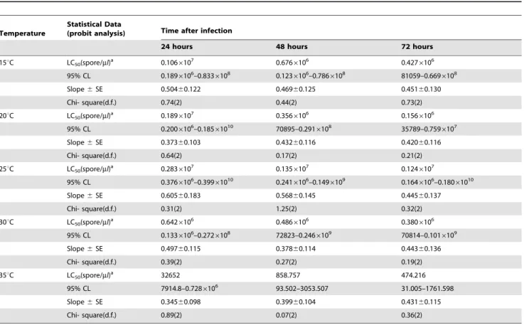
Table 1.Results of probit analysis on the effect ofNosema sp. infection onPlutella xylostellaLC50 stands for Lethal Dose 50 (concentration in water having 50% chance of causing death); F symbolizes F-statistic; d.f. stands for degrees of freedom.
Temperature
Statistical Data
(probit analysis) Time after infection
24 hours 48 hours 72 hours
15uC LC50(spore/ml)a 0.106
6107 0.6766106 0.4276106
95% CL 0.1896106–0.8336108 0.1236106–0.7866108 81059–0.6696108
Slope6SE 0.50460.122 0.46960.125 0.45160.130
Chi- square(d.f.) 0.74(2) 0.44(2) 0.73(2)
20uC LC50(spore/ml)a 0.189
6107 0.3566106 0.1566106
95% CL 0.2006106–0.18561010 70895–0.2916108 35789–0.7596107
Slope6SE 0.37360.103 0.43260.116 0.42060.116
Chi- square(d.f.) 0.64(2) 0.17(2) 0.21(2)
25uC LC50(spore/ml)a 0.283
6107 0.1356107 0.1246107
95% CL 0.3766106–0.39961010 0.2416106–0.1496109 0.1646106–0.18061010
Slope6SE 0.60560.183 0.56860.145 0.44560.137
Chi- square(d.f.) 0.31(2) 1.25(2) 0.32(2)
30uC LC50(spore/ml)a 0.6426106 0.4866106 0.3806106
95% CL 0.1336106–0.2726108 72823–0.2466109 70814–0.1016109
Slope6SE 0.49760.115 0.37860.114 0.44360.136
Chi- square(d.f.) 0.39(2) 0.27(2) 0.19(2)
35uC LC50(spore/ml)a 32652 858.757 474.216
95% CL 7914.8–0.7286106 93.502–3053.507 31.005–1761.598
Slope6SE 0.34560.098 0.39960.104 0.43160.115
Chi- square(d.f.) 0.89(2) 0.07(2) 0.36(2)
a n = 250.
doi:10.1371/journal.pone.0062884.t001
Effect of Nosema on the Number of Emerged Adults and Egg Production of DBM
Adult males and females obtained from the study described in the previous section were counted. Twenty-four hours after adult eclosion, 2 females and 2 males from each treatment were placed in plastic containers (10 cm610 cm68 cm) containing a cabbage
leaf for oviposition and a cotton ball soaked with 10% sugar solution as food. The leaves were checked daily for oviposition, and the number of eggs was recorded until no more eggs were laid.
The experiments had a completely randomized design. The experiments were replicated 5 times spore concentration and per temperature. After the larvae hatched, 10 larvae per treatment
were randomly sampled, placed in plastic containers
(28 cm616 cm610 cm), and reared on fresh cabbage leaves until
death or pupation. As soon as the larvae pupated, they were transferred into labeled 2- cm-wide and 5-cm-deep sterilized vials. The sex of each emerging adult was determined, and the emerged adults from each replicate were placed into plastic containers
through a small hole on the cover and allowed to mate under laboratory conditions as previously described for the parents. This experiment was continued up to the fourth generation. The number of eggs produced and number of emerged adults for each treatment were recorded for 4 consecutive generations.
Statistical Analysis
LC50were analyzed at 24, 48, and 72 h after treatment using
probit analysis [7], and calculated LC50values were considered to
be significant if their 95% confidence intervals did not overlap.
Statistical software POLO-PC was used for LC50 analysis.
Percentage mortality was subjected to analysis of variance (ANOVA), and means were compared using Tukey’s test, with a reference probability of p#0.05; analyses were run on MiniTab software version 16. The effects of temperature, Nosema spore concentration, and generation number, as well as their interac-tions, on the number of eggs produced and number of emerged adults were evaluated separately for each treatment by ANOVA using the General Linear Model. When values of ANOVA were significant, means were separated using Tukey’s range test at
p= 0.05.
Histological Preparations
In this study, a 16103 spore concentration was chosen to investigate the effect of Nosema on the midgut of DBM larvae because it was the intermediate concentration in our experiment. In general, late second-instar larvae were allowed to ingest 103 spores placed on 5 mm cabbage leaf discs. A larva was placed in each well of 24-well plastic culture plates. Larvae were maintained on the Nosemacontaminated discs for 24 h. Larvae that did not consume the entire disc during this period were removed from the
experiment. After larvae ingested the leaf pieces, they were transferred to new plastic containers (10 cm610 cm68 cm)
containing fresh cabbage leaves and then dissected at 24, 48, and 72 h post-treatment, fixed in Bouin’s solution for 24 h, dehydrated in an ethanol series, and embedded in paraffin wax (58uC to 60uC). Sections were cut using a rotary microtome (5mm thickness) and stained with hematoxylin-eosin [8]. After staining, sections were dehydrated, cleared in xylene, and mounted in Canada balsam. Mounted sections were examined using an Olympus BX43 light microscope equipped with an Olympus DP72 10 megapixel camera.
Results
Pathogenicity of Nosema to Larval Stage of P. xylostella
The susceptibility of the second-instar larvae to Nosema was tested by leaf dip bioassay, in which the larvae were allowed to feed on leaf discs treated with 16102, 16103, 16104, and 16105
spore concentrations at 24, 48, and 72 h post-treatment (Table 1). The results showed that this insect was highly susceptible at 24 h after treatment with Nosema at 35uC [50% lethal concentration (LC50), 32652 (spore/mL) of Nosema] (Table 1). The toxicities of Nosemaat 30, 25, 20, and 15uC were 19, 86, 58, and 32 times lower than that at 35uC, respectively. The same trend was observed for the second-instar larvae at 48 and 72 h. However, the LC50[474.2
Figure 2. Effect of temperature on adult emergence and egg production.Graph bars represent mean 6SE of number of adults emerged (a), number of eggs produced by DBM adults (b) developed from Nosema infected DBM larvae at 15u, 20u, 25u, 30u and 35uC. Different letters above error bars indicate significant difference (P,0.05).
doi:10.1371/journal.pone.0062884.g002
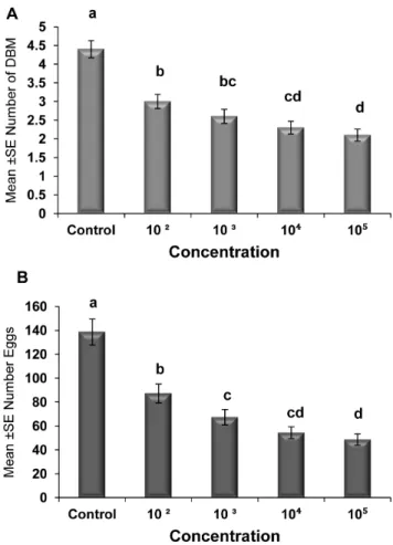
Figure 3. Effect of concentration ofNosemaon adult emergence and egg production.Graph bars represent mean6SE of number of adults emerged (a), number of eggs produced by DBM adults (b) developed from larvae inoculated at different dosages ofNosemasp. spores. Different letters above error bars indicate significant difference (P,0.05).
doi:10.1371/journal.pone.0062884.g003
(spores/mL)] for DBM after 72 h at 35uC was the lowest recorded in this study. Overall, the LC50 values declined with increasing
temperature and durations of exposure to Nosema (length of bioassay).
The results of the laboratory bioassay (leaf dip) showed that all
Nosemaconcentrations tested at different temperatures against the second instar larvae ofP. xylostellacaused various levels of mortality at all temperatures (Figure 1). As expected, the mortality of the second- instar DBM fed with Nosema-treated discs significantly increased with increasing spore concentrations (Figure 1), where the greatest accumulated larval mortality (92%) was observed when 16105 spores were applied at 35uC (F = 93.82, d.f. = 4,
P= 0.0; Figure 1E), and subsequently at 20 C (Figure 1B) and 30uC (.50% Figure 1D), with the lowest mortality (26%) observed
at 25uC (Figure 1C).
The larval mortality was also significantly affected by the time (days) after spore ingestion at 15uC (F = 17.85, d.f. = 3,p,0.05), 20uC (F = 42.50, d.f. = 3, p,0.05), 25uC (F = 25.09, d.f. = 3,
p,0.05), 30uC (F = 37.83, d.f. = 3, p,0.05), and 35uC [F = 73.66, d.f. = 3, p,0.05 (Figure 1)]. However, no significant interaction (p.0.05) occurred between concentration and time (days) for all temperatures in terms of influencing the larval mortality.
Effect of Nosema on the Number of Emerged Adults and Egg Production
The effects of different spore concentrations of Nosemaon the number of emerged adults and eggs produced by DBM adults developed from infected larvae at 15, 20, 25, 30, and 35uC are shown in Tables 2 and 3. The results indicated that all the main effects (temperature, concentration, and generation) and the respective interaction terms were highly significant for both mean numbers of DBM adults and eggs produced (Tables 2 and 3), except the interaction between concentration and generation for emerged adults and the interaction between temperature, concentration and generation for eggs produced. The highest reductions in the number of DBM were most pronounced at temperatures of 35 and 30uC (Figure 2a), and the highest reduction in the number of eggs was noted at 35uC (Figure 2b). The largest reduction in the number of emerged adults and number of eggs laid by infected moths were observed in the larvae exposed to 16105 spores/larvae. Other treatments also signifi-cantly reduced the number of emerged adults and eggs laid (16102, 16103, and16104) compared with that of the control
(Figures 3a and b). The reduction in the number of eggs laid and adults emerged was also noted in the subsequent generations, particularly the third and fourth generations (Figures 4a and b). In general, the number of emerged adults and egg produced showed inverse relationships with spore concentrations at all temperatures (Figure 5a and b). Temperature significantly affected the number
Figure 4. Comparison of adult emergence and egg production through successive generations.Graph bars represent mean6SE of number of adults emerged (a), number of eggs produced by DBM adults (b) developed from Nosema infected DBM larvae through successive generations. Different letters above error bars indicate significant difference (P,0.05).
doi:10.1371/journal.pone.0062884.g004
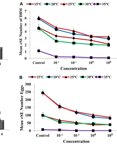
Figure 5. Influence of Nosema concentration and different temperatures on adult emergence and egg production.Data represent the mean6SE of number of adults emerged (a), number of eggs produced by DBM adults (b) developed fromNosemainfected DBM larvae at 15u, 20u, 25u, 30uand 35uC. Vertical bars = S.E.
of emerged adult and egg produced at all concentrations. However, the lowest numbers were observed at 35uC at all concentrations compared with those at other temperatures. Interestingly, at 35uC, the adults failed to emerge both at low and high doses ofNosemaspores, and the number of eggs produced were adversely affected (Figures 5a and b). Progeny production through generations was also significantly (p,0.05) affected by
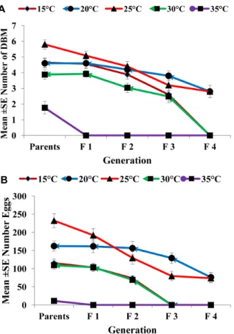
Nosema infection. The production of progeny was markedly affected as the generation number increased (Figures 6a and b). The population size was collapsed after the first and third generations at 35uC and at 15 and 30uC, respectively.
Effect of Nosema on the Histopathology of midgut ofP. xylostellaLarvae
The midgut ofP. xylostellalarvae is a long straight tube, which consists of columnar and goblet cells linked by well-developed border of microvilli. The epithelial cells rest on a basement membrane and muscle fibers (Figure 7A). Sections from larvae that had been fed for 24 h with Nosema-infected leaves showed distended and bulbous distal ends of the epithelium columnar cells (Figure 7B). Although the goblet cells may show some morpho-logical changes, there were no signs of damage at this stage (Figure 7B). Degeneration of the epithelium columnar cells continued, such that after 48 h of ingestion of a Nosema-infected food, the lumen exhibited debris of disrupted cells (Figure 7C).
The goblet cells also showed signs of damage after 48 h, but both types of cells were still attached to the basement membrane. At 72 h post-treatment, the epithelial cells exhibited swelling, and some microvilli were disrupted because of the swelling of the cells and lysis of cytoplasmic material (Figure 7D). In addition, the vacuoles increased in size, and some columnar cells were dislodged and sloughed into the lumen of the midgut (Figure 7D, arrows). This result clearly showed that Nosema infection had caused cytopathological changes in the midgut epithelial cells.
Discussion
The present results showed thatNosemais highly pathogenic to
P. xylostella (Table 1), confirming previously reported data that DBM larval mortality was high even at low concentrations of
Nosema spores [4] and for the gypsy moth (Lymantria dispar L.), where mortality varied between 79% and 99%, independently of the spore concentration [9]. The silkworm has also been reported to show high larvae mortality upon infection byNosemasp [10,11]. In contrast, minimal mortality (3%) was observed in southwestern corn borer Diatraea grandiosella larvae infected with Nosema sp. (isolate 506), even at high doses [12].
Our results showed that the susceptibility of the P. xylostella
larvae to the pathogen was dose-dependent. More than 90% of larvae died by day 4 after treatment at a dose (spore concentration) of 105 (spores/mL) compared with those at lower doses. The mortality ofP. xylostellalarvae at higher doses might be attributed to intestinal damage and bacterial septicemia as a result ofNosema
infection [13]. Temperature had a negative effect on mortality, where the greatest accumulated larval mortality (92%) was
Figure 6. Influence of temperature on adult emergence and egg production ofP. xylostellathrough successive generations.
Data represent the mean6SE of the number of adults emerged (a), number of eggs produced by DBM adults (b) developed fromNosema infected DBM larvae through successive generations. Vertical bars = S.E. doi:10.1371/journal.pone.0062884.g006
Table 2.Result of ANOVA for adults emerged as infected by Nosemainfection at various spore concentrations,
temperatures through five generations.
Effect Df F P- value
Temperature 4 267.1 ,0.05
Concentration 4 92.8 ,0.05
Generation 4 148.6 ,0.05
Temperature6Concentration 16 2.34 ,0.05
Temperature6Generation 16 12.02 ,0.05
Concentration6Generation 16 1.45 .0.05
Temperature6Concentration6Generation642.35,0.05Error500
doi:10.1371/journal.pone.0062884.t002
Table 3.Result of ANOVA for eggs produced by adults infected withNosemaat various spore concentrations, temperatures through five generations.
Effect Df F P- value
Temperature 4 293.2 ,0.05
Concentration 4 109.9 ,0.05
Generation 4 148.6 ,0.05
Temperature6Concentration 16 12.9 ,0.05
Temperature6Generation 16 13.5 ,0.05
Concentration6Generation 16 2.61 ,0.05
Temperature6Concentration6Generation641.01.0.05Error500
doi:10.1371/journal.pone.0062884.t003
observed at 35uC, followed by at 20 and 30uC (.50%), with the
lowest accumulated mortality (26%) observed at 25uC, which was considered to be the optimum temperature. The mortality in
Nosema pyrausta-infectedOstrinia nubilalislarvae was reported to be high at 24uC than 30uC [14]. This suggests that larvae are able to survive longer at low temperatures than at higher temperatures. An increase in mortality over days could have resulted from the increasing damage that spores caused while spreading in the body of the larvae. This eventually led to tissue destruction, and hence the larvae died before pupation or even if they reached the pupation stage, the number of adults emerged is less and their fecundity is low. However, the larvae were unable to bear the impact of the interaction between high temperature and Nosema
infection, and mortality continued to increases over time, indicating an additive effect of Nosema infection, particularly at 30 and 35uC. Therefore, there was high mortality reaching a peak (92%) at 35uC. Nosema sp. spores were found in the midgut of
Antheraea mylittaat the beginning of infection, and then spread to the fat body, and infection was observed in the tracheal epithelium, Malphigian tubules, and gonads during the later stages [15].
These results suggest a clear impact of bothNosemainfection and temperature onP. xylostellaegg production and number of adults emerging. The reduction in the number of egg production may be related to several factors affecting the egg laying process, because
Nosemainfects certain tissues involved in egg production [16]. The high spore concentration of Nosema in the gonads of A. mylitta, Antheraea assamensis, andBombyx moriwas found to affect reproduc-tive potential and fertility [17]. Reduction in the number of eggs deposited by infected females could be a response to the competition between the host and the microsporidia for nutrients [18,19]. In addition, infected females have been suggested to compensate for the loss of nutrients to the microsporidia by
producing fewer eggs [19,20]. Our observations are consistent with the effects of other Nosemaspp., such as N. pyrausta on the European corn borer,Ostrinia nubilalis(Hu¨bner) [21], andNosema
sp. on silkworm [10,22].
Our results also indicate that the impact of bothNosemainfection and temperature on the number of adults emerging from infected larvae occurred over 4 subsequent generations. This reduction in the number of the progeny could have been caused by the larval and pupae mortality as a result of the damage and changes in the organs of the Nosema-infected insects. This effect is passed on through generations, leading to fewer healthy adults and, eventually, population collapse. These findings are in accordance with previous results that indicate similar sub-lethal effects of other microsporidia on insects [23]. For example, the effectiveness of the microsporidium Edhazardia aedis (Kudo) in controlling a semi-natural population ofAedes aegypti(L.) was evaluated over a 2-year period, which led to the successful elimination of a population ofA. aegyptiin Florida [24].
In general, low and high temperatures had adverse effects on the production of progeny of the infected DBM, particularly at
35uC where the progeny failed to emerge. However, high
temperature is not as stable in natural conditions as it is under laboratory conditions; temperature fluctuates within a day, and it is usually cooler at night than in the day. Thus, future studies will be aimed toward the development of an efficient field trial before the possible commercial production of Nosema as a biocontrol agent of DBM in the field.
Since the midgut performs important functions such as digestion and absorption, we presume that the negative effects ofNosema
infection on DBM was partially because of the damage it causes to the midgut epithelium, leading to digestive and food absorption disorders. Subsequently, vacuolization occurred as a result of cell elongation after infection [25]. The disruption of the microvilli and
Figure 7. Histopathological effects ofNosemain onP. xylostellamidgut.General aspects of the midgut larvae (A) and histopathological effects ofNosemaon it (B) after 42 h of treatment, (C) after 48 h of treatment, (D) after 72 h of treatment. In C&D arrows indicate lysis of columnar cells and disrupted columnar cells (stars) are sloughed into the lumen of midgut. V, vacuoles; Lu, lumen; Gc, goblet cell, Cc, columnar cell; Mv, microvilli; Bm, basement membrane. Magnification 40 x.
vacuolization of the intracellular organelles in the midgut epithelial cells were similar to the cytotoxic effects observed in the midgut of
P. xylostella after treatment with Bacillus thuringiensis [26] and
Vairimorpha sp. [27]. All these changes have been reported in several susceptible Lepidoptera [28].
The histopathological changes caused by Nosema infection suggest that Nosema sp. could be fatal to DBM if infected with a sufficient amount of spores. In conclusion, the result of the present study suggests thatNosemainfection has a highly negative impact on DBM, and thus, its utilization could be considered in IPM. However, more studies are still required in this regard.
Acknowledgments
The authors wish to thank MARDI for providing the DBM egg masses and all lab assistants in the Entomology, Histology (MPG2) and Parasitology labs. We would like to thank Editage for providing editorial assistance.
Author Contributions
Conceived and designed the experiments: NK ZA NI MA IA. Performed the experiments: NK ZA NI MA IA. Analyzed the data: NK ZA NI MA IA. Contributed reagents/materials/analysis tools: NK ZA NI MA IA. Wrote the paper: NK ZA HD NI MA IA.
References
1. Zalucki MP, Shabbir A, Silva R, Adamson D, Liu S-S, et al. (2012) Estimating the Economic Cost of One of the World’s Major Insect Pests,Plutella xylostella (Lepidoptera: Plutellidae): Just How Long is a Piece of String? Journal of Economic Entomology 105(4): 1115–1129.
2. Shelton AM, Wyman JA, Cushing NL, Apfelbeck K, Dennehy TJ, et al.(1993) Insecticide resistance of diamondback moth (Lepidoptera: Plutellidae) in North America. Journal of Economic Entomology 86: 11–19.
3. Zhao JZ, Li YX, Collins HL, Gusukuma-Minuto L, Mau RFL (2002) Monitoring and characterization of diamondback moth (Lepidoptera: Plutelli-dae) resistance to spinosad. Journal of Economic Entomology 95: 430–436. 4. Idris AB, Zainal-Abidin BAH, Sajap AS, Noran AM Hussan AK (2004) Some
studies onNosemainfecting DBM in Malaysia. In: Endersby NM, Ridland PM, editors.The management of diamondback moth and other crucifer pests. Proceedings of the Fourth International Workshop, 26–29 November 2001. Melbourne, Australia: Department of Natural Resources and Environment. 295–303.
5. Undeen AH, Va´vra J, (1997) Research Methods for Entomopathogenic Protozoa. In: Lacey, L.A. (Ed.), Manual of Techniques in Insect Pathology. Academic Press, NY, 117–151.
6. Cantwell GE 1970. Standard and Methods for Counting Nosema spores.
American Bee. Journal 110:222–223.
7. Finney DJ (1971) Probit analysis. London, New York, Cambridge University Press.
8. Humason GL (1967) Animal tissue techniques, 2nd ed. Freeman, SanFrancisco, California, 569 p.
9. Goertz D, Pilarska D, Kereselidze M, Solter LF, Linde A (2004) Studies on the impact of twoNosemaisolates from Bulgaria on the gypsy moth (Lymantria dispar L.). Journal of Invertebrate Pathology 87: 105–113.
10. Velide L, Rao AP (2011) Studies on the impact of microsporidiosis on tropical tasar silkwormAnthereae mylittaDrury. Journal of Applied Biosciences 44:2994–
2999.
11. Shabir Ahmad B, Ifat B, Afifa SK (2009).Microsporidiosis of silkworm,Bombyx moriL. (Lepidoptera- bombycidae): A review. African Journal of Agricultural Research 13: 1519–1523.
12. Inglis GD, Lawrence AM, Davis EM (2003) Impact of a Novel Species ofNosema on the Southwestern Corn Borer (Lepidoptera: Crambidae).Journal of Economic Entomology 96(1): 12–20.
13. Fuxa JR (1981) Susceptibility of lepidopterous pests to two types of mortality
caused by the microsporidium Vairimorpha necatrix. Journal of Economic
Entomology 74: 99–102.
14. Solter LF, Onstad DW, Maddox JV (1990) Timing of disease-influenced processes in the life cycle ofOstrinia nubilalisinfected withNosema pyrausta. Journal of Invertebrate Pathology 55: 337–341.
15. Remadevi OK, Sasidharan TO, Bhattacharya J, Vossbrinck CR, Rajan PD (2010)Some pathological effects and transmission potential of a microsporidian isolate (Nosemasp.) from the teak defoliatorHyblaea puera (Lepidoptera:Hyblaei-dae). International Journal of Tropical Insect Science 30(3): 138–144. 16. Jolly MS, Sen SK (1972) Infection ofAntheraea mylitta Drury (Lepidoptera:
Saturniidae) by a microsporidian (Nosemasp.).Indian J. Seric. 11 (1): 52–57. 17. Bansal AK, Saxena NN, Shukla RM, Roy DK, Sinha BRRP, et al (1997) A
technique proposed for estimation of microsporidiosis in grainages. Sericology 37: 11–14.
18. Bauer LS, Nordin OL (1989) Effect ofNosema fumiferanae(Microsporidia) on Fecundity, Fertility, and Progeny Performance of Choristoneura fumiferana (Lepidoptera: Tortricidae). Environmental Entomology 18: 261–264. 19. Goertz D, Golldacka J, Linde A (2008) Two different and sublethal isolates of
Nosema lymantriae(Microsporidia) reduce the reproductive success of their host, Lymantria dispar. Biocontrol Science and Technology 18 (4): 419–430. 20. Diss AL, Kunkel JG, Montgomery ME, Leonard DE (1996) Effects of Maternal
Nutrition and Egg Provisioning on Parameters of Larval Hatch, Survival and Dispersal in the Gypsy MothLymantria disparL. Oecologia, 106: 470–477. 21. Bruck DJ, Lewis LC, Gunnarson RD (2001) Interaction ofNosema pyraustaand
temperature on Ostrinia nubilalis egg production and hatch. Journal of
Invertebrate Pathology 78: 210–214.
22. Rath SS, Prasad BC, Sinha BR (2003) Food utilization efficiency in fifth instar larvae ofAntheraea mylitta(Lepidoptera: Saturniidae) infected withNosemasp. and its effect on reproductive potential and silk production. Journal of Invertebrate Pathology 83: 1–9.
23. Becnel JJ, Andreadis TG (1999) Microsporidia in Insects. In: M. Wittner and
L.M. Weiss, Editors, The Microsporidia and Microsporidiosis, ASM Press,
Washington, D.C, 447–501.
24. Becnel JJ, Johnson MA (2000) Impact of Edhazardia aedis (Microsporidia:
Culicosporidae) on a seminatural population ofAedes aegypti(Diptera: Culicidae). Biological Control 18: 39–48.
25. Salkeld EH (1951) A toxicological study of certain new insecticides as ‘‘stomach poisons’’ to the honey beeApis mellifera. J. Can. Ent. 8339–8361.
26. Wiwat C, Thaithanun S, Pantuwatana S, Bhumiratana A (2000) Toxicity of Chitinase-ProducingBacillus thuringiensisssp.KurstakiHD-1 (G) towardPlutella xylostella.Journal of Invertebrate Pathology 76: 270–277.
27. Haque MdA, Canning EU, Wright DJ (1999) Entomopathogenicity of Vairimorpha sp. (Microsporidia) in the diamondback moth, Plutella xylostella (Lepidoptera: Yponomeutidae). Bulletin of Entomological Research 89: 147– 152.
28. Yu C-G, Mullins MA, Warren GW, Koziel MG, Estruch JJ (1997) The Bacillus thuringiensis vegetative insecticidal protein Vip3A lyses midgut epithelium cells of susceptible insects. Appl. Environ. Microbiol. 63: 532–536.