in Cerebral and Small Mesenteric Arterial Smooth Muscle
Cells of Simulated Microgravity Rat
Jun-Hui Xue1., Lian-Hong Chen2., Hua-Zhou Zhao3., Yong-Dong Pu3
, Han-Zhong Feng4, Yu-Guang Ma4, Jin Ma4, Yao-Ming Chang1,4, Zuo-Ming Zhang1*, Man-Jiang Xie4*
1Department of Aerospace Clinical Medicine, Key Laboratory of Aerospace Medicine of Ministry of Education, Fourth Military Medical University, Xi’an, Shaanxi Province, China,2Department of Chest Surgery, Tangdu Hospital of Fourth Military Medical University, Xi’an, Shaanxi Province, China,3309 Clinical Divisions, Department of General Surgery, General Hospital of PLA, Beijing, China,4Department of Aerospace Physiology, Key Laboratory of Aerospace Medicine of Ministry of Education, Fourth Military Medical University, Xi’an, Shaanxi Province, China
Abstract
Background:The differential adaptations of cerebrovasculature and small mesenteric arteries could be one of critical factors in postspaceflight orthostatic intolerance, but the cellular mechanisms remain unknown. We hypothesize that there is a differential regulation of intracellular Ca2+determined by the alterations in the functions of plasma membrane Ca
Lchannels and ryanodine-sensitive Ca2+releases from sarcoplasmic reticulum (SR) in cerebral and small mesenteric vascular smooth
muscle cells (VSMCs) of simulated microgravity rats, respectively.
Methodology/Principal Findings: Sprague-Dawley rats were subjected to 28-day hindlimb unweighting to simulate microgravity. In addition, tail-suspended rats were submitted to a recovery period of 3 or 7 days after removal of suspension. The function of CaLchannels was evaluated by patch clamp and Western blotting. The function of ryanodine-sensitive Ca2+releases in response to caffeine were assessed by a laser confocal microscope. Our results indicated that
simulated microgravity increased the functions of CaLchannels and ryanodine-sensitive Ca2+releases in cerebral VSMCs, whereas, simulated microgravity decreased the functions of CaLchannels and ryanodine-sensitive Ca2+releases in small mesenteric VSMCs. In addition, 3- or 7-day recovery after removal of suspension could restore the functions of CaLchannels and ryanodine-sensitive Ca2+releases to their control levels in cerebral and small mesenteric VSMCs, respectively.
Conclusions: The differential regulation of CaL channels and ryanodine-sensitive Ca2+ releases in cerebral and small mesenteric VSMCs may be responsible for the differential regulation of intracellular Ca2+, which leads to the altered
autoregulation of cerebral vasculature and the inability to adequately elevate peripheral vascular resistance in postspaceflight orthostatic intolerance.
Citation:Xue J-H, Chen L-H, Zhao H-Z, Pu Y-D, Feng H-Z, et al. (2011) Differential Regulation and Recovery of Intracellular Ca2+in Cerebral and Small Mesenteric Arterial Smooth Muscle Cells of Simulated Microgravity Rat. PLoS ONE 6(5): e19775. doi:10.1371/journal.pone.0019775
Editor:Carlo Gaetano, Istituto Dermopatico dell’Immacolata, Italy
ReceivedDecember 21, 2010;AcceptedApril 5, 2011;PublishedMay 18, 2011
Copyright:ß2011 Xue et al. This is an open-access article distributed under the terms of the Creative Commons Attribution License, which permits unrestricted use, distribution, and reproduction in any medium, provided the original author and source are credited.
Funding:This work was supported by National Natural Science Foundation of China (No. 30800545, 30900283 and 31070768). The funders had no role in study design, data collection and analysis, decision to publish, or preparation of the manuscript.
Competing Interests:The authors have declared that no competing interests exist. * E-mail: [email protected] (M-JX); [email protected] (Z-MZ)
.These authors contributed equally to this work.
Introduction
Postspaceflight orthostatic intolerance has been regarded as one of the major adverse effects of microgravity exposure and there are still no effective countermeasures [1,2]. Human studies from spaceflight research and bed rest have indicated that the altered autoregulation of cerebral vasculature and the inability to adequately elevate peripheral vascular resistance may be the fundamental causes in the occurrence of orthostatic intolerance after spaceflight [3,4]. In the past decades, ground-based animal studies with tail-suspended hindlimb-unweighting rat model, which has been widely used to simulate physiological effects of microgravity [5], have revealed that simulated microgravity induced cerebrovascular adaptations including the increased
myogenic tone, enhanced vasoreactivity, hypertrophic remodeling, and endothelial dysfunction [6,7], whereas, simulated microgravity induced small mesenteric arterial adaptations including the decreased myogenic tone, attenuated vasoreactivity, atrophic remodeling, and endothelial dysfunction [7,8]. These findings suggest that differential adaptations of cerebrovasculature and small mesenteric arteries could be one of critical factors in postspaceflight orthostatic intolerance, but the cellular mecha-nisms remain unknown.
Intracellular Ca2+
in vascular smooth muscle cells (VSMCs) is an important determinant for functional and structural adapta-tions in vasculature [9]. Ca2+influx from the long-lasting
voltage-dependent Ca2+
(L-type, CaL) channels in plasma membrane and
Ca2+
reticulum (SR) are likely to play the essential roles in controlling intracellular Ca2+
[10]. It is known that the increased intraluminal pressure depolarizes VSMCs and then enhances the extracellular Ca2+
influx by opening CaLchannels. The increased
concentra-tion of intracellular Ca2+
([Ca2+
]i) subsequently activates RyRs
and produces the transient local Ca2+release events in micromolar
concentrations (Ca2+ sparks) from SR, which in turn activate
nearby Ca2+
-activated K+
(KCa) channels in plasma membrane,
leading to membrane hyperpolarization, inhibition of CaL
channels, and thereby favoring vasodilation by reducing the Ca2+
influx [11]. Therefore, CaLchannels in plasma membrane
and RyRs in SR are important mediators to control arterial excitation-contraction coupling and subsequent structural remod-eling by handling intracellular Ca2+. It has been demonstrated that
hypertension [12], atherosclerosis [13], diabetes [14], and hypoxia [15] are all associated with the abnormal function of CaLchannels
or RyRs.
Our previous work reported that 28-day simulated microgravity increased the concentration of intracellular Ca2+
in rat cerebral VSMCs [16] associated with the upregulation of CaL channels
[17]. In contrast, there is a report that 14-day hindlimb unloading decreased the level of intracellular Ca2+
in rat small mesenteric VSMCs by reducing the function of ryanodine-sensitive Ca2+
releases associated with the downregulation of RyR2 mRNA and protein expression [18]. We also reported that 28-day simulated microgravity down-regulated the CaL channels in rat small
mesenteric VSMCs [17]. These results suggested that alterations of CaL channels in plasma membrane and ryanodine-sensitive
Ca2+
releases from SR may account for the changes of intracellular Ca2+
in cerebral and small mesenteric VSMCs of simulated microgravity rats. Furthermore, these results also indicated that CaLchannels and ryanodine-sensitive Ca2+releases
may play an important role in the differential adaptations of cerebral and small mesenteric arteries in simulated microgravity rats. However, it is not clear whether the alterations of CaL
channels in plasma membrane are accompanied by the changes of ryanodine-sensitive Ca2+
releases from SR in cerebral or small mesenteric VSMCs of simulated microgravity rats, respectively. In addition, it is not clear whether the function of CaLchannels and
ryanodine-sensitive Ca2+
releases could recover after removal of suspension. The purpose of the present work was 1) to investigate the function of CaL channels by comparing the whole-cell
currents, protein expressions ofa1C-subunit in cerebral and small
mesenteric VSMCs isolated from control rats, simulated micro-gravity rats, and rats after removal of suspension, and 2) to assess the functions of ryanodine-sensitive Ca2+ releases by
measure-ments of intracellular Ca2+
in response to caffeine in cerebral and mesenteric VSMCs isolated from the same groups as described above. Taken together, the present study provided initial evidences that there is a differential regulation and recovery in the functions of CaLchannels and ryanodine-sensitive Ca2+releases of cerebral
and small mesenteric VSMCs from simulated microgravity rats, respectively, which may provide a novel mechanism for cerebral and small mesenteric arterial adaptations during microgravity.
Materials and Methods
All animal procedures described in this study were performed in adherence with the Guide for the Care and Use of Laboratory Animals
published by the US National Institutes of Health (NIH Publication No. 85-23, revised 1996), with approval from Committee on the Ethics of Animal Experiments of the University of Fourth Military Medical University (Permit Number: 10001). All surgery was performed under sodium pentobarbital anesthesia,
and all efforts were made to minimize suffering. Unless otherwise stated, all chemicals and reagents used in this study were obtained from Sigma Chemical Company (St. Louis, Missouri, USA).
Animal model
The tail-suspended, hindlimb-unweighting rat model, which was described in detail previously [5], was used to simulate the cardiovascular effects of microgravity. The present study was divided into two experiments. Experiment I was designed to investigate the functions of CaLchannels and ryanodine-sensitive
Ca2+releases in cerebral and small mesenteric VSMCs isolated
from control rats, simulated microgravity rats, and tail-suspended rats submitted to a recovery period of 3 days after removal of suspension.Experiment IIwas designed to investigate the function of CaL channels in small mesenteric VSMCs isolated from
simultaneous control rats and tail-suspended rats submitted to a recovery period of 7 days after removal of suspension. InExperiment I, a total of 90 male Sprague-Dawley rats, 7–9 weeks of age and weighing,200 g, were randomly assigned into 3 groups (n= 30/ group): 28-day simultaneous control (CON-28 d), 28-day tail-suspension (SUS-28 d), and tail-suspended rats submitted to a recovery period of 3 days at the end of 28-day suspension (SUS+Rec-3 d). InExperiment II, 20 male Sprague-Dawley rats, 7– 9 weeks of age and weighing,200 g, were randomly assigned into 2 groups (n= 10/group): 35-day simultaneous control (CON) and tail-suspended rats submitted to a recovery period of 7 days at the end of 28-day suspension (SUS+Rec-7 d). The SUS rats were maintained in an about230uhead-down tilt position with their hindlimbs unloaded and caged individually in a room maintained at 23uC on a 12:12-h light-dark cycle. The CON rats and rats submitted to a recovery period were maintained in individual cages and were treated similarly except for the tail suspension. All the animals received standard lab chow and water ad libitum. Animals were anesthetized with pentobarbital sodium (50 mg/kg ip) and killed by exsanguinations via the abdominal aorta. The wet weight of left soleus and the length of left femur were measured to confirm the efficacy of deconditioning and monitor any effect on growth.
Isolation of VSMCs
Enzymatic isolation of single VSMC was carried out as previously described [17,19]. Briefly, brain and superior mesen-teric tissues were removed rapidly and placed in 4uC physiological salt solution (PSS). PSS contained (in mM) 137 NaCl, 5.6 KCl, 1 MgCl2, 0.42 Na2HPO4, 0.44 NaH2PO4, 4.2 NaHCO3, and 10
HEPES, equilibrated with 95% O2and 5% CO2at pH adjusted to
7.4 with NaOH. The cerebral arteries (superior, middle, and basilar arteries with the circle of Willis) and small mesenteric arteries with its branches were dissected and cut into 1–2 mm length. The cerebral arterial segments were digested for 18 min (small mesenteric arterial segments for 25 min) at 37uC with the solution contained 4 mg/ml papain (Biochrom, Berlin, Germany), 2 mg/ml dithioerythritol (Amresco, St. Louis, Missouri, USA), 1 mg/ml bovien serum albumin (BSA), and 5 mM taurine in PSS. Vessel segments were then transferred to enzyme-free PSS containing 1 mg/ml BSA and 5 mM taurine at room temperature for 10 min and triturated with a flame-polished pipette to disperse VSMCs. Isolated VSMCs were suspended in Ca2+-free PSS
containing 1 mg/ml BSA and 5 mM taurine and stored at 4uC for use within 8 h.
Electrophysiological measurements
(CEZ-2300, Nihon Kohden Co., Tokyo, Japan) and a version interface (Axon Instruments, Foster City, California, USA). Command-voltage protocols and data acquisition were performed with pCLAMP software (version 8.0, Axon Instruments). Patch pipettes (tip resistance 2–6 MVwhen filled with a pipette solution) were fabricated on an electrode puller (Narishige Instruments, Tokyo, Japan) with borosilicate glass capillary tubing. A coverslip containing the cells was positioned in a 2-ml recording chamber and superfused with the extracellular (bath) solution. All measurements were performed at room temperature (22–24uC). Resting membrane potential (Em) was measured with the current-clamp configuration of the patch-current-clamp technique while the cell was held at zero membrane current [21]. The external (bath) solution contained (in mM) 135 NaCl, 4 KCl, 1 MgCl2, 2 CaCl2,
10 HEPES, and 10 glucose, equilibrated with 95% O2and 5%
CO2 at pH 7.4 adjusted with NaOH. The pipette solution
contained (in mM) 143 KCl, 1 CaCl2, 1 MgCl2, 3 EGTA, and 10
HEPES, equilibrated with 95% O2 and 5% CO2 at pH 7.2
titrated with KOH.
Whole-cell CaL channel currents were measured with the
conventional voltage clamp configuration. Cell capacitance (Cm) and access resistance (Ra) were estimated from the capacitive current transient evoked by applying a 20-mV pulse for 40 ms from a holding potential of260 mV to240 mV. The cell was held at
240 mV and then stepped in 10-mV increments from 230 to +60 mV. Voltage steps were 250 ms in a duration and 2-s intervals were allowed between steps. Currents were filtered at 0.5 kHz and digitized at 4 kHz. Nonspecific membrane leakage and residual capacitive currents were subtracted using the P/4 protocol. Currents were sampled and averaged while the current amplitude was stabilized. Barium (Ba2+
) was used rather than Ca2+
as the charge carrier to increase unitary currents and to minimize Ca2+
-dependent run-down. Currents were normalized to Cm to obtain the current densities. To obtain theI–Vcurve of CaL, the current
densities were plotted against the corresponding command potentials. Two kinds of external solutions were used, i.e.,solution AandB.Solution Awas used while making a gigaohm seal between the recording pipette and cell surface. It contained (in mM) 130 NaCl, 5.4 KCl, 1 MgCl2, 10 BaCl2, 10 HEPES, and 10 glucose,
equilibrated with 95% O2and 5% CO2at pH 7.4 adjusted with
NaOH. After a seal of 2 GVwas obtained, the perfusion fluid was changed tosolution Bbefore current recording. It contained (in mM) 75 Tris-Cl, 50 BaCl2, 10 HEPES, and 10 glucose, equilibrated with
95% O2and 5% CO2at pH 7.4 titrated with Tris base. The pipette
solution contained 150 CsCl, 1 MgCl2, 10 EGTA, 5 HEPES, 5
Na2ATP, and 5 Na2creatine phosphate, equilibrated with 95% O2
and 5% CO2at pH 7.2 titrated with CsOH. In the present study,
extracellular application of 5mM Bay K 8644 (the specific agonist of CaL) and 0.1mM nifedipine (the specific blocker of CaL) were used
to identify the properties of CaLas describe before [16,17].
Evaluation of CaLchannel protein expression by Western blotting
Protein samples of cerebral and small mesenteric arteries were prepared according to the published methods [17]. Briefly, cerebral and small mesenteric arterial specimens were minced into small pieces and homogenized on ice containing tissue protein extraction reagent (T-PER, Pierce, Rockford, Illinois, USA) and protease inhibitor (Halt, Pierce, Rockford, Illinois, USA). Large tissue debris and nuclear fragments were removed by two centrifuge spins (1,000 rpm for 5 min, 12,000 rpm for 15 min) at 4uC and supernatants were obtained. The concentration of protein samples was determined by the bicinchoninic acid method (Pierce, Rockford, Illinois, USA) using BSA as a standard.
Equivalent amounts of proteins from different groups were loaded to adjacent lanes for SDS-PAGE and each sample based on tissue pooled from 4,5 animals. Protein samples were run for 80 min at 30 mA for electrophoretically size-separation using a 8% Tris-Glycine gel (Invitrogen, Carlsbad, California, USA). After size separation, proteins were transferred onto a nitrocellulose membrane at 100 mA for 3 h and blocked with 5% nonfat dry milk in PBS containing 0.1% (w/v) Tween 20 (PBS-T) overnight at 4uC. Subsequently, the membranes were incubated for 3 h with a 1:200 dilution of rabbit polyclonal antibody against amino acids 848–865, which corresponds to the C-terminus site of CaLchannel
a1C-subunit (Alomone Labs, Jerusalem, Israel). The membrane
then incubated for 45 min with Infrared (IR)-labeled secondary antibodies (LI-COR) in PBS-T containing 0.01% SDS. A monoclonal mouse antibody raised against the structural protein b-actin (Sigma) was used as a lane-loading control. The bound antibody was detected by the Odyssey infrared imaging system (LI-COR), and the densities of the immunoreactive doublet bands at 200 and 240 kD associated with anti-a1C subunit of CaL
channel were expressed as a percentage of theb-actin density for each lane. Densitometry analysis of bands was performed by Scion image (Scion, Frederick, MD).
Measurement of intracellular Ca2+
In an attempt to establish whether simulated microgravity affects the function of ryanodine-sensitive Ca2+
releases from SR in cerebral and small mesenteric VSMCs, respectively, we investi-gated the average changes of intracellular Ca2+ fluorescence
intensity in response to caffeine. The intracellular Ca2+
was measured with Ca2+
indicator, Fluo-3-acetoxymethyl ester (Fluo-3/AM, Molecular Probes, Oregon, USA), as previous described [22]. Fresh cerebral and small mesenteric VSMCs were incubated with Fluo-3/AM in a final concentration of 5mM for 30 min at 37uC. After incubation, the Fluo-3/AM-loaded cells on coverslips were washed with the Ca2+
-free balanced salt solution (BSS) in the following composition (mM): 126 NaCl, 5 KCl, 0.3 NaH2PO4, 10
HEPES, 1 MgCl2, 10 glucose, 1 EGTA, equilibrated with 95% O2
and 5% CO2at pH 7.4 adjusted with NaOH [18,23]. The cells
were scanned under a laser confocal microscope (Olympus FV1000, Tokyo, Japan) by illuminating with a krypton/argon laser at 488 nm emitted light and capturing the emitting fluorescence at 526 nm. To ensure efficient quantum capture, the cells were placed on the bottom of a recording chamber and images were recorded after 10,20 s when fluorescence intensity became stable. During continuously scanning, 10 mM caffeine in Ca2+
-free BSS was administrated to the cell and a period of 3 min was recorded. To avoid any laser-induced change in Ca2+
signaling, each cell was scanned only once. Also, only one cell was scanned in each scanning time to avoid cell density-induced changes in fluorescence intensity. The average fluorescence intensity was used to indicate the changes of intracellular Ca2+
and the maximal increase of Ca2+
fluorescence intensity was used to indicate the function of ryanodine-sensitive Ca2+
releases from SR in VSMCs as describe before [24].
Statistical Analysis
Except the data of body weight are given as means6SD, all other data are expressed as means6SE. One-way ANOVA was used to determine the differences of CaLchannel current densities
in different groups, followed by a S-N-K-Post Hoc. Student’st-test was used to determine significant differences of body weights, soleus weights, femur lengths, and the maximal increases of Ca2+
Results
Physical characteristics of experimental animals
As summarized in Table 1, there were no significant differences in either final body weights or the lengths of left femur among CON, SUS, and SUS+Rec groups inExperiment IandExperiment II, suggesting a normal growth rate during and after simulated microgravity. However, after 28-day tail-suspension, the wet weights of left soleus in SUS-28 d rats were about 58.7% less than that in CON-28 d rats, indicating the deconditioning effects of simulated microgravity. In addition, the wet weights of left soleus in SUS+Rec-3 d rats were 24.3% less than that in CON-28 d rats, which means 3-day recovery after removal of CON-28-day tail-suspension could only partially recover the mass reduction of atrophic soleus due to simulated microgravity. There were no significant difference in the wet weights of left soleus between CON-35 d and SUS+Rec-7 d rats, suggesting a 7-day recovery after removal of 28-day tail-suspension could completely recover the mass reduction of soleus.
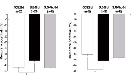
Simulated microgravity depolarized the cerebral VSMCs and hyperpolarized the small mesenteric VSMCs
For cerebral VSMCs, cell capacitance (Cm), an index of cell membrane area, did not show significant differences among CON-28 d (23.260.6 pF, n = 22), SUS-28 d (22.860.6 pF, n = 17), and SUS+Rec-3 d rats (23.660.9 pF, n = 26). Correspondly, there were neither significant changes in Cm of small mesenteric VSMCs among CON-28 d (23.460.9 pF, n = 24), SUS-28 d (23.160.6 pF, n = 23), and SUS+Rec-3d rats (23.860.7 pF, n = 23). These results suggested that simulated microgravity did not affect the cell surface areas of cerebral and small mesenteric VSMCs, which were consistent with our previous report [16,21]. As compared with that in CON-28 d rats, 28-day tail-suspension significantly depolarized the membrane potential of cerebral VSMCs (Fig. 1A) and significantly hyperpolarized the membrane potential of small mesenteric VSMCs (Fig. 1B). However, 3-day recovery after removal of suspension could completely recover the membrane potential to their control levels in cerebral and small mesenteric VSMCs, respectively (Fig. 1). These results suggested simulated microgravity induced a differential regulation of membrane potential in cerebral and small mesenteric VSMCs, respectively, which are in general agreement with previous report [16,21].
Simulated microgravity increased the current densities of CaLchannel in cerebral VSMCs and decreased the current densities of CaLchannel in small mesenteric VSMCs
The typical time- and voltage-dependent inward currents evoked by increasing depolarizations from a holding potential of240 mV were shown in the left panel of Fig. 2. Whole-cell CaLcurrents of
cerebral VSMCs in SUS-28 d rats showed larger inward compo-nents of trace as compared with that recorded in CON-28 d (the left panel of Fig. 2A). In contrast, whole-cell CaL currents of small
mesenteric VSMCs in SUS-28 d rats showed smaller inward components of trace as compared with that in CON-28 d rats (the left panel of Fig. 2B). The mean current-voltage relationship (I–V) curves which were further expressed in terms of current densities were shown in the right panel of Fig. 2. TheI–Vcurves further showed that simulated microgravity significantly increased the CaL
current densities in cerebral VSMCs (the right panel of Fig. 2A) and significantly decreases the CaLcurrent densities in small mesenteric
VSMCs (the right panel of Fig. 2B), respectively. However, 3-day recovery after removal of suspension could completely restore the CaLcurrent densities of cerebral VSMCs to their control levels (the
right panel of Fig. 2A) and partially recover the CaL current
densities in small mesenteric VSMCs (the right panel of Fig. 2B). Only 7-day recovery after removal of suspension could completely restore the CaLcurrent densities of small mesenteric VSMCs to
their control levels (Fig. 2C). These results suggested there are a differential regulation and recovery of CaL current densities in
cerebral and small mesenteric VSMCs, respectively.
Simulated microgravity enhanced the expressions of CaL a1C-subunit in cerebral arteries and reduced the
expressions of CaLa1C-subunit in small mesenteric arteries
The apparent masses of 200 and 240 kD doublet bands were shown in the membrane among CON-28 d, SUS-28 d, and SUS+Rec-3 d rats, which correspond to the predicted sizes of the short and long (or full length) forms of the CaLa1C-subunit protein
in the cerebral and small mesenteric arteries, respectively (Fig. 3). As the internal control, the expressions ofb-actin (42 kDa) were similar in different lanes in the bottom membrane, showing equal loading of proteins. The averaged data were expressed as percentage of the b-actin signal. As compared with that of CON-28 d rats, CaLchannela1C-subunit expressions of SUS-28 d
Table 1.Body weight, the length of left femur, and the wet weight of left soleus in CON, SUS, and SUS+Rec rats.
Body weight (g) Length of left femur (mm) Wet weight of left soleus (mg)
Initial Final
Experiment I
CON-28 d (n = 30) 229.367.5 358.868.5 35.660.8 150.862.4 SUS-28 d (n = 30) 228.668.1 361.369.2 35.460.6 62.362.1**
SUS+Rec-3 d (n = 30) 227.568.8 372.869.9 35.760.5 114.262.5**
Experiment II
CON-35 d (n = 10) 218.7613.6 403.8614.5 38.561.3 155.064.2 SUS+Rec-7 d (n = 10) 226.7612.3 396.3613.1 37.360.9 148.062.2
Values of body weights are means6SD; others are means6SE. CON-28 d: 28-day control rats; SUS-28 d: 28-day tail-suspended rats, SUS+Rec-3 d: tail-suspended rats submitted to a recovery period of 3 days at the end of 28-day suspension. CON-35 d: 35-day control rats; SUS+Rec-7 d: tail-suspended rats submitted to a recovery period of 7 days at the end of 28-day suspension.
**P,0.01vs.CON.
rats increased by ,105% in cerebral arteries and decrease by ,32% in small mesenteric arteries, respectively. However, there were no significant differences in CaL channel a1C-subunit
expressions of cerebral and mesenteric arteries between SUS+ Rec-3 d and CON-28 d rats, which indicated that 3-day recovery could completely restore the CaLchannela1C-subunit expression
in cerebral and small mesenteric arteries to their control levels, respectively. These results suggested that simulated microgravity differentially regulated the expression of CaLchannels in cerebral
and small mesenteric arteries, respectively. Furthermore, 3-day recovery can completely recover the effects of simulated microgravity on CaL channel expression in cerebral and small
mesenteric arteries, respectively.
Simulated microgravity increased the maximal increases of Ca2+ in cerebral VSMCs and decreased the maximal increases of Ca2+in small mesenteric VSMCs in response to caffeine
The present data showed that a high concentration of caffeine (10 mM) evoked a transient peak increase of [Ca2+
]i and was
followed by a sustained increase in [Ca2+]
ithat was above the basal
values in the continued presence of caffeine (Fig. 4 and Fig. 5). In present study, caffeine-evoked increases of [Ca2+
]i depended
essentially on Ca2+
release from the RyRs in SR because the Ca2+
-free solution (removal of Ca2+
from extracellular solution) was used to diminish Ca2+
influx through voltage-dependent Ca2+
channels in plasma membrane. As compared with that of CON-28 d rats, CON-28-day simulated microgravity significantly induced a higher resting [Ca2+
]ilevel in cerebral VSMCs (Fig. 6Aa) and a
lower resting [Ca2+
]ilevel in small mesenteric VSMCs (Fig. 6Ba),
respectively, which were consistent with previous report [16,18]. However, 3-day recovery after removal of suspension could restore the resting [Ca2+]
i to their control level in cerebral and small
mesenteric VSMCs, respectively (Fig. 6Aa and Fig. 6Ba). In addition, the acute applications of 10 mM caffeine evoked a significant augment of the maximal increase of [Ca2+
]iby,64.0%
in cerebral VSMCs isolated from SUS-28 d rats as compared with that of CON-28 d. However, the maximal caffeine-induced increase of [Ca2+]
i in SUS+Rec-3 d rats was similar to that
obtained in CON-28 d rats, indicating a complete recovery of ryanodine-sensitive Ca2+
releases in cerebral VSMCs (Fig. 4 and Fig. 6Ab). In contrast, prolonged suspension resulted in a significant reduction of the maximal caffeine-induced increase of [Ca2+]
iby
,22.7% in small mesenteric VSMCs as compared with that of CON-28 d rats. Correspondly, there were no significant differences in the maximal caffeine-induced increase of [Ca2+]
i between
SUS+Rec-3 d and CON-28 d rats, suggesting a complete recovery of ryanodine-sensitive Ca2+
releases in small mesenteric VSMCs (Fig. 5 and Fig. 6Bb). These results suggested that simulated microgravity differentially regulated the function of ryanodine-sensitive Ca2+
-releases in cerebral and small mesenteric arteries, respectively. Furthermore, 3-day recovery can completely recover the effects of simulated microgravity on ryanodine-sensitive Ca2+
releases in cerebral and small mesenteric arteries, respectively.
Discussion
The principal and novel finding of this work is that simulated microgravity differentially regulates the intracellular Ca2+
by its effects on CaLchannels in plasma membrane and the
ryanodine-sensitive Ca2+ releases from SR, i.e., simulated microgravity
increased the functions of CaLchannels and ryanodine-sensitive
Ca2+
releases in cerebral VSMCs, whereas, simulated micrograv-ity decreased the functions of CaL channels and
ryanodine-sensitive Ca2+releases in small mesenteric VSMCs. In addition,
3-or 7-day recovery after removal of suspension could rest3-ore the functions of CaLchannels and ryanodine-sensitive Ca2+releases to
their control levels in cerebral and small mesenteric VSMCs, respectively. These differential regulations of CaL channels and
ryanodine-sensitive Ca2+
releases in cerebral and small mesenteric VSMCs may be responsible for the altered autoregulation of cerebral vasculature and the inability to adequately elevate peripheral vascular resistance.
Figure 1. Comparisons of membrane potential in cerebral (A) and small mesenteric VSMCs (B) isolated from CON-28 d, SUS-28 d, and SUS+Rec-3 d rats. CON-28 d: 28-day simultaneous control rats; SUS-28 d: 28-day tail-suspended rats, SUS+Rec-3 d: tail-suspended rats submitted to a recovery period of 3 days at the end of 28-day suspension. Values are means6SE with the number of cells recorded in parentheses. *P,0.05vs.CON.
Differential adaptations in cerebral and small mesenteric arteries during real/simulated microgravity
There is a blood pressure gradient from the head to the feet in humans at 1 G in the upright posture. When exposed to microgravity, all gravitational blood pressure gradients are lost. Thus, blood vessels in cerebral circulation are exposed to higher than
normal 1-G blood pressure, whereas vessels in lower body regions are exposed to lower than normal upright 1-G blood pressure. Therefore, the differential adaptations in cerebral and hindquarter arteries during real/simulated microgravity have been postulated to be a basical process of vascular autoregulation in response to sustained elevation or reduction in local transmural pressures, which may Figure 2. Comparison of whole-cell CaLcurrent densities in cerebral (A) and small mesenteric VSMCs (B and C) isolated from CON, SUS, and SUS+Rec rats.CON: simultaneous control rats, SUS: 28-day tail-suspended rats, SUS+Rec: tail-suspended rats submitted to a recovery period of 3 or 7 days at the end of 28-day suspension. Representative recordings in the left panel were used to show the whole-cell CaLcurrents in different groups. The meanI–Vcurves were further expressed in terms of current densities in the right panel. Values are means6SE with the number of cells recorded in parentheses. *P,0.05 and **P,0.01 as compared with control by ANOVA.
contribute to cardiovascular dysfunction and ultimately lead to the post spaceflight orthostatic intolerance [3,7]. In addition, after release from simulated microgravity, the functional and structural adapta-tions in the forebody arteries or hindquarter arteries of rats were partially restored (7-day recovery) and fully recovered (35-day recovery) to their control levels, which means that these changes were reversible within the time frame of previous experiments [7].
Differential regulation of CaL channels in cerebral and small mesenteric arteries of simulated microgravity rats
CaLchannels of the Cav1.2 gene family are the principal Ca2+
influx pathway in VSMCs and participate in multiple physiological functions, including excitation-contraction coupling, cell apoptosis, and cell proliferation in small resistance vessels. CaLchannels are
heteromeric complex composed of a central pore-forming a1C
subunit and regulatoryb,c, anda2dsubunits in VSMCs. The
pore-forminga1C subunit (about 190–240 kDa) is considered to confer
most functional properties to the CaLchannel. It is currently believed
that membrane depolarization of VSMCs associated with elevated blood pressure permits extracellular Ca2+ entry through Ca
L
channels, whereas, membrane hyperpolarization of VSMCs associ-ated with lower blood pressure inhibits extracellular Ca2+
entry
through CaLchannels [12,25]. Therefore, CaLchannels regulate a
tonic level of vascular tone and contribute to the dynamic autoregulation in the vascular beds. The abnormalities of CaL
channels are regarded as part of the extensive biological and morphological adaptations during the pathogenesis of vascular diseases. By whole-cell and Western blotting, the present work demonstrated that simulated microgravity depolarized cerebral VSMCs (Fig. 1A) and then upregulated the function of CaLchannels
in cerebral arteries of rats (Fig. 2A and Fig. 3A), which are in agreement with the reports that hypertensive rats associated with anomalous arterial tone show an increased function of CaLchannels
to sustain the elevated Ca2+influx [12]. In contrast, the present work
also indicated that simulated microgravity hyperpolarized small mesenteric VSMCs (Fig. 1B) and then downregulated the function of CaLchannels in small mesenteric arteries of rats (Fig. 2B and Fig. 3B),
which were in consistent with the reports that inhibition of CaL
channels due to hypotension in shock rats [26]. In addition, the membrane potential (Fig. 1) and the function of CaLchannels (Fig. 2
and Fig. 3) could return to their control levels in cerebral and small mesenteric arteries after 3- or 7-day recovery after removal of suspension, which indicated that these differential changes are reversible when the simulated microgravity were released.
Figure 3. Comparison of CaLa1C-subunit expression by Western blotting in cerebral (A) and small mesenteric arteries (B) isolated from CON-28 d, SUS-28 d, and SUS+Rec-3 d rats.CON-28 d: 28-day simultaneous control rats, SUS: 28-day tail-suspended rats, SUS+Rec-3 d:
tail-suspended rats submitted to a recovery period of 3 days at the end of 28-day suspension. Values are expressed as means 6SE from 4 independent experiments. **P,0.01 and *P,0.05vs.CON by t-test.
Differential regulation of ryanodine-sensitive Ca2+ -releases in cerebral and small mesenteric arteries of simulated microgravity rats
RyRs are tetrameric channel proteins which are located in the SR plasma membrane. Three isoforms of RyRs have been identified as RyR1, RyR2, and RyR3, which are all present in
smooth muscle cells [27]. RyRs are one of the main actors in the generation of Ca2+
signals, transient local releases of Ca2+
, and this Ca2+
events provides the primary pathway for SR Ca2+
release into the cytosol underlying the excitation-contraction coupling. There is an indirect coupling between CaLchannels in the plasma
membrane and RyRs in the SR of arterial VSMCs, i.e., RyRs can Figure 4. Caffeine elicits the increases of intracellular Ca2+fluorescence intensity in cerebral VSMCs isolated from CON-28 d,
SUS-28 d, and SUS+Rec-3 d rats.(A) Representative dot graph of Fluo-3/AM fluorescence recorded showed the typical transient increases of
intracellular Ca2+fluorescence intensity evoked by 10 mM caffeine. Caffeine was present at times shown by the horizontal bar. (B) Summarized data indicated the average changes of the intracellular Ca2+
fluorescence intensity before and during the application of caffeine. Values are means6SE with the number of cells recorded in parentheses. CON-28 d: 28-day simultaneous control rats, SUS-28 d: 28-day tail-suspended rats, SUS+Rec-3 d:
tail-suspended rats submitted to a recovery period of 3 days at the end of 28-day suspension. *P,0.05 as compared with their controls by t-test, respectively.
be opened by elevations in cytosolic Ca2+
by opening CaL
channels, termed Ca2+
-induced Ca2+
release (CICR), and thus can contribute to the overall rise in the concentration of intracellular
Ca2+
[13,28]. Caffeine is commonly used to activate RyRs by increasing the affinity of the Ca2+
activator site for Ca2+
, whereas ruthenium red is a frequently used inhibitor. At concentrations of Figure 5. Caffeine elicits the increases of intracellular Ca2+fluorescence intensity in small mesenteric VSMCs isolated from CON-28 d,
SUS-28 d, and SUS+Rec-3 d rats.(A) Representative dot graph of Fluo-3/AM fluorescence recorded showed the typical transient increases of
intracellular Ca2+
fluorescence intensity evoked by 10 mM caffeine. Caffeine was present at times shown by the horizontal bar. (B) Summarized data indicated the average changes of the intracellular Ca2+
fluorescence intensity before and during the application of caffeine. Values are means6SE with the number of cells recorded in parentheses. CON-28 d: 28-day simultaneous control rats, SUS-28 d: 28-day tail-suspended rats, SUS+Rec-3 d:
.5 mM, Caffeine activates all RyR isoforms to cause Ca2+
release from the SR [27]. The massive release of Ca2+
in response to high mM concentrations of caffeine reflects the amount of calcium within the SR. Structural and functional studies indicate the important role of the SR and any alteration of ryanodine-sensitive Ca2+
-releases from the SR could consequently alter contractile responsiveness of arterial smooth muscle cells in cardiovascular diseases [29]. The present study demonstrated that 28-day simulated microgravity enhanced the function of
ryanodine-sensitive Ca2+
releases (the maximal increases of caffeine-induced [Ca2+
]itransients) in cerebral VSMCs (Fig. 4 and Fig. 6Ab) and
reduced the function of ryanodine-sensitive Ca2+ releases (the
maximal increases of caffeine-induced [Ca2+
]itransients) in small
mesenteric VSMCs (Fig. 5 and Fig. 6Bb). However, 3-day recovery after removal of suspension could completely recovery the function of ryanodine-sensitive Ca2+-releases in cerebral and
small mesenteric VSMCs, respectively (Fig. 6), which also suggested that the changes of ryanodine-sensitive Ca2+
-releases Figure 6. Comparison of resting level of Ca2+fluorescence intensity and maximal increases of Ca2+ fluorescence intensity in
cerebral (A) and small mesenteric VSMCs (B) isolated from CON-28 d, SUS-28 d, and SUS+Rec-3 d rats.Values are means6SE with the number of cells recorded in parentheses. CON: 28-day simultaneous control rats, SUS: 28-day tail-suspended rats, SUS+Rec-3 d: tail-suspended rats submitted to a recovery period of 3 days at the end of 28-day suspension. *P,0.05 as compared with control by t-test.
due to simulated microgravity are reversible when the simulated microgravity were released.
Practical implications of differential regulation of intracellular Ca2+in cerebral and small mesenteric VSMCs during simulated microgravity
Vascular contraction is dependent on an increase in intracel-lular free Ca2+
concentration as a result of rapid Ca2+
release from intracellular stores, chiefly SR, and from Ca2+influx via plasma
membrane CaLchannels. Therefore, the differential regulation of
the intracellular Ca2+
in cerebral and small mesenteric VSMCs of simulated microgravity rats might be an underlying mechanism of microgravity-induced orthostatic intolerance. For cerebral vessels, the upregulation of CaLchannels (Fig. 2 and Fig. 3) accompanied
with the enhanced function of ryanodine-sensitive Ca2+
-releases (Fig. 4 and Fig. 6) may resulted in an increased concentration of intracellular Ca2+
(Fig. 6) for the maintenance of an elevated myogenic ton. The elevated Ca2+
-dependent vascular tone is an important protective mechanism against an increased cerebral perfusion pressure induced by simulated microgravity to reduce the risk of excessive capillary filtration, cerebral edema and possible stroke [6,17]. In contrast, splanchnic and muscular vascular beds are the main contributors to the maintenance of peripheral vascular resistance, so the downregulation of CaL
channels (Fig. 2 and Fig. 3) accompanied with the reduced function of ryanodine-sensitive Ca2+
-releases (Fig. 5 and Fig. 6) in small mesenteric arteries may lead to a decreased concentration of intracellular Ca2+
(Fig. 6) for the maintenance of an attenuated myogenic tone. The attenuated Ca2+-dependent vascular tone is
also an important protective mechanism against a reduced mesenteric perfusion pressure induced by simulated microgravity [6,17]. However, the altered Ca2+-dependent vascular tone might
be an important aspect responsible for the altered autoregulation of cerebral vasculature and the inability to adequately elevate peripheral vascular resistance during postspaceflight orthostatic intolerance. It is supposed that the differential adaptations in the functions of CaLchannels and ryanodine-sensitive Ca2+releases
are an immediate and early adaptive response to microgravity, which are reversible and could recover when the microgravity exposure disappears.
Limitations of the study
First, the present work showed that 3-day recovery after removal of suspension could completely restore the effects of simulated microgravity on the function of ryanodine-sensitive Ca2+
releases from SR in cerebral and small mesenteric VSMCs of rats (Fig. 6). However, 3-day recovery after removal of suspension could completely restore the function of CaLchannels in cerebral VSMCs
(Fig. 2A) and partly restore the function of CaLchannels in small
mesenteric VSMCs (Fig. 2B). Only 7-day recovery after removal of suspension could completely restore the function of CaLchannels in
small mesenteric VSMCs (Fig. 2C). What is the reason for the differential recovery in CaL channels of cerebral and small
mesenteric VSMCs isolated from simulated microgravity rats needs the further investigation. Second, it is notable that two families of Ca2+
release channels exist in the SR, the inositol 1,4,5-trispho-sphate receptors (IP3Rs) and the RyRs. In the present study, the
increased Ca2+
transients were mainly due to activate RyRs because caffeine may either direct or indirect inhibition of IP3Rs [23].
However, activation of IP3Rs has been also reported to evoke
localized Ca2+
transients and contribute to the global elevation of intracellular Ca2+
[30], so it is very interesting to study the role of IP3Rs during simulated microgravity in the further work.
Conclusion
Taken together, the present work showed that simulated microgravity differentially regulated the intracellular Ca2+
deter-mined by its effects on the functions of CaLchannels and
ryanodine-sensitive Ca2+
releases in cerebral and mesenteric VSMCs of rats, respectively. However, 3- or 7-day recovery after removal of simulated microgravity could restore these functions to their control levels in cerebral and mesenteric VSMCs, respectively. Our results suggested that differential regulation of intracellular Ca2+
might be the initiating factors leading to the functional and structural adaptations in the vessel caused by altered distribution of pressures and flows within the vasculature during microgravity.
Author Contributions
Conceived and designed the experiments: J-HX L-HC H-ZZ M-JX. Performed the experiments: J-HX L-HC H-ZZ Y-DP H-ZF. Analyzed the data: JM Y-MC Z-MZ. Wrote the paper: Y-GM M-JX.
References
1. Blomqvist GC (1996) Regulation of the systemic circulation at microgravity and during readaptation to 1G. Med Sci Sports Exerc 28: S9–13.
2. Buckey JC, Jr., Lane LD, Levine BD, Watenpaugh DE, Wright SJ, et al. (1996) Orthostatic intolerance after spaceflight. J Appl Physiol 81: 7–18.
3. Hargens AR, Watenpaugh DE (1996) Cardiovascular adaptation to spaceflight. Med Sci Sports Exerc 28: 977–982.
4. Gazenko OG, Genin AM, Egorov AD (1981) Summary of medical investigations in the U.S.S.R. manned space missions. Acta Astronaut 8: 907–917. 5. Morey-Holton ER, Globus RK (2002) Hindlimb unloading rodent model:
technical aspects. J Appl Physiol 92: 1367–1377.
6. Lin LJ, Gao F, Bai YG, Bao JX, Huang XF, et al. (2009) Contrasting effects of simulated microgravity with and without daily -Gx gravitation on structure and function of cerebral and mesenteric small arteries in rats. J Appl Physiol 107: 1710–1721.
7. Zhang LF (2001) Vascular adaptation to microgravity: what have we learned? J Appl Physiol 91: 2415–2430.
8. Wilkerson MK, Muller-Delp J, Colleran PN, Delp MD (1999) Effects of hindlimb unloading on rat cerebral, splenic, and mesenteric resistance artery morphology. J Appl Physiol 87: 2115–2121.
9. Wray S, Burdyga T, Noble K (2005) Calcium signalling in smooth muscle. Cell Calcium 38: 397–407.
10. Li XQ, Zheng YM, Rathore R, Ma J, Takeshima H, et al. (2009) Genetic evidence for functional role of ryanodine receptor 1 in pulmonary artery smooth muscle cells. Pflugers Arch 457: 771–783.
11. Wellman GC, Nelson MT (2003) Signaling between SR and plasmalemma in smooth muscle: sparks and the activation of Ca2+-sensitive ion channels. Cell Calcium 34: 211–229.
12. Pesic A, Madden JA, Pesic M, Rusch NJ (2004) High blood pressure upregulates arterial L-type Ca2+channels: is membrane depolarization the signal? Circ Res 94: e97–104.
13. Rainbow RD, Macmillan D, McCarron JG (2009) The sarcoplasmic reticulum Ca2+store arrangement in vascular smooth muscle. Cell Calcium 46: 313–322. 14. Dong L, Xie MJ, Zhang P, Ji LL, Liu WC, et al. (2009) Rotenone partially reverses decreased BK Ca currents in cerebral artery smooth muscle cells from streptozotocin-induced diabetic mice. Clin Exp Pharmacol Physiol 36: e57–e64. 15. Zheng YM, Wang QS, Rathore R, Zhang WH, Mazurkiewicz JE, et al. (2005) Type-3 ryanodine receptors mediate hypoxia-, but not neurotransmitter-induced calcium release and contraction in pulmonary artery smooth muscle cells. J Gen Physiol 125: 427–440.
16. Xie MJ, Zhang LF, Ma J, Cheng HW (2005) Functional alterations in cerebrovascular K(+) and Ca(2+) channels are comparable between simulated microgravity rat and SHR. Am J Physiol Heart Circ Physiol 289: H1265–H1276. 17. Xue JH, Zhang LF, Ma J, Xie MJ (2007) Differential regulation of L-type Ca2+
channels in cerebral and mesenteric arteries after simulated microgravity in rats and its intervention by standing. Am J Physiol Heart Circ Physiol 293: H691–H701.
18. Colleran PN, Behnke BJ, Wilkerson MK, Donato AJ, Delp MD (2008) Simulated microgravity alters rat mesenteric artery vasoconstrictor dynamics through an intracellular Ca(2+) release mechanism. Am J Physiol Regul Integr Comp Physiol 294: R1577–R1585.
20. Ma YG, Dong L, Ye XL, Deng CL, Cheng JH, et al. (2010) Activation of cloned BK(Ca) channels in nitric oxide-induced apoptosis of HEK293 cells. Apoptosis 15: 426–438.
21. Fu ZJ, Xie MJ, Zhang LF, Cheng HW, Ma J (2004) Differential activation of potassium channels in cerebral and hindquarter arteries of rats during simulated microgravity. Am J Physiol Heart Circ Physiol 287: H1505–H1515. 22. Chang H, Ma YG, Wang YY, Song Z, Li Q, et al. (2010) High glucose alters
apoptosis and proliferation in HEK293 cells by inhibition of cloned BK(Ca) channel. J Cell Physiol.
23. Hume JR, McAllister CE, Wilson SM (2009) Caffeine inhibits InsP3 responses and capacitative calcium entry in canine pulmonary arterial smooth muscle cells. Vascul Pharmacol 50: 89–97.
24. Peng H, Yaney GC, Kirber MT (2010) Modulation of Ca(2+) release through ryanodine receptors in vascular smooth muscle by protein kinase Calpha. Pflugers Arch 460: 791–802.
25. Moosmang S, Schulla V, Welling A, Feil R, Feil S, et al. (2003) Dominant role of smooth muscle L-type calcium channel Cav1.2 for blood pressure regulation. EMBO J 22: 6027–6034.
26. Zhao Q, Zhao KS (2007) Inhibition of L-type calcium channels in arteriolar smooth muscle cells is involved in the pathogenesis of vascular hyporeactivity in severe shock. Shock 28: 717–721.
27. Brini M (2004) Ryanodine receptor defects in muscle genetic diseases. Biochem Biophys Res Commun 322: 1245–1255.
28. Jaggar JH, Wellman GC, Heppner TJ, Porter VA, Perez GJ, et al. (1998) Ca2+
channels, ryanodine receptors and Ca(2+)-activated K+channels: a functional unit for regulating arterial tone. Acta Physiol Scand 164: 577–587.
29. Essin K, Gollasch M (2009) Role of ryanodine receptor subtypes in initiation and formation of calcium sparks in arterial smooth muscle: comparison with striated muscle. J Biomed Biotechnol 2009: 135249.