P . b . b . 0 2 Z 0 3 1 1 0 5 M , V e r l a g s p o s t a m t : 3 0 0 2 P u r k e r s d o r f , E r s c h e i n u n g s o r t : 3 0 0 3 G a b l i t z
Indexed in EMBASE/Excerpta Medica/Scopus
www.kup.at/kardiologie
Member of the ESC-Editors’ Club
Member of the
Offizielles Organ des
Österreichischen Herzfonds
Homepage:
www.kup.at/kardiologie
Online-Datenbank mit
Autoren- und Stichwortsuche
Mouse Models for Myocardial
Ischaemia/Reperfusion
Conci E, Pachinger O, Metzler B
Journal für Kardiologie - Austrian
Journal of Cardiology 2006; 13
Jetzt in 1 Minute
Früh-erkennung der PAVK: boso
ABI-system 100
PAVK – Die unterschätzte Krankheit
Die periphere arterielle Verschlusskrank-heit (PAVK) ist weitaus gefährlicher und verbreiteter als vielfach angenommen. Die getABI-Studie [1] zeigt, dass 20 % der > 60-Jährigen eine PAVK-Prävalenz aufweisen. Die PAVK wird oft zu spät diagnostiziert. Das liegt vor allem da-ran, dass die Betroffenen lange Zeit be-schwerdefrei sind und eine entsprechen-de Untersuchung daher meist erst in akuten Verdachtsfällen erfolgt. Mit dem Knöchel-Arm-Index („ankle- brachial in dex“ [ABI]) ist die Diagnose einer PAVK durchführbar. Der Knöchel-Arm-Index (ABI) ist ein wesentlicher Marker zur Vorhersage von Herzinfarkt, Schlag-anfall und Mortalität.
PAVK-Früherkennung mit dem boso ABI-system 100: Ein Gewinn für alle. Eine präzise und schnelle, vaskulär orientierte Erst untersuchung.
Der entscheidende Wert für die Dia-gnose der PAVK ist der Knöchel-Arm-Index („ankle-brachial index“ [ABI]). Das boso ABI-system 100 ermittelt die-sen Wert zeitgleich und oszillometrisch an allen 4 Extremitäten. Die eigentliche Messung dauert dabei nur ca. 1 Minu-te. Ein ABI-Wert < 0,9 weist im
Ver-gleich mit dem Angiogramm als Gold-standard mit einer Sensitivität von bis zu 95 % auf eine PAVK hin und schließt umgekehrt die Erkrankung mit nahezu 100 % Spezifität bei gesunden Perso-nen aus.
Das boso ABI-system 100 wurde wei-terentwickelt und ist jetzt optional mit der Messung der Pulswellenge-schwindigkeit ausgestattet.
Optional ist das boso ABI-system 100 ab sofort auch mit der Möglichkeit zur Messung der
Pulswellengeschwindig-keit (ba) verfügbar. Mit der Messung der Pulswellengeschwindigkeit („pulse wave velocity“ [PWV]) kann eine arteri-elle Gefäßsteifigkeit diagnostiziert wer-den. Die Steifigkeit der arteriellen Ge-fäße nimmt mit einer fortschreitenden Arteriosklerose zu, was sich durch eine Erhöhung der Pulswellengeschwindig-keit darstellt. PWV und ABI-Wert er-möglichen eine noch fundiertere Risi-kostratifizierung von kardiovaskulären Ereignissen.
Literatur:
1. http://www.getabi.de
Weitere Informationen:
Boso GmbH und Co. KG Dr. Rudolf Mad
A-1200 Wien
J KARDIOL 2006; 13 (7–8) Ischaemia/Reperfusion Models
239
Mouse Models for Myocardial Ischaemia/Reperfusion
E. Conci, O. Pachinger, B. Metzler
Abstract: The past few years have witnessed a re-markable advance in our understanding of the patho-physiology of coronary heart disease. Myocardial is-chaemia usually occurs on the basis of coronary atherosclerosis. Although the functional conse-quences of depriving the myocardium of its blood sup-ply have been appreciated for many years, the coro-nary heart disease is still the leading cause of morbid-ity and mortalmorbid-ity in the western world. This has fo-cused the attention of physicians on restoring blood flow to the ischaemic region in order to prevent tissue necrosis and regain organ function. Reperfusion of ischaemic tissues is often associated with microvascu-lar dysfunction that is manifested as impaired endo-thelial-dependent dilatation in arterioles and leuko-cyte plugging in capillaries. The availability of a broad
variety of knockout mice provides important clues about the progression of the ischaemia/reperfusion (I/R) injury. Therefore mouse models for I/R are of great importance for the development of new therapeutic strategies for humans.
Kurzfassung: Mausmodelle in der myokardialen Ischämie/Reperfusion. Die letzten Jahre wurden durch bemerkenswerte Fortschritte im Verständnis der Pathophysiologie der koronaren Herzkrankheit ge-prägt. Die myokardiale Ischämie ist hauptsächlich durch die Atherosklerose der Koronargefäße bedingt. Obwohl die funktionellen Folgen von verminderter Blutversorgung am Herzmuskel hinlänglich bekannt sind, ist die koronare Herzkrankheit immer noch die häufigste Todesursache in der westlichen Welt.
Des-
Myocardial Ischaemia and Reperfusion
Transient myocardial ischaemia is defined as a state of myo-cardial impairment due to an imbalance between the level of coronary perfusion and myocardial energy demand. Clinical hallmark is the characteristic chest pain known as angina pec-toris. However, episodes of silent myocardial ischaemia also occur, particularly in individuals with diabetes mellitus and cardiac transplants. Pathophysiologic manifestations of tran-sient ischaemia include electromechanical uncoupling, im-paired ventricular function, which may be manifest as hypo-kinesis or ahypo-kinesis, and altered electrical activity, which may be relevant by an acute injury pattern on the electrocardio-gram, arrhythmus and/or conduction disturbances. If coronary occlusion is removed within approximately 20 minutes after onset, tissue viability is preserved, and its only transient dam-age often results in the phenomenon of stunning, which exhib-its temporary contractile failure of the myocardium, but it is not associated with development of necrosis.
Acute myocardial infarction is the irreversible injury and sequent necrosis in a wavefront from subendocardium to sub-epicardium due to severe and prolonged reduction in coronary perfusion. The major determinants of myocardial infarct size are duration and severity of ischaemia, size of the myocardial area at risk, and magnitude of collateral blood flow available shortly after coronary occlusion. Infarct size also can be influ-enced by heart rate, wall tension and myocardial contractility. Systemic alterations in the adrenergic nervous system and local alterations in the adrenergic receptor-adenylate cyclase system also are operative in influencing the progression of myocardial ischaemic injury.
Myocardial reperfusion is the restoration of coronary blood flow, which either occurs spontaneously or is therapeutically induced, after a period of coronary occlusion. The effects of reperfusion include not only reversible, functional changes, but also irreversible injury. A wide spectrum of deleterious
effects results from reperfusion per se when it is
super-imposed on already ischaemically altered myocardium. Reperfusion also has the potential to salvage ischaemic myo-cardium. The net effect depends upon the severity and dura-tion of the ischaemic insult before reperfusion [1].
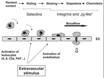
Myocardial ischaemia/reperfusion induces a profound in-flammatory condition with activation of multiple cell types,
including leucocytes and endothelial cells(Fig. 1). Reduced
NO bioavailability signals important patho-physiological
halb richten sich die therapeutischen Bemühungen auf die Wiederherstellung der Perfusion in den ischämi-schen Arealen, um die Entstehung einer Gewebsnekro-se zu vermeiden und um die Funktion des Organs wie-derherzustellen. Die Reperfusion von ischämischem Gewebe geht allerdings oft mit mikrovaskulärer Dys-funktion einher, die sich durch Beeinträchtigung der endothelabhängigen Vasodilatation in Arteriolen und Ansammlung von Leukozyten in den Kapillaren mani-festiert. Die Verfügbarkeit einer großen Anzahl an Knockout-Mäusen ermöglicht wichtige Erkenntnisse über das Zustandekommen der Myokardschädigung durch Ischämie und Reperfusion. Deshalb sind Maus-modelle von großem Interesse für die Entwicklung neuer therapeutischer Strategien beim Menschen.
J Kardiol 2006; 13: 239–44.
Received and accepted: February 28th, 2006.
From the Department of Cardiology, University Hospital of Internal Medicine, Inns-bruck, Austria
Correspondence to: Bernhard Metzler, MD, M. Sc., Department of Cardiology, University Hospital of Internal Medicine, A-6020 Innsbruck, Anichstraße 35; E-Mail: [email protected]
Figure 1: Scheme of involvement of the adhesion molecules in ischaemia/reperfu-sion. Under normal conditions, there are virtually no interactions between the endo-thelium and neutrophils. After coronary ischaemia/reperfusion, the blood vessel’s abil-ity to form NO is severely reduced. With decreased supplies of NO, the coronary ves-sel may constrict. Additional adhesion molecules were expressed on the endothelium and polymorphonuclear leukocytes (PMNs) begin with rolling as the first step in PMN sequestration. As the PMN slows, tighter interactions between CD18 (on PMN) and ICAM-1 (on endothelium) cause the second phase of the PMN-EC interaction called sticking. The final step, the diapedesis, involves the coordination of many factors.
240 J KARDIOL 2006; 13 (7–8)
changes, such as leucocyte adherence and transmigration of mononuclear cells [2]. At least three major pathways may contribute to lethal reperfusion injury. The first mechanism
involves massive calcium overload via Na+/H+ and Na+/Ca2+
exchange during reperfusion, which leads to mitochondrial calcification and contraction band necrosis. A second mecha-nism leads to intracellular accumulation of osmotic active catabolites during ischaemia that may induce massive cell swelling and sarcolemmal rupture when reperfusion provides exposure of the injured cells to abundant extracellular fluid. A third way involves the consequences of a burst of oxygen-based free radical generation. Thus, myocardial reperfusion can cause accelerated progression of cell death in myocytes with prior severe ischaemic damage coupled with salvage of myocytes with less severe ischaemic injury. Some of the known risk factors for cardiovascular disease (hyperchol-esterolaemia, diabetes and hypertension) appear to exaggerate many of the microvascular alterations elicited by ischaemia and reperfusion.
Although much experimental evidence exists in support of the reperfusion component of injury, ischaemia without following reperfusion will cause the destruction of most of the ischae-mic myocardium. This leads to an obvious paradox: the need for re-establishing blood flow at the expense of a profound in-flammatory response [3].
Mouse Types Used in Cardiovascular
Research
Ischaemia/reperfusion research with animal models has being carried out for a considerable time. Knowledge of the patho-genesis and therapeutic intervention of disease conditions and the use of animal models in the research have evolved almost simultaneously. Animal models are designed to be prelimi-nary tools for better understanding of the pathogenesis and improvement in diagnosis, prevention and therapy of heart ischaemic diseases in humans. The evaluation of a risk factor as a single independent variable, with almost complete exclu-sion of other factors, can be performed in animals free of inter-current diseases or abnormalities and with well known genetic characteristics. Furthermore, experiments using animals are the only way to develop and test new diagnostic, preventive and therapeutic procedures for both ethical and practical reasons. The investigator can choose the species, time and method, as well as obtain tissue, serum samples and other materials needed for measurements under optimal conditions, selective circumstances that are difficult, if not impossible, in studies with human subjects.
Attracted by its well-defined genetic map, a number of inves-tigators have begun to use the mouse as an experimental sys-tem for research on cardiovascular diseases. Hundreds of inbred lines have been established and both congenic and recombinant strains are available to facilitate genetic experi-mentation.
Recently, genetic models have been developed in which genes are either overexpressed, deleted or mutated. Such mouse models have considerable advantages, because they overcome
the need to administer factors or their inhibitors, which can be problematic and often difficult to quantify. They also seem to tolerate prolonged monoclonal antibody treatments better.
Beside the use of conventional wild type mice the availability of a broad variety of genetically altered mice in the last years lead to a big rise in interest for these strains.
Knockout Mice
Knockout mice are produced by a laboratory technique called gene targeting. This is the replacement of a gene sequence from the mouse’s own genome with a related one that has been modified to contain a mutation. The replacement occurs by homologous recombination, where two similar DNA sequences line up next to each other and exchange parts. Gene targeting is carried out in mouse embryonic stem cells (ES cells). The aim of this procedure is to get modi-fied ES cells to contribute to the germ line, which gives rise to sperm. Some sperms are produced that carry the desired mutation, and if these fertilise a normal egg, mice develop with one copy of the mutated gene in every cell. Interbreeding such mice will produce some homozygous individuals in the next generation – mice inheriting the mutation from both parents and therefore carrying two copies of the mutant gene. These are knockout mice. Although they are extremely useful in studying gene function, producing and taking care of knockout mice in special facilities is very expensive. Other limitations are the developmental defects, so that many knockout mice die while they are still embryos. The goal of conventional knockout technology is to knock out both alleles so that the gene is entirely absent from all cells. The purpose of the more advanced types of knock-outs, the so called conditional knockknock-outs, in contrast, is to delete a gene in a particular organ, cell type, or stage of devel-opment.
Conditional Knockout Mice
The latest wave of mouse models has advanced beyond
gener-alisedgene knockouts to develop new strategies for precision
engineeringof endogenous genes within specific cell types.
The most successfulapproach has been based on the
genera-tion of mice that harbourfloxed alleles, which contain LoxP
recognition sequencesflanking a critical exon that is required
for the expression orfunction of the gene of interest [4]. These
miceare generated by homologous recombination of targeting
vectorsin embryonic stem cells that bring in the LoxP sites
into the germline. The floxed allele mice express the normal
gene product,because the LoxP sites are located within the
intron sequencesthat are spliced out during RNA processing.
However, the intervening sequences between the LoxP sites
can be excised by the expressionof CRE recombinase, which
is brought into the genetic backgroundof the floxed allele
mice via interbreeding. By controllingthe expression of CRE
recombinase to a specific tissue, for exampleto the ventricular
chamber, it is possible to generate micethat harbour a
ven-tricular-restricted mutation in a gene thatis widely expressed,
thereby allowing a direct examination of the role of a given
J KARDIOL 2006; 13 (7–8) Ischaemia/Reperfusion Models
241
Transgenic Mice
A transgenic mouse contains additional, artificially-intro-duced genetic material in every cell. This often confers a gain of function, for example the mouse may produce a new pro-tein, but a loss of function may occur if the integrated DNA interrupts another gene. A transgenic mouse is a very useful system for studying mammalian gene function and regulation because analysis is carried out on the whole organism. DNA can be integrated by injecting it into the pronucleus of a ferti-lised ovum. The DNA can integrate anywhere in the genome, and multiple copies often integrate in a head-to-tail fashion. There is no need for homology between the injected DNA and the host genome.
Mouse Models for Myocardial Ischaemia
and Reperfusion
In the present review we will describe different mouse models for myocardial ischaemia/reperfusion. With the help of a big variety of genetically manipulated mice the pathophysiologi-cal effects of the targeted genes can be assessed and should bring some new insights about ischaemia/reperfusion.
Open Chest Ischaemia/Reperfusion Model
In 1995 Michael et al. [5] first described the open chest in vivo
mouse model. Mice are anaesthetised with phenobarbital (50 mg/kg, i.p.), fixed in the supine position by taping the extremities and the upper jaw. A midline skin incision from the xiphoid process to the submentum is made. After separat-ing the salivary glands, the muscles overlyseparat-ing the trachea are retracted, and a tracheotomy is performed. A polyethylene tube (No. 90) is carefully inserted into the trachea (about 5 mm from the larynx), taped in place to prevent dislodgement and connected via a loose junction to a rodent ventilator (model 687; Harvard Apparatus). After ventilation (tidal vol-ume 1,2 ml/min., rate 110 strokes/min.) is started by supple-mentation of 100 % oxygen the chest is opened by a lateral cut along the left side of the sternum. An adequate tidal volume results in an adequate inflation of the lungs without over-expression. The whole procedure is aided by a microscope (Olympus SZH 10). With an electrocoagulator, intercostal blood vessels are coagulated. The chest walls are then re-tracted using a 6-0 silk suture for better visualisation of the heart. After removing the pericardial sac and slightly retract-ing the left auricle, the left descendretract-ing artery (LAD) becomes clearly visible. A 1 mm section of a PE-10 tubing is placed on top of the LAD to secure its ligation without damaging the artery. The LAD ligation is done with an 8-0 silk and becomes evident by discoloration of the left ventricle (Fig. 2). To avoid cooling of the animal, it is important to use a heating pad placed under the mouse. After a defined time of LAD occlu-sion, the ligature can be removed by cutting the knot on top of this PE-10 tube and reperfusion can be visually confirmed.
Minimal Invasive Ischaemia/Reperfusion Model
The advantage of this in vivo model with an implantable
de-vice for artery occlusion is to overcome the high level of back-ground inflammation due to surgical trauma associated with
the open chest model, permitting a more accurate and inter-pretable response about the involvement of different cytokines and other mediators in the ischaemia/reperfusion injury of the myocardium [6].
After performing a thoracotomy as described in the open chest
model, an 8-0 Surgipro monofilamentpolypropylene suture
with the U-shaped tapered needle is passed under the LAD. The two ends of the suture are threaded through a 0.5 mm piece of PE-10 tubing, forming a loose snare around the LAD,
and are then threadedthrough the end of a size 3 Kalt suture
needle (Fine Science Tools) and exteriorised through each side of the chest wall. The chest is closed, and the ends of the exteriorised 8-0 suture are then tucked under the skin. The animal is removed from the respirator and is allowed to
breathe 100 % O2 via a nasal cone until full recovery of
con-sciousness.
Ischaemia Without Reperfusion
Similar to the in vivo open chest model for myocardial
ischae-mia/reperfusion the mouse thorax is opened and prepared to make the anterior heart surface visible. Because reperfusion is not demanded, the LAD ligation is done with an 8-0 silk and will not be removed later. Afterwards, the retraction sutures are removed and the chest wall and skin are closed with su-tures, and the animal is allowed to recover. According to the experimental protocol mice can be sacrificed after induction of a transmural myocardial infarction with any desired time of ischaemia. This protocol seems to be particularly interesting for heart failure studies.
Ischaemia/Reperfusion With Ischaemic Precon-ditioning
Miller et al. [7] have been among the first who demonstrated the early phase of ischaemic preconditioning in the
myocar-dium in the in vivo mouse model. We also used this protocol
and showed its highly effectiveness in inducing a significant limitation of the myocardial damage [8]. After dissecting the pericardium an 8-0 silk suture with a U-shaped needle is
passedunder the LAD. The LAD ligation becomes evident by
discoloration of the left ventricle. For the preconditioning
242 J KARDIOL 2006; 13 (7–8)
protocol the knot on top of the 1 mm plastic tube is tighten carefully and loosen after the given time. Great attention should be paid in order to avoid damage of the LAD.
For preconditioning, the mouse undergoes three cycles of 5 min. artery occlusion followed by 5 min. reperfusion, re-spectively. Ten minutes later, the animal undergoes 30 min. of coronary occlusion of the artery, followed by 2 h of reperfu-sion. By this protocol it is possible to demonstrate a reduction of the infarct size of about 50 %. The responsible mechanism is still not fully elucidated. Various possibly involved signal-ling pathways have already been described. Among the most important seem to be reactive oxygen species, isoforms of protein kinase C (PKC) and adenosine.
Langendorf Model (
Ex Vivo
)Animals are usually injected with heparin (1000 U/kg, i. p.) 20 min. prior to the experimental protocol. Once the animal is anaesthetised, the heart is removed and placed in a weigh bath
with some Ca2+-containing buffer. Holding the aorta with two
forceps, the heart is lifted from the buffer and placed on a perfusion cannula that should have buffer running through at a slow rate. After clamping the aorta to the perfusion needle, buffer flow rate may be increased. If the procedure is per-formed successfully, the heart will begin to beat rhythmically and the drops will become clear.
Once the heart is successfully hung, it is perfused (at 37 °C) with Krebs-Henseleit bicarbonate (KHB) buffer containing
(in mM): 118 NaCl, 4.7 KCl, 1.25 CaCl2, 1.2 MgSO4,
1.2 KH2PO4, 25 NaHCO3, 10
N-[2-hydro-ethyl]-piperazine-N’-[2-ethanesulfonic acid] (HEPES) and 11.1 glucose,
equili-brated with 5 % CO2 – 95 % O2.
Measurements of Myocardial Tissue
Damage
Measurement of Cardiac Enzymes
Blood concentration of cardiac enzymes can be used as an index of cardiac cellular damage [9]. Heparin-blood is col-lected when mice are sacrificed for organ removal, and it is usually taken by puncture of the Vena cava inferior or of the Vena portae. Blood concentration of troponin T is measured by using a quantitative rapid assay kit (Roche Diagnostics, Mannheim, Germany).
Creatine kinase and lactate dehydrogenase isoenzyme 1 (LDH-1) may be electrophoretically analysed using commer-cially available kits (Paragon, Fullerton, CA, USA). Gels are scanned with a densitometer and the relative activity of each single isoenzyme fraction is calculated against total enzyme activities.
Measurement of the Infarction Area With Trans-mission Microscope
After reperfusion, hearts are removed and the portion of tissue below the ligation site is fixed in 4 % paraformaldehyde at
4 °C overnight and embedded in paraffin. Sections of 5µm
are cut from the cross area and stained with hematoxilin and eosin (HE) for histological evaluation of tissue damage.
The measurement of the scar area may be done by reviewing these sections with a B×60 microscope (Zeiss, Jena, Ger-many) equipped with a Sony 3CCD camera and a television monitor. A transmission scanning microscope (Bio-Rad), equipped with a 488 nm argon ion laser and Plan Neofluar 10×/0.3 oculars connected with the program START LSM 510 may be used to scan the images. The scar is defined as the region between the last living myocytes and the cardiac mem-brane, and is measured in square micrometers [10]. Despite of the real three-dimensional extension of myocardial infarction, this two-dimensional approach has some practical advan-tages: it allows to distinguish damaged tissue already after a short time frame of ischaemia and to make an accurate meas-urement of the infarction area despite of its small size. This technique is quite easy to perform and is less time consuming than Evans blue staining.
Zingarelli Damage Scoring System
For quantitative histological evaluation of tissue damage the previously published scoring system by Zingarelli et al. [11] can be used. The following criteria are considered: score 0, no damage; score 1 (mild), interstitial oedema and focal necrosis; score 2 (moderate), diffuse myocardial cell swelling and necro-sis; score 3 (severe), necrosis with the presence of contraction bands and neutrophil infiltrate; and score 4 (highly severe), widespread necrosis with the presence of contraction bands, neutrophil infiltrate and haemorrhage. At least ten tissue sam-ples/group should be used for a satisfying statistical analysis.
Assessment of Area at Risk and Infarction Size With Evans Blue and TTC-Staining
After ligation of the coronary artery 1 % Evans blue is per-fused into the aorta and coronary arteries with distribution throughout the ventricular wall. The left ventricle of each heart is excised and weighed. Sections of the ventricle below the site of ligature have uniformly blue areas, surrounding a smaller colourless one, which is the tissue supplied by the ligated vessel. After incubation in 1.5 % triphenyltetrazol-iumchloride (TTC), viable myocardium stains brick red and the infarct appears pale white (Fig. 3). The area of infarction is determined by computerised planimetry using an image analysis software program (OPTIMAS, Bioscan, Redmond, WA). The size of infarction is determined by the following
equations: weight of infarction = (A1 × WT1) + (A2 × WT2) +
(A3 × WT3) + …, where A is the percent area of infarction by
planimetry from subscribed numbers representing sections, and WT is weight of the same numbered sections. Percentage of infarcted LV is (WT of infarction/WT of LV) × 100. Area at risk as percentage of LV is calculated by (WT of LV – WT of LV stained blue)/WT of LV. The weight of LV stained blue is calculated in a similar fashion: sum of products of the percent area of each slice × the weight of the representative slice.
J KARDIOL 2006; 13 (7–8) Ischaemia/Reperfusion Models
243
relatively inaccurate, so that it seems to be much more suitable for bigger animal hearts like those of a pig or a dog.
Transthoracal and Transesophageal Echocardio-graphy
Transthoracic echocardiography may be performed using a Sonos 5500 ultrasound machine (Hewlett Packard Co.) with a 12 MHz phased array transducer and a frame rate of 41/s. The transducer is used at a depth setting of 2 cm to optimise reso-lution. Mice are anaesthetised and placed on a heating pad in a shallow left lateral position and a standard lead II electrocar-diogram is recorded for heart rate (HR) measurement. After a 2-dimensional (2 D) image is obtained in parasternal short axis view at the level close to papillary muscles, a 2 D guided M-mode trace crossing the anterior and posterior wall of the LV is recorded at a sweep speed of 100 mm/s. It is possible to measure digitally following parameters: LV internal end-systolic and end-diastolic diameters (LVESd, LVEDd), exter-nal LV diastolic diameter (ExLVDd), anterior and posterior wall thickness of systole and diastole (Aws th, Awd th, Pws th and Pwd th). Scherrer-Crosbie et al. described a three-dimen-sional echocardiographic assessment of left ventricular wall motion abnormalities after infarction in the mouse heart [12]. Also a transesophageal echocardiographic approach is de-scribed [13].
Magnetic Resonance Imaging (MRI) Assessment of Murine Heart Function
The group around Weiss described an in vivo MRI assessment
technique of the murine heart [14]. High-resolution images may be obtained to confirm position, define regions of
meta-bolic interest or quantify ventricularfunction. It is possible to
determine left ventricular volumesat end diastole and end
sys-tole using a suitable software package. The left ventricular ejection fraction may be calculated from the relative
differ-encein end-diastolic and end-systolic cavityvolumes.
Single Photon Emission Computed Tomography (SPECT)
The importance of myocardial imaging in small animals has led to the development of SPECT systems designed
specifi-Figure 3: Cross sections of the TTC-stained heart. a) Areas at risk are delineated by Evans blue dye in a representative heart with occlusion of the left descending coro-nary artery. All areas that stain blue were therefore not at risk. b) Areas that appear pale white are infarcted. Areas that remain red were not infarcted but were within the area at risk. Transverse sections are from the apex to the base (from left to right side).
244 J KARDIOL 2006; 13 (7–8)
cally for this application, which give good agreement between
in vivo imaging data and post mortem autoradiographic and staining studies. The true size of myocardial perfusion defects can be detected with very high accuracy. This has important implications for the study of cardiac function in mice, and may be useful in phenotyping transgenic mouse models of heart disease.
Electrocardiogram and In Vivo Left
Ven-tricular Pressure-Volume Measurements
Electrocardiographic Measurements
Continuous ECG recordings can be made with a two-lead ECG Haemodynamic Data Acquisition System apparatus (Instrument Services, Maastricht, The Netherlands) to detect arrhythmias. The first lead is positioned in the left lower leg, the second lead in the right upper leg. Recordings are made every 5 seconds in trend save mode with a sample interval of 1 ms and are started at the beginning of the surgical procedure [15].
In Vivo
Left Ventricular Pressure-Volume MeasurementsLeft ventricular function can be assessed by simultaneous measurement of left ventricular pressure and volume. This can be done with a Sigma SA (CDLeycom, Zoetermeer, The Neth-erlands) single segment data acquisition module operating on
a constant excitation current of 30µA and with Cirlab
soft-ware (LUMC, Leiden, The Netherlands) for offline data analysis. A 1.4 Fr Millar pressure-conductance catheter (SPR-719, Millar Instruments, Houston, TX, USA) is used for the LV pressure-volume measurements. The time-varying
ven-tricular volume V(t) may be estimated from V(t) = ρL2 [G(t) –
GP] where ρ (rho) is the mouse specific blood resistance,
L indicates the distance between the sensing electrodes and G(t) the instantaneous conductance.
Conclusion
Due to the fact that coronary heart disease is still the number one killer in the western world, scientists and doctors are encouraged to find new therapeutic strategies. Due to ethical considerations experimental ischaemia/reperfusion (I/R) stud-ies almost exclusively can be performed in laboratory ani-mals. The availability of a broad variety of genetically altered mice in the last years led to a remarkable rise in the interest for experimental mouse models.
References:
1. Braunwald E, Kloner RA. Myocardial reper-fusion: a double-edged sword? J Clin Invest 1985; 76: 1713–9.
2. Pabla R, Buda AJ, Flynn DM, Blesse SA, Shin AM, Curtis MJ, Lefer DJ. Nitric Oxide attenuates neutrophil-mediated myocardial contractile dysfunction after ischemia and reperfusion. Circ Res 1996; 78: 65–72. 3. Braunwald E, Kloner RA. The stunned myo-cardium: prolonged, postischemic ventricular dysfunction. Circulation 1982; 66: 1146–9. 4. Rajewsky K, Gu H, Kühn R, Betz UA, Müller W, Roes J, Schwenk F. Conditional gene tar-geting. J Clin Invest 1996; 98: 600–3. 5. Michael LH, Entman ML, Hartley CJ, Youker KA, Zhu J, Hall SR, Hawkins HK, Berens K, Ballantyne CM. Myocardial ischemia and reperfusion: a murine model. Am J Physiol 1995; 269: H2147–H2154. 6. Nossuli TO, Lakshminarayana V, Baum-garten G, Taffet GE, Ballantyne CM, Michael LH, Entman ML. A chronic mouse model of myocardial ischemia-reperfusion: essential in cytokine studies. Am J Physiol Heart Circ Physiol 2000; 278: H1049–H1055. 7. Miller DL, Van Winkle DM. Ischemic pconditioning limits infarct size following re-gional ischemia-reperfusion in in-situ mouse hearts. Cardiovasc Res 1994; 42: 680–4. 8. Mayr M, Metzler B, Chung Y, McGregor E, Mayr U, Troy H, Hu Y, Leitges M, Pachinger O, Griffiths JR, Dunn M, Xu Q. Ischemic precon-ditioning exaggerates cardiac damage in PKC-δ null mice. Am J Physiol 2004; 287: H946–H956.
9. Metzler B, Lercher-Hammerer A, Dietrich H, Jehle J, Pachinger O, Mair J. Plasma cardiac troponin T closely correlates with infarct size in a mouse model of acute myocardial infarc-tion. Clin Chim Acta 2002; 325: 87–90. 10. Metzler B, Mair J, Lercher A, Schaber C, Hintringer F, Pachinger O, Xu Q. Mouse model of myocardial remodeling after ischemia: role of intercellular adhesion molecule-1. Cardiovasc Res 2001; 49: 399–407. 11. Zingarelli B, Salzman AL, Szabo C. Ge-netic disruption of poly (ADP-ribose) syn-thetase inhibits the expression of P-selectin and intercellular adhesion molecule-1 in myocardial ischemia/reperfusion injury. Circ Res 1998; 83: 85–94.
12. Scherrer-Crosbie M, Steudel W, Hunziker PR, Liel-Cohen N, Ullrich R, Zapol WM, Picard MH. Three-dimensional echocardiographic assessment of left ventricular wall motion abnormalities in mouse myocardial infarction. J Am Soc Echocardiogr 1999; 12: 834–40. 13. Ramani R, Mathier M, Dawson J, McTiernan CF, Feldman AM. Assessment of infarct size and myocardial function in mice using transesophageal echocardiography. J Am Soc Echocardiogr 2004; 17: 649–53. 14. Chacko VP, Aresta F, Chacko SM, Weiss RG. MRI/MRS assessment of in vivo murine cardiac metabolism, morphology, and function at physiological heart rates. Am J Physiol 2000; 279: H2218–H2224.
15. Lips DJ, Van der Nagel T, Steendijk P, Palmen M, Janssen BJ, Van Dantzig JM, De Windt LJ, Doevendans PA. Left ventricular pressure-volume measurements in mice: com-parison of closed-chest versus open-chest approach. Basic Res Cardiol 2004; 99: 351–9.
This review describes the different mouse I/R models and the techniques for quantification of I/R damage. Beside the
con-ventional in vivo mouse model for I/R, the open chest mouse
model, also newer ones, such as the closed chest I/R model are described. Further the novel quantification of I/R damage by help of a transmission microscope is explained. Also known techniques established in clinical practice in humans, such as echocardiography and MRI, are described for mice. The ad-vantages and disadad-vantages of the different approaches also were discussed. The given company names for the different technical devices should serve just as a proposal. We know that there are a number of equal technical devices manufac-tured by different companies. This review should help the interested scientists to choose the most suitable mouse I/R model to clarify their specific questions.
Die neue Rubrik im Journal für Kardiologie:
Clinical Shortcuts
In dieser Rubrik werden Flow-Charts der Kardiologie kurz und bündig vorgestellt
Zuletzt erschienen:
Interventionelle kathetergestützte
Diagnostik der Synkope
Aortenklappenimplantation (TAVI)
J Kardiol 2015; 22 (5–6): 132–4.
J Kardiol 2014; 21 (11–12): 334–7.
Einsatz einer perioperativen Blockertherapie
Kardiologische Rehabilitation nach
zur Reduktion von Morbidität und Mortalität
akutem Koronarsyndrom (ACS)
J Kardiol 2015; 22 (1–2): 38–40.
J Kardiol 2015; 22 (9–10): 232–5.
Besuchen Sie unsere Rubrik
P
聺
Medizintechnik-Produkte
boso ABI-system 100 Boso GmbH & Co KG IntelliSpace Cardiovascular
Philips Austria GmbH, Healthcare
BioMonitor 2
BIOTRONIK Vertriebs-GmbH CT TAVI Planning mit
syngo.CT Cardiac Function-Valve Pilot Siemens AG Österreich
STA R Max
Stago Österreich GmbH