INTRODUCTION
T
he emergence of new minimally invasivetechniques for parathyroidectomy in the 1990s allowed surgeons to perform a traditional surgical procedure with a technique that allows less trauma, better surgical exposure and better dissection. In the hands of experienced surgeons, a minimally invasive procedure should achieve at least the same results, with the major advantage of reducing invasive trauma and improving aesthetic outcome. Laparoscopy contributed to these results, led to minimally invasive neck surgery to be developed and several new techniques arose. The logical sequence of the surgical procedures of the future should be, following decreasing invasiveness criteria, the execution of surgeries through single access, thus preventing issues inherent to incisions1-9.
The technique of minimally invasive video assisted parathyroidectomy (MIVAT), developed by Miccoli, has become the most widespread9,10. With
such new techniques, however, many doubts arose about the safety of minimally invasive surgery, and new
studies were published comparing open surgery with endoscopic one11-13. The axillary approach was then
used as an alternative to hide the scar, but another trocar was needed to access the thyroid gland, usually through the Axillo-Bilateral-Breast Approach (ABBA), creating a wide dissection and increasing complications risks14-16.
With the idea of natural orifice surgery (NOTES), Witzel et al.17 performed a sublingual
trans-oral access for thyroidectomy in a study with animals in 2007. Benhidjeb et al.18 followed the study and used
the same technique in cadavers, making the first cases in humans19,20. With single portal surgery consecrated
as the surgery of the moment, transaxillary single-port21 thyroidectomy seems to be more plausible.
After a previous study, which aimed to establish a standard for transaxillary single-port thyroidectomy in cadavers, our team continued to develop this technique, now for transaxillary single-port unilateral parathyroidectomy. Thus, the objective of this study was to develop and improve the surgical technique of parathyroidectomy, using a single transaxillary access.
1 - Federal University of the State of Rio de Janeiro (UNIRIO), Department of General and Specialized Surgery, Professional Masters in Videoendos-copic Techniques, Rio de Janeiro, Rio de Janeiro State, Brazil. 2 - Charité Hospital, Center for Innovative Surgery, Berlin, Brandenburg, Germany. 3 - North Fluminense State University (UENF), Department of Post-Graduation, Campos, Rio de Janeiro State, Brazil.
evaluate its viability and reproduction. Method: we performed ten subtotal parathyroidectomies through a transaxillary TriPort access in cadavers. The technique consisted of access through the axillary fossa, creating a subcutaneous tunnel to the anterior cervical region, for handling of the thyroid gland and dissection and resection of the parathyroid glands. Results: all surgeries were successful. The mean time of surgery was 65 minutes (57-79 min), with uncomplicated identification of all anatomical structures. There was no need for comple-mentary incisions in the cervical region. Conclusion: the transaxillary single-port subtotal parathyroidectomy technique was feasible and reproducible, suggesting an alternative for minimally invasive cervical surgery.
METHODS
We carried out the study at the Anatomical Institute of the Federal University of the State of Rio de Janeiro (UNIRIO), in the years 2013 and 2014, using ten fresh frozen cadavers prior to their formalization. Inclusion criteria were cadavers of both genders, middle-aged, without previous neck surgery, and BMI <30.
In the operative technique, we placed the cadaver in dorsal decubitus with the upper limb extended to 270° and flexed at the elbow with the hand under the head. A monitor sit at the head of the cadaver, and the surgeon, as well as the assistant, remained below the ipsilateral arm. The procedure started with a 2cm incision in the axillary sulcus ipsilateral to the parathyroid to be resected, followed by dissection of the subcutaneous tissue with Kelly’s forceps, placement of the TriPort and insulation with CO2 (4 to 8 mmHg) (Figure 1).
We then proceeded to the subcutaneous tunnel, with blunt dissection with a 30° endoscope, 10mm in diameter, and cutting until reaching the neck, above the pectoral muscle, passing over the clavicle (Figure 2).
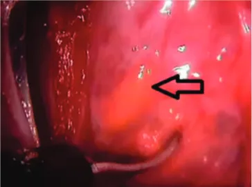
Then we opened the platysma muscle and bluntly separated the pre-thyroid muscles, identifying the thyroid gland. An ascending dissection of the thyroid gland followed with scissors, rotating it medially on its longitudinal axis with an apprehension forceps for identification of the recurrent laryngeal nerve and parathyroids (Figures 3 and 4).
We then apprehended the parathyroid followed by its dissection and section with scissors. We removed the whole gland through the incision, protected by the TriPort, and performed wound synthesis with intradermal suture (Figure 5).
Figure 1. Two-cm incision in the axillary sulcus.
Figure 2. Making of the subcutaneous tunnel.
RESULTS
The transaxillary parathyroidectomy was successful in all cases. It was possible to visualize the two glands on the studied side, as well as the recurrent laryngeal nerve. The mean time of preparation of the subcutaneous tunnel for parathyroid access was 35.1 minutes (29-42) (Table 1).
The mean surgery time was 65 minutes (57-79) (Table 2).
DISCUSSION
The present study shows the feasibility of neck surgery, such as thyroidectomy and parathyroidectomy, through a single access. Since Gagner6 performed an
a 2cm incision in the anterior cervical region, is the most widespread and employed procedure for minimally invasive surgery, described in 1997 for parathyroidectomy for primary hyperparathyroidism. It has been observed that it could also be used in thyroid surgeries and in 2002, Miccoli et al.10 carried out a
multicenter study with 336 patients. There were with seven transient recurrent laryngeal nerve lesions and one permanent lesion (0.3%), nine cases of transient hypoparathyroidism, and two cases of permanent hypoparathyroidism (0.67%). The disadvantages of this surgery are the scar, even if small, in the neck, the contraindication in large glands, and the impossibility to perform lymphadenectomy in the central compartment, which suggests that the main indication for MIVAT are benign diseases.
In order to conceal the surgical scar, the transoral, sublingual approach to thyroidectomy, despite anatomical complexity, was relatively easy and added parathyroidectomy and thyroidectomy
Figure 5. Final aspect of the scar.
Table 1. Time for the subcutaneous tunnel
Cadaver Tunnel time
1 42min
2 39min
3 38min
4 40min
5 35min
6 33min
7 34min
8 30min
9 29min
10 31min
Table 2. Operative time
Cadaver Operative Time
1 79min
2 74min
3 68min
4 70min
5 63min
6 62min
7 62min
8 57min
9 57min
to NOTES techniques17-20. It seems to be a promising
surgery, since the glands have their embryonic origins at the base of the tongue and migrate to the anterior cervical region, being a natural path to go through during surgery. However, it requires studies with larger samples to demonstrate its real importance.
Lee et al.21 devised the single-port axillary
endoscopic thyroidectomy. However, in their study, they used an adapted system, with an Alexis® and a glove, in whichhe inserted three 5mm trocars, in the first, third and fifth fingers. To create the subcutaneous space, they used an acrylic bar and CO2 gas inflating at a pressure of 4 to 6 mmHg. They used a flexible 5mm endoscope, as well as a Sonosurg scissors. Thyroid resection began at the upper pole toward the lower pole with identification and preservation of the recurrent laryngeal nerve and parathyroids. The gland was withdrawn and a suction drain was inserted. The results were good and there were no complications. In the article, they noted that the 5mm range helped to avoid the clash of instruments by creating two imaginary planes, upper plane for the camera and lower plane for the instruments. Another important point detected was the use of instruments of different lengths, preventing the surgeon from colliding with his forceps.
More recently, Kang et al.22 showed the
feasibility and safety of robotic surgery in transaxillary thyroidectomy. There are, however, limitations to robotic surgery due to the high cost and specific training, thus not being accessible to all endoscopic surgeons. There is also a need for larger incisions for the robotic arms. With the new generation of robots and patentsexpiration, there may be cost reduction and accommodation for single-port surgery.
Lee et al.23 studied 259 patients, 96 in the
endoscopic group and 163 in the robotic group, comparing the two techniques. Both groups had similar operative times, as well as length of hospital stay and blood loss, but the number of lymph nodes removed was higher in the robotic group (P.004). In a meta-analysis comparing robotic with endoscopic surgery, Lin et al.24 did not observe statistical difference
regarding operative time and conversions for open surgery. However, the robotic arm obtained a greater number of complications, the authors suggesting
that there is no clinical benefit for robotic surgery, when compared with the endoscopic one, in the accomplishment of thyroidectomies.
In a study by Phillips et al.25, using the same
access for thyroidectomy in animals and cadavers, they observed that it was possible to perform the procedure, with identification of all anatomical structures and preservation of the parathyroid glands. Based on this work, we performed this study aimed at the excision of the parathyroid glands, preferentially for adenomas.
In our study, the use of the TriPort gave us greater freedom of movement with the tweezers, even in a 2cm incision. This access is possible both for thyroidectomy and for parathyroidectomy, avoiding a scar in the cervical region and transferring it to the axillary region. One can extend the scar if the size of the surgical specimen is large, without hampering aesthetics. It is a procedure requiring a team of professionals with experience in Single-Port surgery, since the tweezers work in parallel with the camera and there is collision between them. Laparoscopic tweezers of different sizes can minimize the problem of collisions by keeping the surgeon and the assistant a little further from each other. The space is restricted, but sufficient to work with safety, enabling, with clarity, the identification of noble structures.
In addition to not leaving a visible scar, another advantage is that there is no need for neck hyperextension, a position that causes postoperative pain and is limited in patients with cervical pseudoarthrosis. Among the disadvantages of the technique is its use in patients with lesions larger than 4cm, since it is difficult to dissect and remove the specimen through the subcutaneous tunnel, and in those with disease in multiple glands, due to the need for bilateral dissection. However, since most of the primary hyperparathyroidisms is from single glands, the techniqueis perfectly feasible.
REFERENCES
1. Kalloo AN, Singh VK, Jagannath BS, Niiyama H, Hill SL, Vaughn CA, et al. Flexible transgastric peritone-oscopy: a novel approach to diagnostic and thera-peutic interventions in the peritoneal cavity. Gastro-intest Endosc. 2004;60(1):114-7.
2. Zorron R, Palanivelu C, Galvão Neto MP, Ramos A, Salinas G, Burghardt J, et al. International mul-ticenter trial on clinical natural orifice surgery--NOTES IMTN study: preliminary results of 362 pa-tients. Surg Innov. 2010;17(2):142-58.
3. Lehmann KS, Ritz JP, Wibmer A, Gellert K, Zornig C, Burghardt J, et al. The German registry for natural orifice translumenal endoscopic surgery: report of the first 551 patients. Ann Surg. 2010; 252(2):263-70.
4. Meining A, Feussner H, Swain P, Yang GZ, Lehmann K, Zorron R, et al. Natural orifice transluminal endoscopic surgery (NOTES) in Europe: summary of the working group reports of the Euro-NOTES meeting 2010. Endoscopy. 2011;43(2):140-3. 5. Muenscher A, Dalchow C, Kutta H, Knecht R. The
endoscopic approach to the neck: a review of the literature, and overview of the various techniques. Surg Endosc. 2011;25(5):1358-63.
6. Gagner M. Endoscopic subtotal parathyroidectomy in patients with primary hyperparathyroidism. Br J Surg. 1996;83(6):875.
7. Hüscher CS, Chiodini S, Napolitano G, Recher A. Endoscopic right thyroid lobectomy. Surg Endosc. 1997;11(8):877.
8. Gagner M, Inabnet WB 3rd. Endoscopic thy-roidectomy for solitary thyroid nodules. Thyroid. 2001;11(2):161-3.
9. Miccoli P, Pinchera A, Cecchini G, Conte M, Bendinelli C, Vignali E, et al. Minimally invasive, video-assisted parathyroid surgery for primary hyperparathyroidism. J Endocrin Invest. 1997;20(7):429-30.
10. Miccoli P, Bellantone R, Mourad M, Walz M, Raffaelli M, Berti P. Minimally invasive video-assisted thyroidectomy: multiinstitutional experience. World J Surg. 2002;26(8):972-5.
11. Ikeda Y, Takami H, Sasaki Y, Takayama J, Kuriha-ra H. Are there significant benefits of minimally invasive endoscopic thyroidectomy? World J Surg. 2004;28(11):1075-8.
12. Inabnet WB 3rd, Jacob BP, Gagner M. Minimally invasive endoscopic thyroidectomy by a cervical approach. Surg Endosc. 2003;17(11):1808-11. 13. Oertli D, Harder F. Surgical approach to thyroid
nodules and cancer. Baillieres Best Pract Res Clin Endocrinol Metab. 2000;14(4):651-66.
14. Bärlehner E, Benhidjeb T. Cervical scarless endoscopic thyroidectomy: axillo-bilateral-breast approach (ABBA). Surg Endosc. 2008;22(1):154-7. 15. Shimazu K, Shiba E, Tamaki Y, Takiguchi S,
Taniguchi E, Ohashi S, et al. Endoscopic thyroid cervical anterior, para manuseio da glândula tireoide e dissecção e ressecção das paratireoides. Resultados: todas as cirurgias foram realizadas com sucesso. O tempo médio de cirurgia foi 65 minutos (57-79 min), com identificação, sem dificuldades, de todas as estru-turas anatômicas. Não houve necessidade de incisões complementares na região cervical. Conclusão: a técnica de paratireoidectomia subtotal transaxilar single-port foi viável e reprodutível, sugerindo uma alternativa para a cirurgia cervical minimamente invasiva.
surgery through the axillo-bilateral breast approach. Surg Laparosc Endosc. 2003;13(3):196-201. 16. Koh YW, Park JH, Kim JW, Lee SW, Choi EC.
En-doscopic hemithyroidectomy with prophylactic ipsilateral central neck dissection via an unilateral axillo-breast approach without gas insufflation for unilateral micropapillary thyroid carcinoma: pre-liminary report. Surg Endosc. 2010;24(1):188-97. 17. Witzel K, von Rahden BH, Kaminski C, Stein HJ.
Transoral access for endoscopic thyroid resection. Surg Endosc. 2008;22(8):1871-5.
18. Benhidjeb T, Wilhelm T, Harlaar J, Kleinrensink GJ, Schneider TA, Stark M. Natural orifice surgery on thyroid gland: totally transoral video-assisted thy-roidectomy (TOVAT): report of first experimental results of a new surgical method. Surg Endosc. 2009;23(5):1119-20.
19. Karakas E, Steinfeldt T, Gockel A, Westermann R, Kiefer A, Bartsch DK. Transoral thyroid and para-thyroid surgery. Surg Endosc. 2010;24(6):1261-7. 20. Richmon JD, Pattani KM, Benhidjeb T, Tufano RP.
Transoral robotic-assisted thyroidectomy: a pre-clinical feasibility study in 2 cadavers. Head Neck. 2011;33(3):330-3.
21. Lee D, Nam Y, Sung K. Single-incision endoscopic thyroidectomy by the axillary approach. J Laparo-endosc Adv Surg Tech A. 2010; 20(10):839-42.
22. Kang SW, Lee SC, Lee SH, Lee KY, Jeong JJ, Lee YS, et al. Robotic thyroid surgery using a gasless, transaxillary approach and the da Vinci S system: the operative outcomes of 338 consecutive pa-tients. Surgery. 2009;146(6):1048-55.
23. Lee J, Lee JH, Nah KY, Soh EY, Chung WY. Com-parison of endoscopic and robotic thyroidectomy. Ann Surg Oncol. 2011;18(5):1439-46.
24. Lin S, Chen ZH, Jiang HG, Yu JR. Robotic thyroidectomy versus endoscopic thyroidectomy: a meta-analysis. World J Surg Oncol. 2012;10:239. 25. Phillips HN, Fiorelli RK, Queiroz MR, Oliveira
AL, Zorron R. Single-port unilateral transaxillary totally endoscopic thyroidectomy: a survival ani-mal and cadaver feasibility study. J Minim Access Surg. 2016;12(1):63-7.
Received in: 22/09/2016
Accepted for publication: 15/12/2016 Conflict of interest: none.
Source of funding: none.
Mailing address:
Rossano Kepler Alvim Fiorelli E-mail: [email protected]