©Revista Brasileira de Fisioterapia
RELIABILITY AND VALIDITY OF THORACIC KYPHOSIS MEASUREMENTS
USING THE FLEXICURVE METHOD
T
EIXEIRAFA
1& C
ARVALHOGA
1,21 Post-Graduate Program in Physical Education, Catholic University of Brasília, Brasília, DF – Brazil 2 Medical Department, Parliament Chamber, Brasília, DF - Brazil
Correspondence to: Fellipe Teixeira, QNA 12, casa 06, Taguatinga, CEP 72110-120, Brasília, DF - Brazil Received: 03/07/2006 - Revised: 20/03/2007 - Accepted: 15/05/2007
ABSTRACT
Background: Thoracic hyperkyphosis is one of the most common postural abnormalities. It is defined as increased thoracic curvature in the sagittal plan of the vertebral column. Normal kyphosis may range from 20º to 50º according to Cobb’s radiographic method. The radiographic method is the most popular kyphosis measuring method, but because it is an expensive method and it exposes the individual to radiation, it is not the most appropriate method for periodic patient follow-up. Routine clinical examinations such as physiotherapeutic evaluation of thoracic kyphosis need to be valid, reliable, sensitive, practical and cheap. Objective: To investigate the comparative validity and the intra and inter-rater reliability of thoracic kyphosis measurements using the flexicurve method. Method: This was a cross-sectional study in which the thoracic kyphosis of 56 people was evaluated from sagittal radiography of the thoracic column using Cobb’s method and by means of the flexicurve method, by two evaluators. Results: The intraclass correlation coefficient (ICC) between the measurements from the Cobb and flexicurve methods was 0.906. For diagnosing thoracic hyperkyphosis, the sensitivity was 85% and the specificity was 97%. Conclusion: The flexicurve method was shown to be a suitable quantitative clinical method for measuring the curvature of thoracic kyphosis.
Key words: kyphosis; posture; flexicurve method; Cobb’s method; validity; reliability; flexicurve.
INTRODUCTION
Only one in every three vertebral deformities detected by radiography is diagnosed clinically1. Of the several postural alterations, thoracic hyperkyphosis is among the most common ones. Thoracic hyperkyphosis is the increase of the thoracic curvature in the sagittal plane, and indication for treatment is based on kyphosis angular measurement2. Normal kyphosis ranges from 20 to 50º when assessed by Cobb’s radiographic method3. Radiographic methods are the most commonly used for kyphosis measurement. However, because they are expensive and expose individuals to radiation, they are not the most appropriate methods for periodic patient follow-up or for screening purposes3.
There are some instruments available for clinical measurement of the thoracic kyphosis. A flexible ruler, named
flexicurve, has been used to measure the spine curvatures
on the sagittal plane. This instrument allows a fast, non-expensive, and non-invasive assessment of the curvatures in the clinical setting and in field studies with large populations4. Takahashi and Atsumi5 were the first to describe the flexicurve. Milne and Lauder6 described the first method of utilization of the flexicurve in the clinical setting for kyphosis measurement through the kyphosis index (KI). This protocol
used centimeters (cm) as the measurement unit. Burton7 described another method for angular measurement of the lumbar spine through the flexicurve. The lordosis angle was found by drawing the tangent of the traces that were obtained with the flexicurve. Lovell, Rothstein & Personius8 used the flexicurve to develop a method for kyphosis evaluation by using a 2º degree polynomial for lumbar lordosis in which the linear measures were transformed in angular measures. Routine clinical examinations, such as physical therapy evaluation of the thoracic kyphosis should be valid, reliable, sensitive, practical and cheap. The objective of this study was to verify the concurrent validity and intra and inter-rater reliabilities of the thoracic kyphosis measurements using the Flexicurve method.
METHODS
A
B
Exclusion criteria were: to have a lateral curvature (thoracic scoliosis) as determined by the Adams’8 test that made the realization of the assessment by the Flexicurve method impossible, diseases or deficiencies that did not allow orthostatic posture during the exam, or conditions that contraindicated X-ray exams. The presence of such characteristics was verified in the medical records from the medical department of the public institution. The study was approved by the Ethics Committee of the Catholic University of Brasilia (protocol number 059/2005).
Before the initial assessment, the consent form was read and explained to every volunteer. After signing the informed consent form, each volunteer was invited to enter the radiography room and get in position for the the x-ray exam. Women wore a robe open at the back to undergo the examination, and men underwent the exam bare-chested. All volunteers were bare feet. In order to avoid the thoracic kyphosis image to be overlapped by the upper limbs, the shoulder and elbow were positioned at 90º of flexion. Two trained physical therapists performed the measurement of the thoracic kyphosis using the Flexicurve method. They placed the device directly on the skin of the volunteers. Evaluator nº 2 performed two consecutive measurements. Immediately after the Flexicurve assessment, a lateral radiography of the thoracic spine at the same posture was taken.
Each volunteer had appropriate protection against irradiation and was oriented to remain still while the X-ray was taken. The Cobb’s angle was calculated for the thoracic kyphosis according to Bradford et al.9, and this calculation was made by a single evaluator that did not have any information about the data collected with the Flexicurve method.
To determine the angle of the dorsal kyphosis through the Flexicurve method, a 60 cm flexible ruler (Trident®), millimeter paper, adhesive tape, pen, and a specific formula written on the Microsoft Excel were used. Thoracic kyphosis measurement by the Flexicurve method consists in the following procedure:
Localization and marking of the C7 and T12 spinal processes
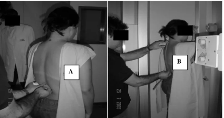
Figure 1 (A) shows the determination of the landmarks according to Field10. For the present study, a plumb marker was attached to the adhesive tape in order to determine the agreement between the placement of the external marker and the internal bone structure identified in the radiography. Agreement was found in 100% of the cases.
Molding of the flexible ruler over the volunteer’s torso The tip of the flexible ruler was positioned on the C7 spinal process. The rule was then molded in the format of the kyphotic curvature down to the spinal process of T12 (Figure 1B). The flexible ruler was then marked on the points
Xtotal H
Xmiddle
Xtotal H
Xmiddle
Figure 2. Illustration of Xtotal (distance between C7 and T12), Xmiddle
(distance between H line and T12) and H (distance between the Xtotal line and the vertex of the curve) measures from the Flexicurve Method.
Figure 1. A) Skin markers of C7 and T12. B) Flexicurve rule molding.
corresponding to C7 and T12, previously marked on the volunteer’s spine.
Transcription of the format of the dorsal spine to the millimeter paper
After being taken off of the subject’s back the flexible ruler maintained the torso’s format and was immediately placed over a millimeter paper. The contour of the ruler on the side it was placed over the subject’s spine was drawn in the millimeter paper and C7 and T12 points were marked.
Definition of the X total, Xmiddle and H
After the contour of the thoracic kyphosis was transferred to the millimeter paper, a straight line connecting the points equivalent to C7 to T12 was traced. Another straight line, perpendicular to the first, was traced between the C7-T12 points, to find the point of greater distance from the curve to the line between C7 and T12.
=180/PI()*(ATAN(H*XTOTAL*( -3*XMIDDLE+2*XTOTAL) /XMIDDLE/(XTOTAL^2+XMIDDLE^22*XTOTAL*XMIDDLE)) -ATAN(3*H*(XTOTAL -2*XMIDDLE)/XMIDDLE^2/
(XTOTAL^2+XMIDDLE^22*XTOTAL*XMIDDLE)*XTOTAL^2 -2*H*(XTOTAL^2-3*XMIDDLE^2)/XMIDDLE^2/
(XTOTAL^2+XMIDDLE^2-2*XTOTAL*XMIDDLE)*XTOTAL+ H*XTOTAL*(-3*XMIDDLE+2*XTOTAL)/XMIDDLE/
(XTOTAL^2+XMIDDLE^2-2*XTOTAL*XMIDDLE)))
= SE(OU(1/3*XTOTAL*(-3*XMIDDLE+2*XTOTAL)/ (XTOTAL -2*XMIDDLE)<0;1/3*XTOTAL*( -3*XMIDDLE+ 2*XTOTAL)/(XTOTAL -2*XMIDDLE)>XTOTAL;
XTOTAL=2*XMIDDLE)
= SE(OU(1/3*XTOTAL*(-3*XMIDDLE+2*XTOTAL)/ (XTOTAL -2*XMIDDLE)<0;1/3*XTOTAL*( -3*XMIDDLE+ 2*XTOTAL)/(XTOTAL -2*XMIDDLE)>XTOTAL;
XTOTAL=2*XMIDDLE)
Groups Kyphosis angle - mean Standard Deviation
Evaluator 1 44.9º ± 8.17
Evaluator 2 (first) 41.4º ± 8.65
Evaluator 2/2 43.6º ± 7.5
Cobb 42.8º ± 9.9
Table 1. Means and standard deviations of the dorsal Kyphosis angle
obtained with the Flexicurve method b by valuators 1, 2 e 2/2 e and with the Cobb radiographic method.
Angular calculus through a 3º degree polynomial After the Xtotal, Xmiddle and H distances had been determined in centimeters on the millimeter paper, the values were typed in a program written on Microsoft Excel to calculate the thoracic kyphosis angle. The specific mathematical formula used is described as follows:
In order to confirm if data are correct, the following formula was used:
Three distinct analyses were performed to compare the Flexicurve method with the Cobb’s angle. The first analysis included results from the first physical therapist (evaluator 1), the second analysis included the first measurement of the second physical therapist (evaluator 2), and the third analysis included the mean of the two measurements made by evaluator 2 (evaluator 2/2).
Statistical procedures were:
1) Calculation of the Intraclass Correlation Coefficient (ICC) between angular measures obtained with the Flexicurve method and with the Cobb’s method;
2) Sensitivity, specificity, positive predictive value (PV+) and negative predictive value (PV-) for the diagnosis of hyperkyphosis or normal kyphosis;
3) Influence of Xtotal, Xmiddle and H values on the determination of the angle obtained by the Flexicurve method;
4) ICC between measurements made by the evaluators 1 and 2 (inter-rater reliability);
5) ICC between both set of measurements of evaluator 2 (intra-rater reliability).
The confidence level was established at 95%. Analyses were done in the program SPSS 13.0 for windows.
RESULTS
Means and standard deviations (SD) for the thoracic kyphosis are presented in Table 1.
ICC values were calculated for Xtotal, Xmiddle, H (Table 2) and the angular measures obtained by evaluators 1 and 2 (inter-rater reliability).
The ICC between groups was also analyzed. Results demonstrated that two measurements are necessary for good concurrent validity values (Table 3).
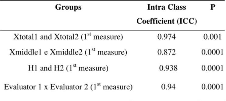
Table 2. Intra Class Coefficient (ICC) for Xtotal, Xmiddle and between
evaluators 1 and 2.
Groups Intra Class
Coefficient (ICC) P
Xtotal1 and Xtotal2 (1st measure) 0.974 0.001
Xmiddle1 e Xmiddle2 (1st measure) 0.872 0.0001
H1 and H2 (1st measure) 0.938 0.0001
Evaluator 1 x Evaluator 2 (1st measure) 0.94 0.0001
Table 3. Intra Class Coefficient (ICC) of kyphosis angle between
groups.
Groups Intra Class
Coefficient (ICC) p
Evaluator 1 x Cobb 0.528 0.003
Evaluator 2 x Cobb 0.589 0.001
Evaluatro 2/2 x Cobb 0.906 0.0001
The intra-rater ICC was 0.87 and the ICC between evaluators (evaluator 1 and evaluator 2 - first measurement) was 0.94. The sensitivity and specificity of the Flexicurve method to detect patients with hyperkyphosis (>50º) and patients with normal kyphosis (20 - 50º)11 were also analyzed. When the mean of the two measurements made by the second evaluator was used (evaluator 2/2), the sensitivity and specificity for detection of hyper-kyphosis were 85% and 97% respectively.
DISCUSSION
0.589 for evaluator 2. However, when the mean of two assessments performed by evaluator 2/2 was considered, the ICC value was of 0.906, which indicates a strong concurrent validity obtained by the flexicurve in relation to the Cobb’s method. The explanation of such a fact is similar to that of other clinical instruments of measurement. When using the adipometer for example, two measurements are necessary in order to decrease possible measurement error. Therefore, comparisons between the two methods were made with the measurements of evaluator 2/2.
Lundon, Li & Bibershtein12 studied three different methods for clinical measurement of kyphosis. Twenty-six subjects were evaluated by three different examiners with three different instruments: the Kyphosis Index (KI) obtained with flexicurve and the DeBrunner’s kyphometer compared to the Cobb’s angle obtained with radiographs. There was a higher inter-rater and intra-rater reliability with the kyphometer compared to the flexicurve. The analysis of variance demonstrated that there was no statistically significant difference between the information obtained with three analyses. The cost of the flexible ruler, however, is much lower than the cost of the kyphometer. The authors indicated the flexicurve as a good qualitative instrument for the measurement of the thoracic kyphosis, in contrast to our findings. With the Flexicurve method, the flexible ruler can be considered a quantitative instrument for measuring the angle of thoracic kyphosis.
Hart & Rose13 have studied the agreement level between radiological measurement and the flexicurve associated to the method of drawing tangents. Data from a single evaluator were used and the ICC was 0.87. In this validity study only six individuals comprised the sample used specifically for lumbar lordosis measurements. This small sample renders results obtained by Hart & Rose unclear, in opposition to the results of the present study with 56 patients that demonstrated an ICC value of 0.906.
Salisbury & Porter14 obtained a correlation r= 0.79 between the flexicurve associated to the method of drawing tangents and ultra-sound for lumbar flexion and r= 0.69 for lumbar extension. Results were worse than ours (r= 0.866). Strong correlation should not be taken as strong accordance. Correlations only indicate if obtained values increase or decrease in similar proportions.
Compared to other clinical instruments for measurement of dorsal kyphosis, the Flexicurve method presented good results. D’Osualdo, Scherano & Iannis15 used the archometer to measure thoracic kyphosis and demonstrated excellent correlation between evaluators, with r= 0.98. Results were better than ours (r= 0.888). However, they found considerable disagreement between the radiological measure and the archometer, in opposition to our findings with the Flexicurve method (ICC = 0.906 and r= 0.862).
In relation to intra-rater agreement for other instruments, Korovesis et al.16 found an ICC of 0.84 for the measures of
dorsal kyphosis obtained by the DeBrunner’s kyphometer. Mannion et al.17 studied the dorsal kyphosis with the spinal
mouse® and the ICC between two evaluators was 0.83.
Results of the present study demonstrated that the inter-rater ICC was 0.94, indicating better agreement than that obtained for other instruments.
Lovell, Rothstein & Personius8 have observed that the ICC between evaluators was 0.54 for lumbar lordosis with the flexicurve associated to the use of a 2º degree polynomial. However, a different method of angular determination was used. In opposition to the present study, the authors observed that measurements made with the flexicurve should always be performed by the same evaluator to avoid errors between evaluators. Results of this study demonstrate excellent agreement between two evaluators (ICC = 0.94).
Walker, Rothstein & Finucane18 reported an intra-rater ICC of 0.90 for the flexicurve associated to the drawing of tangents to measure lumbar lordosis in a sample of 31 healthy youngsters. Hart & Rose13 studied the reliability of the Flexicurve (tangent drawing) for a single evaluator. They obtained an ICC of 0.97 for 23 pairs of repeated measurements, which indicates excellent agreement for the same evaluator over several measurements.
In this method the “H measure” is the measure with highest influence in the estimated angle obtained by calculation through the 3º degree Flexicurve method. Apparently it is not associated to the individual’s height, but to the arch of the dorsal spine. In average, for each change of one centimeter the H value, the angle can be altered up to 11.95º (± 0.246). According to Lovell, Rothstein & Personius8, each millimeter (mm) of change in the H value can cause a change of up to 10º in the angle of the flexicurve. The effect is a consequence of the use of a formula based on a 2º degree polynomial, which implies that the vertex of the arch will always be at half the distance between the end points. This approach considers the curvature of the spine to be always a perfect arch, and this can be a strong source of error.
Caine, McConnell & Taylor19 have demonstrated that the maximal curvature of the spine may be found at different locations of the arch, therefore, certain categories of kyphotic curvatures are not well represented by the kyphosis index (KI). The curvature of the thoracic and lumbar spine is almost never a perfect arch with vertexes at the middle of the arch. This demonstrates that the calculation to obtain the tangent of the kyphosis angle (or even lordosis) should be based in 3º degree polynomials as used at this study. This approach corrects the angular value even if the curve’s vertex is not at the half-distance of the arch.
Results of the present study are better than results reported by Hart & Rose8. These authors described an ICC of 0.86 compared to an ICC of 0.90 in the present study. According to the results of this study and of previous studies demonstrating the difficulties to validate a non-invasive method to measure kyphosis (or even lordosis), is the use of the described protocol is suggested. Thus the 3º degree Flexicurve method should be used with C7 and T12 as references, two measurements should be performed subsequently, and the mean between them should be calculated. Such procedures help to reduce the errors between evaluators and to approximate the value obtained with the 3º degree Flexicurve method to the Cobb’s angle.
This study required many x-ray exams and this fact limited the sample size. Although the mean age of the studied group was high, mean kyphotic curvature of the subjects included in the study was within normal limits (20 – 50º). Therefore, the same factor that could have been a limitation of the study was controlled by characteristic of the studied group.
New studies may and should be done to validate the measures obtained with the 3º degree Flexicurve method for specific populations such as elders and children, for example. These studies would identify the limits of measurement of the 3º degree Flexicurve method proposed at the present study. Results demonstrate that the Flexicurve method is as reliable and valid method to measure thoracic kyphosis. It is a method of easy utilization in the clinical setting and in the context of human posture research.
REFERENCES
1. Pluijm S, Tromp AM, Smit JH, Deeg DJH, Lips P. Consequen-ces of vertebral deformities in older men and women. J Bone Miner Res. 2000;15:1564-72.
2. Poolman R, Been H, Ubags L. Clinical outcome and radiographic results after operative treatment of Scheuermann’s disease. Eur Spine J. 2002;11:561-9.
3. Willner S. Spinal pantograph: a non-invasive technique for describing kyphosis and lordosis in the thoraco-lumbar spine. Acta Orthop Scand. 1981;52:525-9.
4. Hinman MR. Comparison of thoracic kyphosis and postural stiffness in younger and older women. Spine J. 2004;4(4): 413-7.
5. Takahashi E, Atsumi H. Age diferences in thoracic form as indicated by thoracic index . Hum Biol. 1955;27(2):65-74.
6. Milne JS, Lauder IJ. The relationship of kyphosis to the shape of vertebral bodies. Ann hum biol. 1976;3:173-9.
7. Burton AK. Regional lumbar sagittal mobility: Measurement by flexicurves. Clin biomech. 1986;1:20-6.
8. Lovell F, Rothstein J, Personius W. Reliability of clinical measu-rements of lumbar lordosis taken with a flexible rule. Phys Ther. 1989;69(2):96-102.
9. Bradford DS, Lonstein JE, Moe JH, Ogivie JW, Winter RB. Escoliose e outras deformidades da coluna: o livro de moe. 2ª ed. São Paulo: Santos; 1994.
10. Field D. Anatomia palpatória. 2ª ed. São Paulo: Manole; 2001.
11. Fon G, Pitt M, Thies A. Thoracic kyphosis: range in normal subjects. A J R. 1980;134:979-83.
12. Lundon K, Li A, Bibershtein S. Interrater and intrarater reliabi-lity in the measurement of kyphosis in postmenopausal women with osteoporosis. Spine. 1998;23(18):1978-85.
13. Hart DL, Rose SJ. Reliability of a non-invasive method for measuring the lumbar curve. J Ortho Sports Phys Ther. 1986;8:180-4.
14. Salisbury P, Porter R. Measurement of lumbar sagittal mobility. A comparison of methods. Spine. 1987;12(2):190-3.
15. D’osualdo F, Scherano S, Iannis M. Validation of clinical measurement of kyphosis with a simple instrument, the arcometer. Spine. 1997;22:408-13.
16. Korovessis P, Petsinis G, Papazisis Z, Baillousis A. Prediction of thoracic kyphosis using the De Brunner kyphometer. J Spin Disor. 2001;14(1):67-72.
17. Mannion A, Knecht, K, Balaban JE, Grob D. A new skin-surface device for measuring the curvature and global and segmental ranges of motion of the spine: realiability of measurements and comparison with data reviewed from the literature. Eur Spine J. 2004;13:122-36.
18. Walker ML, Rothstein JM, Finucane SD, Lamb RL. Relation-ships between lumbar lordosis, pelvic tilt, and abdominal muscle performance. Phys Ther. 1987;67(4):512-6.