ABSTRACT
Introduction: By clinical definition, mouth breathers use the mouth as their main air pathway during breathing. This results in modifications to tongue and head positioning and may have an influence on craniofacial mechanics during development. Bringing the head forward is also common among mouth breathers and may lead to misalignments in adjacent segments of the human body. Objective: To evaluate neck (cervical) range of motion (ROM) among mouth-breathing children and compare this with a group of nose-breathing children. Method: Ten mouth-breathing children of both sexes aged 6.90 ± 1.37 years and ten nose-breathing children aged 7.70 ± 1.42 years participated in this study. The ROM for neck flexion, extension and protrusion of the head were evaluated. Student’s t test for independent samples was used for the statistical analysis, considering p< 0.05 as the statistical significance level. Results: The mouth-breathing children had a significantly smaller ROM for neck extension (59.0°± 10.79°), compared with the nose-breathing group (72.9°± 8.82°) (p= 0.001). The ROM for flexion and protrusion was not statistically different between groups (59.0°± 10.79°). Conclusion: The mouth-breathing children presented smaller neck extension ROM than the nose-breathing children did, but for protrusion and flexion ROM there was no difference between the groups.
Key words: mouth breathing; cervical spine; joint arthrometry.
RESUMO
Mensuração da Amplitude de Movimento Cervical em Crianças Respiradoras Orais
Introdução: Por definição clínica, respiradores orais (RO) utilizam a boca como maior via de acesso de ar durante a respiração. Isso resulta em alterações na posição da língua e cabeça e pode influenciar a mecânica craniofacial durante o desenvolvimento. A anteriorização da cabeça também é comum em RO, podendo levar a desalinhamentos em segmentos adjacentes do corpo humano. Objetivos: Avaliar a amplitude de movimento (ADM) cervical em crianças RO e comparar com crianças respiradoras nasais (RN). Métodos: Dez crianças RO, de ambos os sexos, com idade de 6,90 ± 1,37 anos e dez RN, de ambos os sexos, com idade de 7,70 ± 1,42 anos, participaram do estudo. O Cervical Range of Motion (CROM) foi utilizado para medir a ADM de flexão, extensão e
protrusão da cabeça. Para a análise estatística foi utilizado o teste t Student para amostras independentes, considerando nível de
significância estatística o valor de p< 0,05. Resultados: Crianças RO apresentam uma ADM de extensão cervical significativa
-mente menor (59,0° ± 10,79°) quando comparadas ao grupo RN (72,9° ± 8,82° ) (p= 0,001). A ADM de flexão e protrusão não foi
estatisticamente diferente entre os grupos. Conclusão: As crianças RO apresentaram menor ADM de extensão cervical do que as crianças RN, no entanto, em relação às ADM de protusão e flexão, não há diferença entre os grupos.
Palavras-chave: respirador bucal; coluna cervical; artrometria articular.
MEASUREMENT OF NECK RANGE OF MOTION AMONG
MOUTH-BREATHING CHILDREN
N
eivaPD
1& K
irKwooDrN
21 Physical Therapy Clinic, Pontifícia Universidade Católica de Minas Gerais, Belo Horizonte, MG - Brazil.
2 Physical Therapy Department, Universidade Federal de Minas Gerais, Belo Horizonte, MG - Brazil.
Correspondence to: Patrícia Dayrell Neiva, Rua Equador, 118, Apto 902, São Pedro, CEP 30330-390, Belo Horizonte, MG – Brasil, e-mail: [email protected]
INTRODUCTION
The act of breathing through the nose is inherent to the human being, who is, at birth, a physiological
nasal breather1. When air passes through the nose, three
distinct functions are performed: heating, moisturizing,
and filtering, called air-conditioning functions of the up
-per airway2. Anatomical and functional integrity of the
upper airway allows nasal breathing to be physiological, establishing an air flow resistance of 50% of the total
airway resistance1. According to Moss’s functional matrix
theory, nasal breathing allows not only the functions of suction, mastication and swallowing, but also adequate
growth and development of the craniofacial complex2.
Any factor that impedes air passage through the nose will allow the access way to be replaced by the mouth.
Oral breathing is an adaptive feature with a multifacto
-rial etiology, and its persistence may be harmful3,4. In
the literature, there is little consensus on the definition
of mouth-breather (MB)5,6. Some studies consider as
mouth-breathers those individuals which present upper airway mechanical obstruction, others as those with the simple habit of breathing through the mouth or those individuals that breathe through the mouth for periods of time or spend a certain amount of time with the mouth
open (open mouth posture - OMP)7. However, studies
conducted by otorhinolaryngologists6, taking into account
dental-craniofacial alterations in general, classify as Mouth
Breathing Syndrome any postural alterations8,9, daytime
sleepiness, migraines, night-time anxiety, enuresis, frequent tiredness, problems at school, and bruxism. MB children display vertical increase of the lower third of the face, narrow maxillary arch, high-arched palate, changes in
hyoid bone position10, short upper lip and inverted lower
lip, labial incompetence without passive lip closure, hy
-potonia of the masticatory muscles, changes in tongue
position at rest and while swallowing6. Mouth breathing
is a clinical condition common in school-aged children, and some studies already relate this clinical entity to the persistence of postural alterations8.
Forward head posture characterized by lower cervi
-cal spine flexion and occipital extension is a common
clinical finding in MB children11. Adoption of this pos
-ture may be influenced by airway interference in the
craniofacial development12-14. Although the literature is
controversial regarding the association of head posture
and cranial morphology15,16, a progressive increase of
the craniovertical angle and a forward head posture were
observed in patients with upper airway obstruction13,17.
Head posture is defined by the craniovertical angle, which is the angle between the vertical (gravitational) line and the line formed by two points marked on the face of the
individual. This method was described by Vig et al.13.
Head extension decreases the value of this angle, and
head flexion increases it. Vig et al.13 observed a
two-degree reduction in the craniovertical angle two months after the removal of the adenoids in MB children. In a
longitudinal study, Wenzel et al.18 observed that, with
the decrease in nasal resistance after corticoid therapy, there was a decrease in the craniovertical angle, that is, the reversal of upper airway obstruction minimized forward head posture.
Range of motion (ROM) measures are routinely in
-cluded in vertical and especially cervical spine assessments, a conduct justiied by the relationship between ROM and the functional limitations of patients with cervical pain. Cervical spine ROM is obtained in physical therapy practice by measuring the movement of the head in relation to the trunk19. The different instruments used, as well as the lack of systematic procedures presented by the studies, reduce data reproducibility and contribute to a strong variation in
the active and passive ROM values of the cervical spine20.
Therefore, the aim of this study was to measure the cervical ROM in 5-12 year olds with MB diagnosis, and compare these measures with those of nose-breathing (NB) children.
MATERIALS AND METHODS
An observational study was carried out. We assessed twenty children of both genders, aged 5 to 12 years, of
which ten were clinically diagnosed as MB by the oto
-rhinolaryngologist through nasopharyngoscopy, and ten
were NB. The NB group did not undergo nasopharyngos
-copy. Diagnosis was made by the otorhinolaryngologist through a clinical observation of the airway, closed lip position, normal occlusion, normal facial dimensions and a questionnaire applied to relatives by a physical therapist. This questionnaire assessed the absence of harmful oral habits, sleep disorders, intense sialorrhea and night-time snoring. Children with a diagnosis of cerebral palsy or the inability to assume the orthostatic position were excluded from the study. There was no randomization, and three participants were excluded from the study. All participants signed the informed consent approved by the Committee of Research Ethics of the Pontifícia Universidade Católica of Minas Gerais (CEP 2004/129).
The Cervical Range of Motion (CROM) instrument
(Performance Attainment Associates, 958 Lydia Drive, Ro
the sagittal plane. The anterior inclinometer measures the
ROM of lateral flexion on the frontal plane. These incli
-nometers are gravitational. For the rotation measures, the inclinometer is magnetic and moves along the transversal plane. For the protrusion and retraction measures, a ruler is used21,22. The protrusion and retraction are described as a forward head motion on the sagittal plane. Anatomically, it consists of a combination of lower cervical flexion and
upper cervical extension19. The CROM is an easy-to-use
instrument and its intertest and intratest reproducibility was established with excellent results, especially in flexion
and extension measurements20,22.
For data collection and measurement standardization,
the children were instructed to sit on a chair with stan
-dardized seat adjustment, positioning the hip and knee at ninety degrees and preventing thoracic spine movement. The head was aligned at neutral (zero degree) rotation and lateral lexion, and the participants were asked to ix their gaze at eye level. The tester stood to the left of the child, stabilizing the scapulas with their right hand in order to avoid thoracic rotation.
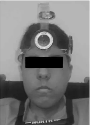
The tester taught the participant how to actively per
-form the head flexion, extension and protrusion movements (Figure 1). After the learning session, a one-minute rest was allowed, after which the movements were recorded.
Flexion and extension (degrees) were recorded first, fol
-lowed by head protrusion measurement (centimeters; cm) (Figure 2). Three movements were performed for each measure.
Next, weight and height measurements were taken in order to determine the Body Mass Index (BMI). The purpose of this measure was to characterize the NB and MB groups.
STATISTICAL ANALYSIS
The Kolmogorov-Smirnov test was applied in order to determine whether the variable studied had a normal distribution.
To analyze the anthropometric differences and the variables investigated between the groups, the Student
t-test was used for independent samples, considering a sig
-niicance level of 95% (p < 0.05). The statistical analyses were performed with Statistical Package for Social Sciences (SPSS, Chicago, IL, USA) 11.0.
RESULTS
The ive male and ive female MB children presented an average age of 6.9 ± 1.37 years and BMI of 18.54 ± 7.18. The ive male and ive female NB children had an average age of 7.70 ± 1.42 and BMI of 21.72 ± 5.14. The differences between the groups were not signiicant.
Regarding the cervical extension measure, the MB
Figure 2. Side view of correctly positioned cervical range of motion instrument (CROM).
Figure 1. Cervical range of motion instrument (CROM) correctly positioned on a mouth-breathing child.
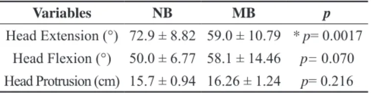
children presented signiicantly lower cervical extension ROM (59.0º ± 10.79º) (p = 0.001) when compared to the
-tistically signiicant difference in lexion ROM between the MB group (58.1º ± 14.46º) and the NB group (50.0º ± 6.77º) (p = 0.07) and in cervical protrusion between the MB group (16.26º ± 1.24º) and the NB group (15.7º ± 0.94º) (p = 0.20).
Table 1. Cervical ROM of nasal breathers (NB) and mouth breathers (MB).
Variables NB MB p
Head Extension (°) 72.9 ± 8.82 59.0 ± 10.79 * p= 0.0017
Head Flexion (°) 50.0 ± 6.77 58.1 ± 14.46 p= 0.070
Head Protrusion (cm) 15.7 ± 0.94 16.26 ± 1.24 p= 0.216
Results in Mean ± Standard Deviation * p< 0.05 to compare NB and MB groups.
DISCUSSION
The results from the study showed a decrease in cer
-vical extension ROM in MB children when compared to the NB children. These indings are in accordance with
a study by Farah & Tanaka23, which assessed individu
-als of both genders with myofunctional alterations using goniometry. The lexion and extension ROM values varied between 47.88º ± 10.58º and 57.34º ± 11.88º, respectively. Despite the fact that the measuring instruments were not the same, the measurement principles of the studies were similar, strengthening the indings of extension ROM loss in MB children.
Extension ROM limitation in MB may be attributed to the imbalance between the muscular activity of neck lexors and extensors. While assessing surface electromyography of the sternocleidomastoid and trapezius muscles in MB individu
-als, Ribeiro et al. 24 found a higher electrical activity during relaxation and a lower electrical activity during maximal voluntary contraction, when compared to NB individuals. The hyperactivity of the sternocleidomastoid and trapezius upper ibers decreases the length-tension curve of these muscles, yielding a shortening of the neck extensors, thus limiting cervical spine extension24. Together with muscular hyperactivity, head extension and lower mandible position may be determining factors in the craniofacial morphology of growing individuals with nasal obstruction13.
While studying the development of the cervical lordosis in MB children aged 8, 11, and 15, Hellsing et
al. 17 observed a decrease in lordosis as age increased.
Similarly, Muto et al.25 used cephalometry to assess 10
young NB individuals aged 20 to 35 with full dentition, no bad occlusion and no mastication disorders. The authors observed less cervical lordosis, but a 4 mm increase in pharyngeal space with head extension. Similar associations between craniovertical angulation and smaller pharyngeal diameter in normal individuals have already been shown
in experimental studies in which the individuals were assessed with their head positioned at different degrees of flexion and extension. The percentage of increase in pharyngeal diameter with head extension depends on the way the individuals extend their head. The results showed that the airway became wider when the extension occurred in the high cervical segment. These findings may justify the compensatory forward head posture that the MB children adopt to facilitate breathing. By means of clinical observations, it is well established that cervical spine movement decreases with age due to degenerative changes26.
There is no consensus in the literature with regard to normal lexion/extension ROM values of the cervical
spine. Dvorak & Panjabi 26,27 describe ROM by cervical
spine segments, with higher ranges between 15 and 20º in the C5-C6 and C6-C7 segments, and lower ranges between the C1-C2 and C2-C3 segments (5 to 10º). Total ROM is
around 110º, with extension ROM (75o) being higher than
lexion ROM (35o). Furthermore, ROM values for active
lexion (35º ± 70º) and active extension (50º ± 93º) dif
-fer from those for passive lexion (59º ± 76º) and passive extension (53º ± 77º)28.
The different instruments used to measure the cer
-vical ROM may justify the differences in the described ranges. All measuring instruments must be capable of indicating correct and reliable values, thus insuring their validity and reproducibility, respectively. In the present study, the CROM was used as the measuring instrument because it is easy to handle, low-cost, and has good clinical practice acceptance. Moreover, the literature shows that the CROM presented reliable intratester and intertester
results22. The instrument is placed on the patient’s head,
and the tester does not need to move the instrument to take measurements, thus avoiding errors caused by handling and manual adjustments. A previously trained tester took all the cervical movement measurements for the present study.
We determined the children’s age group based on
previous studies8,29 that also worked with this age group;
however, we cannot exclude interferences by craniofacial and motor development changes, which tend to increase in the pre-pubertal phase.
One of the limitations of the measurement system is the dificulty in keeping the children in a static position, which hampered instrument reading. However, the children
were asked to ix their gaze at eye level. Therefore, pos
-sible errors such as reading dificulty and imprecision, as well as the effort and the erroneous perception of the end of ROM, were mitigated30,31.
CONCLUSION
The present study observed a statistically signiicant decrease in cervical extension ROM between MB children compared to NB children. It is believed that their head forward posture causes biomechanical damage between the extensor and lexor neck muscles, limiting extension ROM.
The use of simple measuring instruments should be included in physical therapy practice in order to help physi
-cal therapists systematize their intervention.
REFERENCES
1. Saffer M, Rasia Filho AA, Lubianca Neto JF. Efeitos sistêmicos da obstrução nasal e da respiração oral persistente na criança. Rev AMRIGS. 1995;39(3):179-82.
2. Lessa FC, Enoki C, Feres MF, Valera FC, Lima WT, Matsumoto MA. Breathing mode influence in craniofacial development. Rev Bras Otorrinolaringol. 2005;71(2):156-60.
3. Weckx LLM, Weckx LY. Respirador bucal: causas e conse-quências. Rev Bras Med. 1995;52(8):863-74.
4. Ferrugini AM, Valle ACF, Soares CF, Schettino CS, Croce LSS, Leite ICG. Crescimento e desenvolvimento craniofacial. Jornal Bras de Fono. 2002;3(11):135-9.
5. Aragao W. Aragao’s function regulator, the stomatognathic system and postural changes in children. J Clin Pediatr Dent. 1991;15(4):226-31.
6. Di Francesco RC, Passerotii G, Paulucci B, Miniti A. Res-piração oral na criança: repercussões diferentes de acordo com o diagnóstico. Rev Bras Otorrinolaringol. 2004;70(5):665-70. 7. Lourenco EA, Lopes KC, Pontes A Jr, Oliveira MH, Um
-emura A, Vargas AL. Comparison between radiological and nasopharyngolaryngoscopic assessment of adenoid tissue vol -ume in mouth breathing children. Rev Bras Otorrinolaringol. 2005;71(1):23-7.
8. Yi LPS, Weckx LLM. Avaliação postural em crianças de 5 a 12 anos que apresentam respiração oral. Fisioter Mov. 2003;16(3):29-33.
9. Lima L, Baraúna MA, Sologurem M, Canto R, Gastaldi A. Postural alterations in children with mouth breathing as -sessed by computerized biophotogrammetry. J Appl Oral Sci. 2004;12(3):232-7.
10. Tavares CA, Braga IP, Silva HJ. Alterações posturais nos re -spiradores orais. J Bras de Fono. 2002;3(12):233-6.
11. Fujiki PR. Influência da hipertrofia adenoideana no crescimento e desenvolvimento craniodentofacial. Ortodontia. 1999;32(1): 70-7.
12. Tecco S, Festa F, Tete S, Longhi V, D’Attilio M. Changes in head posture after rapid maxillary expansion in mouth-breathing girls: a controlled study. Angle Orthod. 2005;75(2):171-6. 13. Vig KW. Nasal obstruction and facial growth: the strength of
evidence for clinical assumptions. Am J Orthod Dentofacial Orthop. 1998;113(6):603-11.
14. Fields HW, Warren DW, Black K, Phillips CL. Relationship be -tween vertical dentofacial morphology and respiration in adoles -cents. Am J Orthod Dentofacial Orthop. 1991;99(2):147-54. 15. Valera FC, Trawitzki LV, Anselmo-Lima WT. Myofunctional
evaluation after surgery for tonsils hypertrophy and its cor -relation to breathing pattern: A 2-year-follow up. Int J Pediatr Otorhinolaryngol. 2006;70(2):221-5.
16. Shanker S, Fields HW, Beck FM, Vig PS, Vig KWL. A Longi -tudinal assessment of upper respiratory function and dentofa -cial morphology in 8- to 12-year-old children. Semin Orthod. 2004;10(1):45-53.
17. Hellsing E. Changes in postural EMG activity in the neck and mastigatory muscles following obstruction of the nasal airways. Eur J Orthod. 1986;8:247-53.
18. Wenzel A, Henriksen J, Melsen B. Nasal respiratory resistance and head posture: effect of intranasal corticosteroid (Budes -onide) in children with asthma and perennial rhinitis. Am J Orthod. 1983;84(5):422-6.
19. Nordin M, Frankel L. Biomecânica básica do sistema músculo esquelético. 3ª ed. São Paulo: Guanabara Koogan; 2003. 20. Tousignant MBL, O’Donoughue S. Validity study for cervical
range of motion (CROM) goniometer for cervical flexion and extension. Spine. 2000;25:324-30.
21. Jordan K. Assessment of published reliability studies for cervi -cal spine range of motion measurement tools. J Manipulative Physiol Ther. 2000;23:180-95.
22. Capuano-Pucci DRW. Intratester and intertester realibility of the cervical range of motion device. Arch Phys Med Rehabil. 1991;72(4):338-41.
23. Farah EA, Tanaka C. Postura e mobilidade da coluna cervical e do tronco em portadores de alterações miofuncionais orais. Rev Reg Araçatuba Assoc Paul Cir Dent. 1997;51(2):171-5. 24. Ribeiro EC, Marchiori SC, Silva AM. Electromyographic analy
-sis of trapezius and sternocleidomastoideus muscles during nasal and oral inspiration in nasal- and mouth-breathing children. J Electromyogr Kinesiol. 2002;12(4):305-16.
25. Muto T, Takeda S, Kanazawa M, Yamazaki A, Fujiwara Y, Mi -zoguchi I. The effect of head posture on the pharyngeal airway space (PAS). Int J Oral Maxillofac Surg. 2002;31(6): 579-83. 26. Panjabi M, Dvorak J. Age e gender related normal motion of
the cervical spine. Spine. 1992;17:393-8.
27. Dvorak J. Clinical validation of functional flexion/extension radiographs of the cervical spine. Spine. 1992;18(1):120-7. 28. Bogduk N, Mercer S. Biomechanics of the cervical spine I: Normal
kinematics. Clin Biomech (Bristol, Avon). 2000;15(8): 633-48. 29. Krakauer LH, Guilherme A. Relationship between mouth breath
-ing and postural alterations of children: a descriptive analysis. Int J Orofacial Myology. 2000;26:13-23.
30. Wendy R. Intertester reliability of cervical range of motion device. J Orthop Sports Phys Ther. 1992;15(3):147-50. 31. Bredenkamp H. Validity study of head and neck flexion-exten