Analysis of the film thickness of a root canal sealer following three
obturation techniques
Análise da espessura da linha de cimento endodôntico em três
técnicas de obturação
Gustavo André De Deus* Fábio Martins**
Ana Carolina Machado Rocha Lima** Eduardo Diogo Gurgel-Filho*** Claudio Ferreira Maniglia**** Tauby Coutinho-Filho*****
ABSTRACT:The aim of this study was to obtain a quantitative analysis of the film thickness of a root canal sealer formed after filling by three different techniques. Thirty human maxillary incisors were selected and access cavities were prepared using high-speed diamond stones and water spray. A size #15 K-Flexofile was introduced in the canal of each specimen until it was seen just at the apical foramen. The working length was determined to be 1 mm short of that position and the canals were prepared to an apical size of #45 K-Flexofile. Copious irrigation with 5.25% NaOCl (sodium hypochlorite) was used during and after instrumentation. The samples were divided into three groups and obturated as follows: G1 - lateral condensation, G2 - lateral condensation with an accessory cone, and G3 - continu-ous wave of condensation. The samples were evaluated in the cervical, middle and apical thirds. The film thickness of the root canal sealer was measured through a microscopic evaluation. Statistical analysis was obtained using the Wilcox test. Statistical analysis showed significant differences between G3 and G1, G3 and G2 (p£0.05). In general, the lowest film thickness was observed in the continuous wave of condensation (G3). Lateral condensation with an ac-cessory cone (G2) and lateral condensation (G1) demonstrated poorer results in this study, showing a higher film thickness. The small film thickness of the sealer obtained by the continuous wave of condensation technique may in-crease the clinical performance of this technique.
DESCRIPTORS:Dental cements; Endodontics; Root canal obturation; Root canal therapy.
RESUMO:O objetivo deste estudo foi realizar uma análise quantitativa da espessura da linha de cimento endodôntico formada por três técnicas de obturação. Trinta incisivos centrais superiores humanos foram selecionados e acessados de modo convencional. Uma lima nº 15 K-Flexofile foi usada para a verificação da patência foraminal e para determi-nação do comprimento de trabalho, que foi estabelecido a 1 mm aquém do forame apical. Os canais foram preparados até a lima nº 45. Hipoclorito de sódio a 5,25% foi usado durante toda a instrumentação. Os dentes foram divididos em 3 grupos e obturados pelos seguintes critérios: G1 - condensação lateral; G2 - compressão hidraúlica e G3 - onda de condensação. Os dentes foram seccionados e analisados nos terços cervical, médio e apical. A espessura do filme de ci-mento foi determinada por um processo semi-automático de análise e processaci-mento digital de imagens realizado no software KS 400. O tratamento estatístico foi realizado com teste de Wilcox, que revelou diferenças significantes entre G3 e G1 e entre G3 e G2, sendo p£0,05. Não houve diferenças estatisticamente significantes entre G1 e G2. De um modo geral, a compressão hidraúlica (G2) e condensação lateral (G1) revelaram uma maior espessura do filme de ci-mento. A menor espessura do filme de cimento obtida pela técnica de onda de condensação tende a melhorar a perfor-mance clínica dessa técnica em relação às outras testadas.
DESCRITORES:Cimentos dentários; Endodontia; Obturação do canal radicular; Tratamento do canal radicular.
INTRODUCTION
Although gutta-percha is not considered an ide-al filling materiide-al, it still represents the first choice for a solid core filling for the root canal system,
* MSc.
** Specialized in Endodontics; *****PhD, Department of Endodontics, School of Dentistry, Rio de Janeiro State University. *** PhD; ****Doctorate Student – Department of Endodontics, School of Dentistry, State University of Campinas.
producing the best clinical performance when as-sociated to a root canal sealer2,5. The excellent
capa-city of lateral and apical sealing, leakage indexes and also its ability to promote a three dimensional filling.
Gutta-percha does not provide an apical seal to ink penetration when used without a root canal se-aler5,8
. Upon this confirmation of the necessity of the presence of a root canal sealer, investigations about the performance of the sealer were conduc-ted throughout the years. The physical properties of different canal sealers were analyzed and a strong tendency to increase their adhesiveness was observed during the 1970’s12
. It has also been confirmed that leakage may occur within the sea-ler or by its dissolution, either in the interface bet-ween sealer and dentine, or betbet-ween sealer and the gutta-percha9. Another aspect to consider is that
areas filled by a sealer are more vulnerable17
. The presence of a root canal sealer, in any filling tech-nique, reduces clinical leakage5,8
. Moreover, the root canal sealer is capable of filling imperfections and increasing the adaptation of the gutta-percha filling. Although many studies have indicated the undeniable necessity of a root canal sealer, its con-firmed solubility implies the necessity to limit its presence to a thin film thickness1,17. Sealer film
thickness may be of particular relevance to con-ventional techniques that involve solid core fil-lings.
The aim of this study was to obtain a quantita-tive analysis of the film thickness of a root canal sealer formed after filling by three different tech-niques.
MATERIALS AND METHODS
For the present work, thirty maxillary central incisors were selected from the Tooth Bank of the Rio de Janeiro State University. Standard access cavities were made and the patency of each canal was confirmed by inserting a #20 file through the apical foramen before and after completion of the root canal preparation. The working length was de-termined at 1 mm short of the apex and the canals were shaped manually using a crown-down tech-nique and stainless steel Flexofiles®
(Dentsply-Ma-illefer, Ballaigues, Switzerland) and Gates Glidden burs (#6, #5, #4, #3).
The coronal and middle thirds of each canal were preflared using Gates Glidden drills (Dent-sply-Maillefer, Ballaigues, Switzerland), #6, #5, #4 and #3. The middle and apical thirdswere prepa-red with Flexofiles®
(Dentsply-Maillefer,
Ballai-gues, Switzerland), #60, #55, #50 and #45 using a balanced force technique as described by Roaneet al.16
, 1985.The canals were irrigated between each file with 2 ml of freshly prepared 5.25% solution of sodium hypochlorite using a syringe and 27-gauge needle. All the teeth received a final flush of 10 ml of 17% EDTA (pH 7.7) (Biodinâmica, Ibiporã, PR, Brazil) followed by 10 ml of 5.25% sodium hypoch-lorite to remove the smear layer23
.
The 30 teeth were randomly divided into three equal groups and obturated as follows: Group 1: lateral condensation technique; Group 2: lateral condensation technique with an accessory cone4
, and Group 3: warm gutta-percha condensation technique3,10,17,18,19
combined with the Obtura II System (Obtura Corp., Fenton, MO, USA) in the backfilling phase (i.e. continuous wave of conden-sation).
A zinc-oxide eugenol sealer (Endofill, Dentsply, Petrópolis, RJ, Brazil) was mixed manually accor-ding to the recommendations of the manufacturer and used for all groups. A #40 file was used to pick up the measured spoon ofsealer(0.25 ml) two ti-mes from the mixing pad and placed into the canal whilst rotating it counterclockwise.
In the lateral condensation group (G1), a #45 master gutta-percha cone(Diadent Group Interna-tional, Chongchong Buk Do, Korea) was coated with a measured spoon of sealer (0.25 ml) and pla-ced in the canal to the full working length. Lateral compaction was achieved in each canal by using accessory MF gutta-percha cones (medium fine) (Dentsply-Maillefer, Ballaigues, Switzerland) and the endodontic finger spreader size B ( Dentsply-Maillefer, Ballaigues, Switzerland). A heated ins-trument was used to remove the excess gutta-percha.
In the lateral condensation with an accessory cone (G2), the tip of a medium sized non-standar-dized gutta-percha cone with a 0.6 taper (Diadent Group International, Chongchong Buk Do, Korea) was trimmed back until tug-back was achieved in the full working length. The trimmed gutta-percha cone was coated with a measured spoon of sealer (0.25 ml). A heated instrument was used to remove the excess gutta-percha and then vertical force was applied with a plugger (1.0 mm, Dentsply-Ma-illefer, Ballaigues, Switzerland) to compact the gutta-percha in the coronal third of the canal4,17
. In the warm gutta-percha group (G3), the tip of a medium sized non-standardized gutta-percha De Deus GA, Martins F, Lima ACMR, Gurgel-Filho ED, Maniglia CF, Coutinho-Filho T. Analysis of the film thickness of a root canal
cone with a 0.6 taper (Diadent Group Internatio-nal, Chongchong Buk Do, Korea) was trimmed back until tug-back was achieved in the full wor-king length. The trimmed gutta-percha cone was coated with a measured spoon of sealer (0.25 ml). At the level of the cementum-enamel junction the gutta-percha was scared off with the tip an activa-ted heat carrier (Touch’n Heat model 5004, Analy-tic Technology, Redmond, WA, USA). After deacti-vating the heat carrier, the cooling instrument was removed from the canal, bringing out an increment of gutta-percha. Vertical force was applied with a size 11 plugger (1.1 mm diameter, Dentsply-Mail-lefer, Ballaigues, Switzerland) to compact the gut-ta-percha in the coronal third of the canal. This procedure was repeated twice, first to a level 3-4 mm deeper than the cementum-enamel juncti-on and vertically cjuncti-ondensing the gutta-percha in the middle third of the canal using a size 7 plugger (0.7 mm diameter, Dentsply-Maillefer, Ballaigues, Switzerland), and secondly to a level 4 mm short of the full working length and vertically condensing the gutta-percha in the apical portion of the canal using a size 5 plugger (0.5 mm diameter, Dentsply-Maillefer, Ballaigues, Switzerland). Back-filling of the rest of the canal space was achi-eved by injecting warm gutta-percha using the Obtura II System (Obtura Corp., Fenton, MO, USA), each time injecting a 4-5 mm segment and condensing the gutta-percha with a prefitted plug-ger3,17,18,19
.
The samples were then stored in 100% humi-dity and at 37ºC for 2 weeks. After that,each sam-ple was sectioned longitudinally using a low-speed saw (Isomet, Buhler Ltd., Lake Bluff, NY, USA) with a diamond disc (Æ 125 mm x 0,35 mm x 12,7 mm – model 330C) while constantly irrigating with water in order to prevent overheating. The cuts were made in different points: one located at the cervical third, the other two at the middle and apical thirds respectively. Subsequently, the sam-ples were embedded in an epoxy resin cylinder (Arazyn 1.0, Ara Química, SP, Brazil) to facilitate their manipulation and improve the preparation result. The margins adjoining the epoxy resin and tooth were sealed with cyanoacrylate (Super Bon-der gel, Loctite, Itapevi, SP, Brazil).
Specific sandpapering (NETOT 4050014, Stru-ers, DK) for materialographic preparation was per-formed. The purpose of materialographic specimen preparation is to obtain a surface that is free from
scratches and deformation. To achieve this result the samples were properly ground to remove da-mages or deformed surface material, while intro-ducing as little new deformation as possible, thus preparing the sample surface for polishing. To re-move deformations from fine grinding and obtain a surface that is highly reflective, the specimens were polished before they were examined under the microscope. Polishing was accomplished with diamond paste with 4-1 mm roughness (SAPUQ 40600235, Struers, DK).
The samples were examined under a microsco-pe (Axiscomicrosco-pe, Carl Zeiss Vision Gmbh, Hallbergmo-os, Germany). For each sample, a sequence of pho-tographs with increases of 50 X and 200 X was taken, the dentinal wall/filling material interface being always the observation focus. The negatives were scanned by a 35 mm/medium format film scanner (SprintScan 120, Polaroide, NY, USA) as tiff images (tagged image file format) with 1,200 dpi.
For image analysis and processing, the KS 400
Image System 4.0 (Carl Zeiss Vision Gmbh, Hall-bergmoos, Germany) was used. Through a seg-mentation process on pixels shade of digitized ima-ge, the software allowed us, by a semi-automated process, to obtain measurements made in areas with higher film thickness of the root canal sealer. In order to rationalize and automate the work, a protocol (macro) was developed and used to analy-ze all images.
Through this digital image analysis and proces-sing by the KS 400 Image System 4.0 (Carl Zeiss Vision Gmbh, Hallbergmoos, Germany), about 20 measurements for each observed field examined were obtained.A Wilcox test was used to determine whether there were significant differences between the groups.
RESULTS
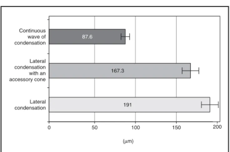
With the aid of the digital analysis and proces-sing imaging, numerical data were obtained from the measurements performed. The data obtained from the apical, middle and cervical cross-secti-ons, and a general average for each technique, are displayed in Graphs 1 and 2. Statistical analysis was obtained using the Wilcoxtest (Table 1).
Cervical third
gre-at lack of homogeneity of the filling mgre-aterial in all techniques. Particularly in this area it was
possi-ble to observe smaller layers of root canal sealer (Figures 1 and 2). The results revealed film thick-ness varying from 15 to 101mm, presenting an ave-rage post of 29mm forGroup 3, 53mm for Group 2 and 97mm for Group 1. The statistical analysis de-monstrated significant differences between groups (p£0.05).
Middle third
In the same manner as for the cervical third, the analysis of the performed cuts of the middle third samples demonstrated a generalized lack of homo-geneity of filling material in all techniques (Figu-re 3). Group 1 clearly confirmed a g(Figu-reater p(Figu-resence of root canal sealer. The measurements revealed layers of root canal sealer varying between 30 and 161mm, presenting an average post of 47 mm for Group 3, 97 mm for Group 2 and 141 mm for Group 1. Statistical analysis established signifi-cant difference between the groups (p£0.05).
Apical third
The observation of samples obtained in this area demonstrated the greatest quantities of root canal sealers in all techniques (Figure 4). Measure-ments revealed sealer layers with thickness var-ying between 121 and 399mm, obtaining an avera-ge post of 337mm for Group 1, 352mm for Group 2 and 187mm for Group 3. There was no statistical difference between Group 1 and 2 (p > 0.05). Howe-ver, statistically significant difference was obser-ved for Group 3 in relation to the previous groups (p£0.05).
DISCUSSION
A fundamental factor for this study is the fact that a thin film thickness sealer should be expec-ted to wet the surface better than a thick film thickness sealer and thus provide a better
se-al7,15,17. This study was based on the necessity to
carry out a technique that reduces this material to a small layer. Many authors documented the im-perative utilization of a root canal sealer and its fundamental requirements17. Grossman6 (1981)
and other authors9,12,21
observed a diversity of cha-racteristics, including solubility, flow, setting time, power of compression, radiopacity, and adhesion properties of the root canal sealers. With the intro-duction of warm gutta-percha techniques, authors have emphasized the necessity to reexamine the De Deus GA, Martins F, Lima ACMR, Gurgel-Filho ED, Maniglia CF, Coutinho-Filho T. Analysis of the film thickness of a root canal
sealer following three obturation techniques. Pesqui Odontol Bras 2003;17(2):119-25.
TABLE 1 - Wilcox test for significant differences for sealer film thickness in different thirds (ns: not signifi-cant, s: significant).
Apical Middle Cervical
Lateral condensation vs.lateral condensa-tion with an accessory cone
ns (p>0.05)
s (p£0.05)
s (p£0.05)
Lateral condensation vs.continuous wave of condensation
s (p£0.05)
s (p£0.05)
s (p£0.05)
Lateral condensation with an accessory conevs.continuous wave of condensation
s (p£0.05)
s (p£0.05)
s (p£0.05) GRAPH 1 -General average of film thickness of sealer in each technique analyzed.
in flu en ce of the film thick ness of the se a ler to-wards the api cal se a ling pro du ced by the fil ling ma te ri al1,11,13,14,22
.
The analy sis of the sam ples in this study sho -wed that the thick ness of the la yer of the se a ler in an in ter fa ce bet we en fil ling and the den ti nal walls va ri ed both ac cor ding to the groups and to the analy zed crosssec ti ons. As ex pec ted, re sults pre -sen ted va ri a ti on in the film thick ness wit hin the same sam ple, due to are as par ti ally ins tru men ted or are as with ac cu mu la ti on of de bris.
The lack of mass uni for mity, the com pro mi sed jux ta po si ti on of the gut taper cha/se a ler and den -ti nal walls unit and the gre a ter ex ten si on of the root ca nal se a ler in ter fa ce were re flec ted in the
analy ses of sam ples in Group 1 and also Group 2, though in a smal ler in ten sity. For the hydra u lic com pres si on and warm gut ta-per cha (Groups 2 and 3), a non-stan dar di zed me di um cone with a 0.6 ta per, which pre sents gre a ter ta per than the stan dar di zed co nes, was used as the mas ter co-ne3,4,14,17. The smal ler la yer of the root ca nal se a ler
at the cer vi cal and midd le thirds can be used to ex pla in su pe ri or adap ta ti on of the me di um lar ge ac -ces sory cone to the co ni cal pat tern of the ca nal pre pa ra ti on4,14,17
.
Void are as were fre quently ob ser ved in Group 1. This is evi den ce for a gre a ter sus cep ti bi-lity of this group to le a ka ge. This is also sup por ted by stu di es that re af firm the gre at im por tan ce of a
FIGURE 2 - Cross-sec ti on of cervical third sho wing a
clo se adap ta ti on of gut ta-per cha (GP) to den ti nal wall (D). The ar rows in di ca te a lack of gut ta-per cha.
FIGURE 3 - Cross-sec ti on of midd le third sho wing a film
thick ness of se a ler (S) with 77 µm. Inter fa ce bet we en gut ta-per cha (GP) and den ti nal wall (D). La te ral con den-sa ti on with an ac ces sory cone, 200 X.
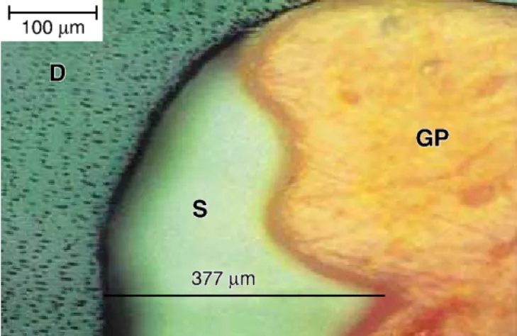
FIGURE 4 - Cross-sec ti on of api cal third sho wing the
hig hest film thick ness of se a ler (S) fol lo wed in this study (337µm). Inter fa ce bet we en gut taper cha (GP) and den -ti nal wall (D). La te ral con den sa -ti on, 200 X.
FIGURE 1 - Cross-sec ti on of cervical third sho wing a
root canal sealer. In an experiment performed by Hataet al.8
(1992) for example, the authors indi-cated that the lack of sealer interface would result in a greater marginal leakage in their samples. Among such investigations, Bamiduro et al.1
(1992) tested the hypothesis that an apical sea-ling promoted by thermoplasticized gutta-percha techniques depends on the thickness of the layer of the root canal sealer. The degree of microleaka-ge was based on the depth of ink penetration at the apical third.
In our study, the samples from Group 3 (warm gutta-percha condensation technique) presented a smaller interface of root canal sealer, and the pre-sence of such unfulfilled spaces by the sealer was significantly smaller. Gutta-percha, after being he-ated, is plasticized from 3 to 5 mm from its heating locus. After heating, dynamic vertical condensati-on is performed, in a technique known as warm gutta-percha3
. This dynamic process of heating and condensing must end 5 mm from the apex17
. The warm gutta-percha technique, when evalua-ted by scanning electron microscopy, demonstra-ted a very close adaptation between dentinal wall, sealer, and gutta-percha20
. At the deepest point of penetration, 3 mm from the apex, a wall-to-wall adaptation was observed. The film thickness of se-aler ranged from 40 to 100mm10
. Our results
pre-sented a greater interval of 15 to 200 mm for the warm gutta-percha condensation technique, which nevertheless offered the best results in all three areas analyzed. It has been shown that devi-ations, undercuts, projections and resorptions many times disable the contact of the instrument to the dentinal walls, creating imperfections in the following filling procedure, especially in Group 1 and 2. On the other hand, considering the homo-geneity factor, samples in Group 3 demonstrated a small incidence of voids, maybe because it promo-tes a higher degree of compaction of gutta-percha cones7
.
CONCLUSIONS
The continuous wave of condensation promoted the lowest film thickness, which may increase the clinical performance of this technique.
ACKNOWLEDGEMENTS
The authors thank the Department of Science and Engineering of Materials (DCMM), Pontifical Catholic University of Rio de Janeiro (PUC-RJ), for the essential technical assistance in this study, es-pecially in memorianto Eng. Maria de Fátima Lo-pes.
REFERENCES
1. Bamiduro R, Ogtenbi G, Shen O. Effect of different sealers on thermoplasticized gutta-percha root canal obturations. J Endod 1992;18:363-6.
2. Brayton S, Davis S, Goldman M. Gutta-percha root canal fillings. Oral Surg Oral Med Oral Pathol 1973;35:226-31. 3. Buchanan LS. The continuous wave of obturation
techni-que: ‘centered’ condensation of warm gutta-percha in 12 seconds. Dent Today 1996;15:60-2,64-7.
4. De Deus QD. Endodontia. 5thed. Rio de Janeiro: Medsi; 1992.
5. Evans J, Simon J. Evaluation of the apical seal produced by injected thermoplasticized gutta-percha in the absence of smear layer and root canal sealer. J Endod 1986; 12:101-7.
6. Grossman LI. Endodontic practice. 10thed. Philadelphia: Lea & Febiger; 1981.
7. Gutmann L. Adaptation of injected thermoplasticized gut-ta-percha in the absence of the dentinal smear layer. Int Endod J 1993; 26:87-9.
8. Hata I, Kawazoe S, Toda T, Weine F. Sealing ability of ther-mafil with or without sealer. J Endod 1992;18:322-36. 9. Hovland J, Dumsha C. Leakage evaluationin vitroof the
root canal sealer cement Sealapex. Int Endod J 1985; 18:179-82.
10. Lifshitz J, Schilder H, Pameijer C. Scanning electron mi-croscope study of the warm gutta-percha technique. J Endod 1983;19:17-24.
11. Maniglia CA, Biffi JC, Carvalho L. Aspecto da guta-percha após a obturação pela técnica da condensação lateral e ul-tra-sônica. Rev Assoc Paul Cirur Dent 1997;51:69-74. 12. Mc Comb D, Smith C. Comparison of the physical
properti-es of polycarboxilate base and conventional root canal sea-lers. J Endod 1976;3:228-35.
13. Michanowicz M, Andrew E, Nicholas P. Low-temperature (70°C) injection gutta-percha: a scanning electron micros-copic investigation. J Endod 1986;12:64-7.
14. Nguyen N. Filling root canals.In:Cohen S, Burns R. Path-ways of the pulp. 6thed. Missouri: Mosby; 1994. p.219-71. 15. Orstavik D. Seating of gutta-percha points: effects of
sea-lers with varying film thickness. J Endod 1982;8:213-8. 16. Roane JB, Sabala CL, Ducanson MG. The “balanced force”
concept for instrumentation of curved canals. J Endod 1985;11:203-9.
17. Ruddle C. Filling root canals.In:Cohen S, Burns R. Path-ways of the pulp. 7thed. Missouri: Mosby; 1994. p.285-7. 18. Schilder H. Filling root canals in three dimensions. Dent
Clin North Am 1967 Nov;723-44.
19. Schilder H. Vertical compaction of warm gutta-percha.In:
Gerstein H. Techniques in clinical endodontics. Philadelp-hia: WB Saunders; 1983. p.76-98.
20. Torabinejad M. Scanning electron microscopic study of root canal obturation using thermoplasticized gutta-per-cha. J Endod 1978;4:245-50.
21. Walton RE, Torabinejad M. Principles and practice of Endodontics. 2nded. Philadelphia: Saunders; 1996.
22. Weller R, Kimbrough F, Anderson R. A comparison of ther-moplastic obturation techniques: adaptation to the canal walls. J Endod 1997;23:703-6.
23. Yamada RS, Armas A, Goldman M, Lin PS. A scanning elec-tron microscopic comparison of a high volume final flush with several irrigating solutions: Part 3. J Endod 1983; 9:137-42.