Ascites and Encapsulating Peritonitis in Endometriosis:
a Systematic Review with a Case Report
Endometriose com ascite e peritonite encapsulante: uma
revisão sistemática com descrição de um caso clínico
Thais Fontes de Magalhães
1Kathiane Lustosa Augusto
1,2Livia Ponte Mota
3Arthur Ribeiro da Costa
3Rainardo Antonio Puster
3Leonardo Robson Pinheiro Sobreira Bezerra
11Department of Gynecology, Universidade Federal do Ceará,
Fortaleza, CE, Brazil
2Department of Gynecology, Universidade de Fortaleza, Fortaleza,
CE, Brazil
3Department of Internal Medicine, Universidade Federal do Ceará,
Fortaleza, Brazil
Rev Bras Ginecol Obstet 2018;40:147–155.
Address for correspondence Thais Fontes de Magalhães, MD, Departmento de Ginecologia, Universidade Federal do Ceara, Rua Alexandre Barauna 949, Fortaleza, CE, 60430-160, Brazil (e-mail: thaisfdemagalhaes@gmail.com).
Keywords
►
endometriosis
►
ascites
►
peritonitis
►
systematic review
►
treatment
Abstract
Endometriosis can have several different presentations, including overt ascites and
peritonitis; increased awareness can improve diagnostic accuracy and patient outcomes.
We aim to provide a systematic review and report a case of endometriosis with this unusual
clinical presentation. The PubMed/MEDLINE database was systematically reviewed until
October 2016. Women with histologically-proven endometriosis presenting with clinically
signi
fi
cant ascites and/or frozen abdomen and/or encapsulating peritonitis were included;
those with potentially confounding conditions were excluded. Our search yielded 37 articles
describing 42 women, all of reproductive age. Ascites was mostly hemorrhagic, recurrent
and not predicted by cancer antigen 125 (CA-125) levels. In turn, dysmenorrhea,
dyspar-eunia and infertility were not consistently reported. The treatment choices and outcomes
were different across the studies, and are described in detail. Endometriosis should be a
differential diagnosis of massive hemorrhagic ascites in women of reproductive age.
Palavras-chave
►
endometriose
►
ascite
►
peritonite
►
revisão sistemática
►
tratamento
Resumo
A endometriose pode ter várias apresentações, incluindo ascite e peritonite, que são
apresentações incomuns. O aumento da conscientização sobre essa doença pode melhorar
a precisão diagnóstica e os resultados das pacientes. Nosso objetivo é fornecer uma revisão
sistemática e relatar um caso de endometriose com esta apresentação clínica incomum. O
banco de dados PubMed/MEDLINE foi revisado sistematicamente até outubro de 2016.
Foram incluídas mulheres com endometriose demonstrada histologicamente, com presença
de ascite clinicamente signi
fi
cativa e/ou abdômen congelado e/ou peritonite encapsulante;
foram excluídas aquelas com comorbidades que pudessem provocar confusão. A pesquisa
selecionou 37 artigos que descrevem 42 mulheres, todas em idade reprodutiva. A ascite foi
principalmente hemorrágica, recorrente, e não indicada pelos níveis de antígeno associado
ao câncer 125 (AC-125). Por sua vez, a dismenorreia, a dispareunia e a infertilidade não foram
relatadas de forma consistente. As escolhas e os resultados do tratamento foram diferentes
entre os estudos, e são descritos em detalhes. A endometriose deveria ser um diagnóstico
diferencial de ascite hemorrágica maciça em mulheres em idade reprodutiva.
received
September 12, 2017 accepted
December 20, 2017 published online March 19, 2018
DOIhttps://doi.org/ 10.1055/s-0038-1626700. ISSN 0100-7203.
Copyright © 2018 by Thieme Revinter Publicações Ltda, Rio de Janeiro, Brazil
Introduction
Endometriosis is defined as the presence of endometrial tissue outside of the uterine cavity, which, like the eutopic endometrium, responds to hormonal stimuli.1Although the symptoms related to endometriosis may be associated with the location of the implant, they are unrelated to the extent of the disease, and a variety of clinical presentations have been described, including the presence of a small amount of peritonealfluid.2It is very rare, however, for endometriosis to present as overt ascites, and the presence of such a condition should prompt the investigation of differential diagnoses, such as malignancy.3The occurrence of encapsu-lating peritonitis, described as the encapsulation of bowel loops by a thickfibrinogenous case, is even rarer, with only five cases previously described in the medical literature.3–8
Women who present with ascites due to endometriosis often undergo multiple diagnostic procedures, including invasive ones, before the underlying cause for ascites is found.9 This results in increased patient risks and health care costs.10General and emergency physicians, as well as gynecologists, should be aware of endometriosis as a poten-tial cause of ascites and encapsulating peritonitis to prompt-ly diagnose it.
With this in mind, this study aims to bring attention to the case of a woman presenting with encapsulating peritonitis and multiple episodes of massive ascites. We also provide a systematic review of the literature regarding the association between histologically-proven endometriosis and clinically-significant ascites, frozen abdomen or encapsulating perito-nitis, including detailed characteristics of their presenta-tions, proposed treatments and outcomes.
Methods
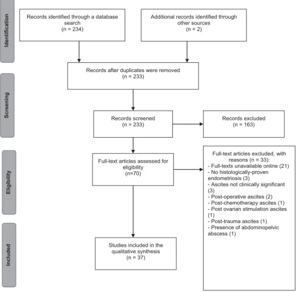
For this systematic review, we used the protocol outlined by the Preferred Reporting Items for Systematic Reviews and Meta-analyses (PRISMA) Statement (►Fig. 1). The electronic PubMed database was searched until October 2016 using the detailed strategy described in►Table 1. The case of a woman with endometriosis, recurrent ascites and encapsulating peritonitis observed at our clinic is also described. The review has been approved by the Ethics in Research Com-mittee under the protocol number 006.01.2017.
Eligibility Criteria
Original articles, clinical trials, case series and case reports of women of any age with histologically-proven endometriosis, presenting with clinically significant ascites and/or frozen abdomen and/or encapsulating peritonitis, published in English, Portuguese, French or Spanish, were eligible for this review.
Because other causes that may occur concurrently with endometriosis can cause the clinical presentations de-scribed, we excluded papers describing patients with: cancer of the ovaries, appendix or peritoneum, or other intra-abdominal cancers; tuberculosis; ovarian hyperstimulation syndrome; ovarian induction or other known causes of
massive ascites; and ascites beginning in the immediate or early post-operative period of exploratory laparotomy/lapa-roscopy. Animal studies; articles published in languages other than the aforementioned ones; and reviews of the literature were also excluded.
Study Selection
The references retrieved were independently screened by two investigators, KA and TM. Initially, the screening was made by title and abstract; then, the full-text versions of the selected papers were obtained, and each article was reviewed forfinal inclusion. If a consensus could not be reached, another author (LB) made thefinal decision regarding inclusion.
Data Extraction
A standardized table was used for data extraction on each selected paper, and information regarding thefirst author, the country of origin, the journal and year of publication, the study type, patient/sample age, patient origin or ethnicity, the clinical presentation, the proposed treatment, and the outcome were included. The characteristics of the ascitic fluid and data regarding the presence or absence of encap-sulating peritonitis were also recorded. Data extraction was performed independently by two investigators (KA and TM).
Case Description
A 28-year-old woman presented to the internal medicine clinic at our institution with wasting syndrome, increased abdominal girth, progressive shortness of breath, dark stools and decreased appetite. She also complained of progressive abdominal and thoracic pain during menses, as well as cyclic dyspareunia that had startedfive years before. She denied infertility or any other chronic medical conditions. Her cancer antigen 125 (CA-125) values were 107.8 and 889.6 on two measurements performed when she was an internal medicine inpatient.
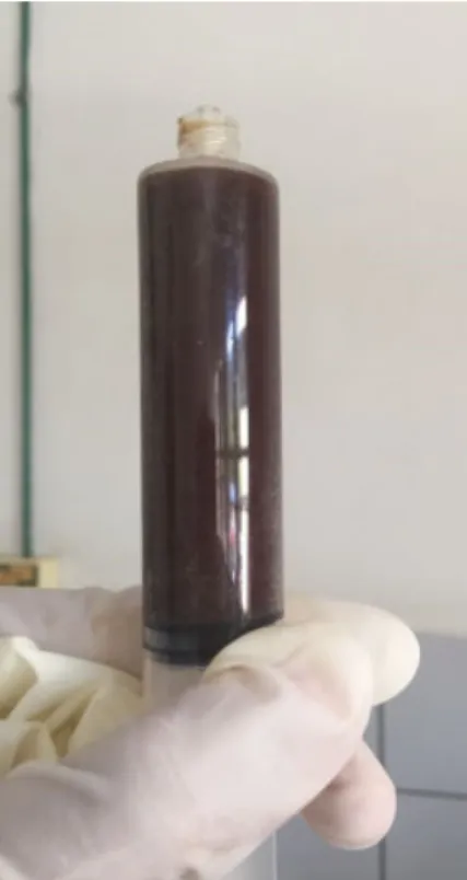
Abdominal ultrasound and contrasted computed tomog-raphy (CT) studies showed loculated, cystic-like ascites pro-ducing a mass effect. A paracentesis procedure revealed a thick, hemorrhagicfluid with low albumin, high cellularity and large concentration of red blood cells (►Fig. 2). Thefluid analysis was negative for bacterial growth or cancer cells. During the diagnostic laparoscopy, multiple adhesions and peritoneal lesions were noticed, and the patient’s abdomen was described as“frozen,”due to the presence of encapsu-lating peritonitis, which did not enable the separation of the peritoneal layers and the confection of the pneumoperito-neum. Eight liters offluid were removed. Histopathology of a peritoneal biopsy taken at this time described chronic peri-tonitis and scarce hemosiderin deposits.
nodules, and apparently hemorrhagic bilateral ovarian cysts. The chest CT and the colonoscopy at that time were normal. A new laparoscopy was performed, and a biopsy con-firmed the diagnosis of endometriosis. Due to peritoneal adhesions, it was not possible to access the pelvic and abdominal organs. This patient was treated with goserelin acetate, with good clinical response. At the six-month follow-up, she was asymptomatic and had regained a considerable amount of weight.
Results
Study Characteristics
Our systematic review yielded 37 articles describing 42 patients with clinically significant ascites, encapsulating peritonitis, or frozen abdomen. One author had a series of three cases, while another had a series of four cases; all of the remaining articles reported only one case each.11,12 The publication dates ranged from 1978 to 2016.
Patient Characteristics and Clinical Presentation The ages of the women who fulfilled the eligibility criteria ranged from 20 years to 47 years.13,14
A total of 8 women in the included articles were described as “black”; 3, as “African-American”; and 2, as “Afro-Caribbean.”12,13,15–22One patient described herself as
“African;” another patient, as“caucasian;” a third one, as “Hispanic;”and another one, as“negro.”23–26Seven studies
did not describe ethnicity, but reported that the patients were Nigerian (n¼3), Malay (n¼2), Brazilian (n¼1) or Japanese (n¼1).3,6,8,27–30 Ethnicity or origin was not
de-scribed at all for the remaining 16 patients.5,7,11,14,31–42
Records identified through a database search
(n = 234)
Screening
Included
Eligi
b
il
ity
Identificatio
n Additional records identified through other sources
(n = 2)
Records after duplicates were removed (n = 233)
Records screened (n = 233)
Records excluded (n = 163)
Full-text articles assessed for eligibility
(n=70)
Full-text articles excluded, with reasons (n = 33): - Full-texts unavailable online (21) - No histologically-proven endometriosis (3)
- Ascites not clinically significant (3)
- Post-operative ascites (2) - Post-chemotherapy ascites (1) - Post ovarian stimulation ascites (1)
- Post-trauma ascites (1) - Presence of abdominopelvic abscess (1)
Studies included in the qualitative synthesis
(n = 37)
Fig. 1 Flow diagram describing the steps in the study selection for inclusion in this systematic review.
Table 1 Detailed search strategy used in the advanced tool of the PubMed/MEDLINE database
Search Keywords
#1 peritonealfibrosisORencapsulating peritoneal sclerosisORsclerosing encapsulating peritonitis ORabdominal cocoonORfrozen abdomenORascites
#2 endometriosisORendometriomaOR endometrioticORhemosiderophage
The ascites was of acute onset in 8 women, and gradual in 24 patients (►Supplementary Material 1). The type of onset was not reported in six cases.
In most patients, the ascites was described as“ hemor-rhagic” and“recurrent,” but descriptions such as“yellow,” “clear yellow,” “brownish green”and“loculated”were also observed.3,6,8,13,25,32,33Ascites was present but not charac-terized infive cases.5,14,15,19,34
The volume of asciticfluid was not shown in some studies, but there are reports of 4.2 L, 4.8 L, 5.0 L and 7 L.13,29,36,41There was associated pleural effusion in eleven patients.3,12,18,19,21,23,32,33,40
Liver involvement by endometriosis was cited in four cases, including one with cysts and another with non-specified focal lesions.3,34
Twenty-two articles reported CA-125 levels. Normal levels were observed in six patients. The biomarker was elevated (>35 U/mL) in 14 patients, ranging from 49 U/mL to>5,000 U/mL. One case had normal CA-125 levels at first, but they became elevated (455 U/mL) after ascites recurrence.12
The most common main clinical presentation was abdo-minal distension, which was sometimes accompanied by other symptoms such as abdominal pain, abdominal tender-ness, abdominal mass, shortness of breath, signs of hypovole-mia, weight loss, nausea or vomiting, asthenia, malaise, cachexia or loss of appetite. In 25 of the 42 women described, at least one symptom of the classic dysmenorrhea, dyspareunia and infertility triad was reported. Of these, dysmenorrhea was the most common, and it was reported in 20 women.8,11–13,15,16,18,20,22–24,27,30–32,36,38–41 Infertility was
reported in 11 patients.11,18,19,22–24,29–31,40,41 Dyspareunia
was reported in 4 patients.15,22,24,36 All three symptoms were present in two studies.22,24 However, in 15 articles (describing 17 patients), the presence or absence of dysmenor-rhea was not mentioned at all.5–7,12,14,19,21,25,26,28,29,33–35,37A
total of 33 studies (with 38 patients) were regarding the presence or absence of dyspareunia,3,5–8,11–14,16–21,25–35,37–42
and 25 studies (with 28 patients) were regarding the presence or absence of infertility.3,5–8,11–17,21,25–28,32–39Fifteen articles
did not mention if any of these three symptoms were present or absent in the cases they reported.5–7,12,14,21,25,26,28,33–35,37
Besides our patient, only five cases of encapsulating peritonitis due to endometriosis have been described in the medical literature.3,5–8
Treatments Used and Outcomes
The treatment choices for the patients included in this review involved hormonal therapies, surgery, anti-infl am-matory drugs (steroidal or non-steroidal) or a combination of
Fig. 2 Thick, hemorrhagic asciticfluid sample.
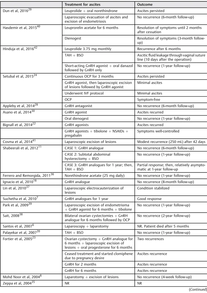
Table 2 Treatment choices and respective outcomes in each study. Outcomes written as described in each article
Treatment for ascites Outcome
Dun et al, 201629 Leuprolideþoral norethindrone Ascites persisted
Laparoscopic evacuation of ascites and excision of endometriosis
No recurrence (6-month follow-up)
Hasdemir et al, 201540 Leuprorelin acetate for 6 months Resolution of symptoms until 2 months
after cessation
Dienogest Resolution of symptoms (3-month
follow-up)
Hinduja et al, 201642 Leuprolide 3.75 mg monthly Recurrence after 6 months
TAHþBSO Asciticfluid leakage through vaginal suture
line (10 days after the operation)
Short-acting GnRH agonistþoral danazol
followed by GnRH only
No recurrence (1-year follow-up)
Setubal et al, 201524 Continuous OCP for 3 months Ascites persisted
GnRH agonist, then laparoscopic excision of lesions followed by GnRH agonist
Minimal ascites
Underwent IVF protocol Minimal ascites
OCP Symptom-free
Appleby et al, 201428 GnRH antagonist No recurrence (6-month follow-up)
Asano et al, 201430 GnRH agonist Ascites recurred
Oral dienogest No recurrence (1-year follow-up)
Bignall et al, 201422 GnRH agonists Ascites recurred
GnRH agonistsþtiboloneþNSAIDsþ
pregabalin
Symptoms well-controlled
Cosma et al, 201441 Laparoscopic excision of lesions Modest recurrence (250 mL) after 42 days
Shabeerali et al, 201211 CASE 1: GnRH analogue No recurrence (6-month follow-up)
CASE 2: Subtotal abdominal
hysterectomyþBSO
No recurrence (1-year follow-up)
CASE 3: GnRH analogues for 1 year; then,
TAHþBSO
Partial response; then, relatively asympto-matic at 1-year follow-up
Ferrero and Remorgida, 201136 Norethindrone acetate (25 mg daily) No recurrence (1-year follow-up)
Ignacio et al, 201018 GnRH analogue No recurrence (8-month follow-up)
Lin et al, 201037 Laparoscopic electrocauterization of
lesions
Condition stabilized
Suchetha et al, 20107 GnRH analogues for 1 year Good response
Park et al, 200939 Laparoscopic excision of endometrioma
þGnRH agonist for 6 monthsþtibolone
No recurrence (1-year follow-up)
Sait, 200838 Bilateral ovarian cystectomiesþGnRH
analogue for 6 months followed by OCP
No recurrence (2-year follow-up)
Santos et al, 20076 Laparoscopyþlaparotomy NR. Patient died after 5 months
Palayekar et al, 200720 TAHþBSO No recurrence (1-year follow-up)
Fortier et al, 200523 Ovarian cystectomyþGnRH analogue for
6 monthsþlaparoscopic excision of
lesionsþoral progesterone for 6 months
Two recurrences
Ceased treatment and started clomiphene due to pregnancy desire
Ascites recurrence
GnRH for 2 months Ascites recurrence
GnRH for 6 months Ascites recurrence
Mohd Noor et al, 20048 Laparotomyþexcision of lesions No recurrence (4-week follow-up)
Zeppa et al, 200435 NR NR
them. Ascites recurrence or persistence was frequent. Three articles did not report how the patients were managed (►Table 2).3,14,35
The hormonal therapies used included long-acting go-nadotropin-releasing hormone (GnRH) agonists (namely goserelin and leuprolide), short-acting GnRH agonists, GnRH antagonists, progestational hormones (specifically
dienogest, medroxyprogesterone and norethindrone), estra-diol, conjugated estrogens, synthetic combined hormones (namely tibolone), steroids with antigonadotropic and anti-estrogenic activity (danazol), and oral contraceptive pills (OCPs).
Other conservative treatments used were isolated pred-nisolone, which did not prevent ascites recurrence, and Table 2 (Continued)
Treatment for ascites Outcome
Cheong and Lim, 20033 NR NR
Jeanes et al, 200234 Double hysterectomyþleft
oophorect-omy followed by estradiol for 6 months followed by goserelin
No recurrence (3-year follow-up)
Moffatt and Mitchell, 200219 Leuprolideþpremarin Recurrence after 4 weeks
TAHþBSOþleuprolide No recurrence (9-month follow-up)
Bhojawala et al, 200017 TAHþRSO No recurrence (6-week follow-up)
Samora-Mata and Feste, 199925 TAHþRSO NR
Myneyyirci-Delale et al, 199812 Laparoscopic excision of lesions followed
by GnRH agonist for 6 months; then,
excision of new ovarian cystþdanazol
daily for 6 months followed by norethin-drone acetate
Recurrence after 1 year; then, no recur-rence (3-year follow-up)
Laparotomyþmonthly depo provera
injections; then, TAHþBSO
Recurrence after 3 years; then, no recur-rence (6-month follow-up)
Appendectomy and left ovarian wedge
resectionþlupron
No recurrence
Lysis of adhesionsþLSO followed by
lupron followed by norethindrone acetate NR
Frigerio et al, 19975 TAHþBSOþappendectomyþ
omen-tectomy
No recurrence (3-year follow-up)
Mejia et al, 199716 Laparotomy No recurrence (15-month follow-up)
Flanagan and Barnes, 199621 LaparotomyþGnRH agonist for 6 months Recurrence (twice in 1 year)
Prednisolone 30 mg daily Recurrence
Leuprorelin for 5 months Recurrence
Myers et al, 199533 TAHþBSOþlysis of adhesions No recurrence (8-month follow-up)
Jose et al, 199432 LSO Recurrence after 1 year
Laparotomyþdanazol NR
Schlueter an McClennan, 199413 Leuprolide acetate monthly No recurrence (3-month follow-up)
Williams and Wagaman, 199115 Medroxyprogesterone acetate Recurrence after 1 month
Depot lupron for 3 months Some ascites
TAHþBSO Recurrence after 3 months
Tenckhoff catheter placed for 2 weeks No recurrence (9-month follow-up)
Chichareon and Wattanakitkrailert,
198831
TAHþBSO No recurrence (6-month follow-up)
Olubuyide et al, 198827 Oral norethisterone No recurrence (1-year follow-up)
Naraynsingh et al, 198526 Depo provera for 6 months No recurrence (4-year follow-up)
Cantor et al, 197914 NR NR
nonsteroidal anti-inflammatory drugs (NSAIDs) in combina-tion with pregabalin and hormonal therapies, which were effective in controlling the symptoms.
The surgical procedures involved drainage of the ascites, excision or electrocauterization of the endometriotic lesions through laparotomy or laparoscopy, lysis of adhesions, abdom-inal hysterectomy, uni- or bilateral salpingo-oophorectomy, ovarian cystectomy, omentectomy, appendectomy, placement of Tenckhoff catheter and/or ovarian wedge resection.
After the initial treatment, due to the desire to achieve pregnancy, one woman underwent in vitro fertilization (IVF), with minimal ascites afterwards.24Another patient started clomiphene, with ascites recurrence.23
A summary of the treatments used for ascites and its respective outcomes, including the time until recurrence, is shown in►Table 2.
Discussion
All patients shown in this review were of childbearing age, likely due to hormonal levels and occurrence of menses. This is, indeed, the most common age range for the presentation of endometriosis in general, which can also rarely occur in older women.43 Endometriosis most commonly affected women of African descent, but it was present in patients of multiple other ethnicities, implying that this diagnosis should not be restricted to patient origin, and must be suspected if the clinical presentation is suitable.
Fluid accumulation was most commonly progressive, but acute onset of symptoms has also been described. The reasons for such a presentation remain unclear, but can be related to the rapid accumulation offluid and to the sponta-neous rupture of a cyst.37
Endometriosis-associated ascites is rare, and encapsulat-ing peritonitis is even less common. Since we excluded women with potentially confounding conditions, it appears that endometriosis itself is the cause of such clinical pre-sentations. It has been hypothesized that the peritoneal irritation caused by endometriosis results in extensivefi bro-sis and inflammation, further optimizing the microenviron-ment for more secondary implants, which in turn exacerbate inflammation. In fact, this theory could also explain the high rate of recurrence of ascites due to endometriosis (►Table 2), ultimately resulting in encapsulating peritonitis, which is described in our case and in four other ones.
Abdominal distension related to ascites was the most common clinical sign, but non-specific signs and symptoms such as malaise and weight loss were also described (►Supplementary Material 1). Additionally, most women had symptoms suggestive of endometriosis, but these were not always reported, bringing attention to the high level of suspicion needed to diagnose this condition. Not all women present with classic symptoms; however, in order for diag-nostic accuracy to be improved, physicians in general (in-cluding emergency room physicians) need to ask about them specifically during history-taking.
Moreover, in some cases, ascitic volume was large and related to pleural effusion, resembling Meigs syndrome.
Great volumes can be caused by the rapid production of fluid, in association with the obstruction of subdiaphrag-matic lymph vessels. In turn, pleuralfluid may be due to the transdiaphragmatic flow of ascites through the lymphatic channels, as has been proposed by Meigs et al,44or due to local reactive inflammation.38
The fluid was generally hemorrhagic, but could have different aspects, such as clear yellow or green-brownish color. On this matter, Bernstein proposed a mechanism by which chocolate cysts would rupture into the peritoneal cavity, leading to irritation and ascites formation; corre-spondingly, other explanations relate to excessive ovarian transudation, superficial endometriosis, open endometriosis lesions or angiogenesis.45–47
Part of the included studies also measured and reported serum CA-125 levels. This biomarker is known to not be accurate for the diagnosis of endometriosis in general; likewise, it was not reliable in the diagnosis of endometriosis presenting with ascites. Although no statistical comparisons were made, it is clear that the values were not intimately correlated to the characteristics of ascites. Furthermore, endometriosis caused CA-125 levels>5,000 U/mL in one case, which commonly indicates malignancy; this further contributes to the need of including endometriosis in the list of differentials of suspected malignancies due to massive ascites.
Several treatment choices were observed in the included articles; the options were similar to the therapies available for endometriosis in general. For the specific treatment of women presenting with ascites, no specific protocol exists, and empirical data does not evidently favor one therapy over another (►Table 2).
Among the strengths of our review are the fact that only articles with histologically proven endometriosis were se-lected; the exclusion of patients with conditions that com-monly cause ascites; and the systematic approach. It is limited, however, in that articles in only four languages were included, and in the fact that some older articles initially screened could not be included because they were not available online, even for purchase. Further research is needed to better define optimal diagnostic and therapeutic approaches in women with unusual presentations of endometriosis.
Conclusion
Contributors
Magalhaes TF, Augusto KL, Mota LP, Costa AR, Puster RA, and Bezerra LRPS contributed with the project and inter-pretation of data, writing of the article, critical review of the intellectual content andfinal approval of the version to be published.
Conflicts of Interest
The authors have no conflicts of interest to declare.
References
1 Chen P, Wang DB, Liang YM. Evaluation of estrogen in endome-triosis patients: Regulation of GATA-3 in endometrial cells and effects on Th2 cytokines. J Obstet Gynaecol Res 2016;42(06): 669–677. Doi: 10.1111/jog.12957
2 Vinci G, Arkwright S, Audebourg A, et al. Correlation between the clinical parameters and tissue phenotype in patients affected by deep-infiltrating endometriosis. Reprod Sci 2016;23(09):1258-–1268. Doi: 10.1177/1933719116638188
3 Cheong EC, Lim DT. Massive ascites–an uncommon presentation of endometriosis. Singapore Med J 2003;44(02):98–100 4 Obaid O, Alhalabi D, Ghonami M. Intestinal obstruction in a
patient with sclerosing encapsulating peritonitis. Case Rep Surg 2017;2017:8316147
5 Frigerio L, Taccagni GL, Mariani A, Mangili G, Ferrari A. Idiopathic sclerosing peritonitis associated withflorid mesothelial hyper-plasia, ovarianfibromatosis, and endometriosis: a new disorder of abdominal mass. Am J Obstet Gynecol 1997;176(03):721–722. Doi: 10.1016/S0002-9378(97)70581-7
6 Santos VM, Barbosa ER Jr, Lima SH, Porto AS. Abdominal cocoon associated with endometriosis. Singapore Med J 2007;48(09): e240–e242
7 Suchetha S, Rema P, Mathew AP, Sebastian P. Endometriosis with massive hemorrhagic ascites. Indian J Cancer 2010;47(02): 224–225. Doi: 10.4103/0019-509X.63004
8 Mohd Noor NH, Zaki NM, Kaur G, Naik VR, Zakaria AZ. Abdominal cocoon in association with adenomyosis and leiomyomata of the uterus and endometriotic cyst : unusual presentation. Malays J Med Sci 2004;11(01):81–85
9 Gupta D, Hull ML, Fraser I, et al. Endometrial biomarkers for the non-invasive diagnosis of endometriosis. Cochrane Database Syst Rev 2016;4:CD012165. Doi: 10.1002/14651858.CD012165 10 Liu E, Nisenblat V, Farquhar C, et al. Urinary biomarkers for the
non-invasive diagnosis of endometriosis. Cochrane Database Syst Rev 2015;(12):CD012019. Doi: 10.1002/14651858.CD012019 11 Shabeerali TU, Rajan R, Kuruvilla AP, et al. Hemorrhagic ascites:
are we missing endometriosis? Indian J Gastroenterol 2012;31 (04):195–197. Doi: 10.1007/s12664-012-0221-1
12 Muneyyirci-Delale O, Neil G, Serur E, Gordon D, Maiman M, Sedlis A. Endometriosis with massive ascites. Gynecol Oncol 1998;69 (01):42–46. Doi: 10.1006/gyno.1998.4953
13 Schlueter FJ, McClennan BL. Massive ascites and pleural effusions associated with endometriosis. Abdom Imaging 1994;19(05): 475–476
14 Cantor JO, Fenoglio CM, Richart RM. A case of extensive abdominal endometriosis. Am J Obstet Gynecol 1979;134(07):846–847. Doi: 10.1016/0002-9378(79)90958-X
15 Williams RS, Wagaman R. Endometriosis associated with massive ascites and absence of pelvic peritoneum. Am J Obstet Gynecol 1991;164(1 Pt 1):45–46. Doi: 10.1016/0002-9378(91)90621-W 16 Mejia EM, Alvarez OA, Lee M. Endometriosis with massive bloody
ascites. J Am Board Fam Pract 1997;10(01):59–61
17 Bhojawala J, Heller DS, Cracchiolo B, Sama J. Endometriosis presenting as bloody pleural effusion and ascites-report of a
case and review of the literature. Arch Gynecol Obstet 2000; 264(01):39–41. Doi: 10.1007/PL00007484
18 Ignacio MM, Joseph N, Hélder F, Mamourou K, Arnaud W. Massive ascites, pleural effusion, and diaphragmatic implants in a patient with endometriosis. Eur J Obstet Gynecol Reprod Biol 2010;149 (01):117–118. Doi: 10.1016/j.ejogrb.2009.10.017
19 Moffatt SD, Mitchell JD. Massive pleural endometriosis. Eur J Cardiothorac Surg 2002;22(02):321–323. Doi: 10.1016/S1010-7940(02)00277-4
20 Palayekar M, Jenci J, Carlson JA Jr. Recurrent hemorrhagic ascites: a rare presentation of endometriosis. Obstet Gynecol 2007;110(2 Pt 2):521–522
21 Flanagan KL, Barnes NC. Pleuralfluid accumulation due to intra-abdominal endometriosis: a case report and review of the litera-ture. Thorax 1996;51(10):1062–1063. Doi: 10.1136/thx.51.10.1062 22 Bignall J, Arambage K, Vimplis S. Endometriosis: a rare and interesting cause of recurrent haemorrhagic ascites. BMJ Case Rep 2014;2014:bcr2013010052. Doi: 10.1136/bcr-2013-010052 23 Fortier D, Dedecker F, Gabriele M, Graesslin O, Barau G. [Endometriosis with ascites and pleural effusion: a case report]. Gynecol Obstet Fertil 2005;33(7-8):508–510. Doi: 10.1016/j.gyobfe.2005.05.014 24 Setubal A, Sidiropoulou Z, Soares S, Barbosa C. Endometriosis and
ascites: a strategy to achieve pregnancy. J Minim Invasive Gynecol 2015;22(06):1104–1108. Doi: 10.1016/j.jmig.2015.05.013 25 Samora-Mata J, Feste JR. Endometriosis ascites: a case report. JSLS
1999;3(03):229–231
26 Naraynsingh V, Raju GC, Ratan P, Wong J. Massive ascites due to omental endometriosis. Postgrad Med J 1985;61(716):539–540. Doi: 10.1136/pgmj.61.716.53
27 Olubuyide IO, Adebajo AO, Adeleye JA, Solanke TF. Massive ascites associated with endometriosis in a Nigerian African. Int J Gynae-col Obstet 1988;27(03):439–441. Doi: 10.1016/0020-7292(88) 90127-0
28 Appleby R, Saroya H, Postgate A, Meer Z. A young woman with abdominal distension. BMJ Case Rep 2014;2014:bcr2014203726. Doi: 10.1136/bcr-2014-203726
29 Dun EC, Wong S, Lakhi NA, Nehzat CH. Recurrent massive ascites due to mossy endometriosis. Fertil Steril 2016;106(06):e14. Doi: 10.1016/j.fertnstert.2016.07.1119
30 Asano R, Nakazawa T, Hirahara F, Sakakibara H. Dienogest was effective in treating hemorrhagic ascites caused by endometrio-sis: a case report. J Minim Invasive Gynecol 2014;21(06):1110-–1112. Doi: 10.1016/j.jmig.2014.04.014
31 Chichareon SB, Wattanakitkrailert S. Endometriosis with ascites. Acta Obstet Gynecol Scand 1988;67(02):187–188. Doi: 10.3109/ 00016348809004198
32 Jose R, George SS, Seshadri L. Massive ascites associated with endometriosis. Int J Gynaecol Obstet 1994;44(03):287–288. Doi: 10.1016/0020-7292(94)90185-6
33 Myers TJ, Arena B, Granai CO. Pelvic endometriosis mimicking advanced ovarian cancer: presentation with pleural effusion, ascites, and elevated serum CA 125 level. Am J Obstet Gynecol 1995;173(3 Pt 1):966–967
34 Jeanes AC, Murray D, Davidson B, Hamilton M, Watkinson AF. Case report: hepatic and retro-peritoneal endometriosis presenting as obstructive jaundice with ascites: a case report and review of the literature. Clin Radiol 2002;57(03):226–229. Doi: 10.1053/ crad.2001.0667
35 Zeppa P, Vetrani A, Cozzolino I, Palombini L. Endometrial glands in ascites secondary to endometriosis. Diagn Cytopathol 2004;30 (02):131–132. Doi: 10.1002/dc.10390
36 Ferrero S, Remorgida V. Endometriosis presenting with hemor-rhagic ascites. Arch Gynecol Obstet 2011;283(06):1429–1430. Doi: 10.1007/s00404-010-1796-3
38 Sait KH. Massive ascites as a presentation in a young woman with endometriosis: a case report. Fertil Steril 2008;90(05):2015.e17-–2015.e19
39 Park BJ, Kim TE, Kim YW. Massive peritonealfluid and markedly elevated serum CA125 and CA19-9 levels associated with an ovarian endometrioma. J Obstet Gynaecol Res 2009;35(05): 935–939. Doi: 10.1111/j.1447-0756.2009.01122.x
40 Hasdemir PS, Ikiz N, Ozcakir HT, Kara E, Guvenal T. Endometriosis associated with relapsing ascites and pleural effusions. J Obstet Gynaecol 2015;35(04):419. Doi: 10.3109/01443615.2014.948823 41 Cosma S, Ceccaroni M, Benedetto C. A pseudoneoplasticfinding of
deep endometriosis: laparoscopic triple segmental bowel resec-tion. Wideochir Inne Tech Malo Inwazyjne 2014;9(03):463–467. Doi: 10.5114/wiitm.2014.41617
42 Hinduja I, Kapadia K, Udwadia F, Bhilawadikar R, Adhe A, Zaveri K. Unusual presentation of endometriosis with haemorrhagic ascites - A case report. J Obstet Gynaecol 2016;36(01):133–134. Doi: 10.3109/01443615.2015.1030605
43 Streuli I, Gaitzsch H, Wenger JM, Petignat P. Endometriosis after menopause: physiopathology and management of an uncommon condition. Climacteric 2017;20(02):138–143. Doi: 10.1080/ 13697137.2017.1284781
44 Meigs JV, Armstrong SH, Hamilton HH. A further contribution to the syndrome offibroma of the ovary withfluid in the abdomen and chest, Meigs’syndrome. Am J Obstet Gynecol 1943;46:19–37. Doi: 10.1016/S0002-9378(16)40440-0
45 Bernstein JS, Perlow V, Brenner JJ. Massive ascites due to endo-metriosis. Am J Dig Dis 1961;6:1–6. Doi: 10.1007/BF02239240 46 Sherer DM, Eliakim R, Abulafia O. The role of angiogenesis in the
accumulation of peritonealfluid in benign conditions and the development of malignant ascites in the female. Gynecol Obstet Invest 2000;50(04):217–224. Doi: 10.1159/00001032