Resting-State Activities of the Amygdala in Han Chinese
Sufang Li1, Qihong Zou2, Jun Li1, Jin Li1, Deyi Wang1, Chaogan Yan1, Qi Dong1, Yu-Feng Zang1,3* 1State Key Laboratory of Cognitive Neuroscience and Learning, Beijing Normal University, Beijing, China,2MRI Research Center and Beijing City Key Lab for Medical Physics and Engineering, Peking University, Beijing, China,3Center for Cognition and Brain Disorders, Affiliated Hospital, Hangzhou Normal University, Hangzhou, Zhejiang, China
Abstract
Background:Prior research has shown that the amygdala of carriers of the short allele (s) of the serotonin transporter (5-HTT) gene (5-HTTLPR) have a larger response to negative emotional stimuli and higher spontaneous activity during the resting state than non-carriers. However, recent studies have suggested that the effects of 5-HTTLPR may be specific to different ethnic groups. Few studies have been conducted to address this issue.
Methodology/Principal Findings: Blood oxygenation level dependent (BOLD) functional magnetic resonance imaging (fMRI) was conducted on thirty-eight healthy Han Chinese subjects (l/l group, n = 19; s/s group, n = 19) during the resting state and during an emotional processing task. Compared with the s/s group, the l/l group showed significantly increased regional homogeneity or local synchronization in the right amygdala during the resting state (|t|.2.028,p,0.05, corrected), but no significant difference was found in the bilateral amygdala in response to negative stimuli in the emotional processing task.
Conclusions/Significance:5-HTTLPR can alter the spontaneous activity of the amygdala in Han Chinese. However, the effect of 5-HTTLPR on the amygdala both in task state and resting state in Asian population was no similar with Caucasians. They suggest that the effect of 5-HTTLPR on the amygdala may be modulated by ethnic differences.
Citation:Li S, Zou Q, Li J, Li J, Wang D, et al. (2012) 5-HTTLPR Polymorphism Impacts Task-Evoked and Resting-State Activities of the Amygdala in Han Chinese. PLoS ONE 7(5): e36513. doi:10.1371/journal.pone.0036513
Editor:Angela Sirigu, French National Centre for Scientific Research, France
ReceivedNovember 16, 2011;AcceptedApril 2, 2012;PublishedMay 4, 2012
Copyright:ß2012 Li et al. This is an open-access article distributed under the terms of the Creative Commons Attribution License, which permits unrestricted use, distribution, and reproduction in any medium, provided the original author and source are credited.
Funding:This study was supported by the Program for Changjiang Scholars and Innovative Research Team in University (PCSIRT), the 111 Project from the Ministry of Education of China (B07008), and NSFC (81020108022, 30770594). The funders had no role in study design, data collection and analysis, decision to publish, or preparation of the manuscript.
Competing Interests:The authors have declared that no competing interests exist.
* E-mail: [email protected]
Introduction
Previous studies have revealed a critical role of the serotonin (5-HT) neurotransmitter system in the development of emotional circuitry and the onset of affective disorders [1–4]. The serotonin transporter (5-HTT) plays a crucial role in the reuptake of serotonin at brain synapses and it has been widely recognized as the target site of selective serotonin reuptake inhibitors (SSRIs), which have been recognized as both depressant and anti-anxiety drugs [5]. One common polymorphic variant of the 5-HTT-linked polymorphic region (5-HTTLPR, SLC6A4) is located on the chromosome 17q11.1–q12 [6] which is a variable repeat sequence resulting in two common alleles: the short (s) variant and the long (l) variant. The s allele of the 5-HTTLPR polymorphism, which affects the promoter of the 5-HTT gene, causes reduced uptake of the neurotransmitter serotonin into the pre-synaptic cells in the brain [3]. Many studies suggest that the s allele of 5-HTTLPR may confer genetic risk for affective disorder [7], such as major depressive disorder [8–10].
Advances in noninvasive functional neuroimaging techniques provide a unique opportunity to explore and evaluate the functional impact of brain-relevant genetic polymorphisms more rapidly and with greater sensitivity than existing behavioral
assessments [11]. Several blood oxygenation level dependent (BOLD) functional magnetic resonance imaging (fMRI) studies have shown that 5-HTTLPR s carriers have higher evoked activity to negatively valenced stimuli [12–15] and higher resting-state cerebral blood flow (CBF) [16,17] in the amygdala than non-carriers, suggesting a potential underlying mechanism by which the s allele confers affective disorder.
relationship between the intrinsic activity of the amygdala and the 5-HTTLPR polymorphism in Caucasians [16,17,33]. Two of these studies found that s carriers had higher CBF than non-carriers [16,17] and the third study failed to detect significant differences between different genotype groups [33]. This informa-tion may be critical in interpreting the different manifestainforma-tions of responses to aversive stimuli in l and s allele carrier groups and have a very important role in helping us to understand the brain’s ongoing activity between the occurrences of negative external stimuli [17]. However, all subjects of the three resting-state fMRI studies were Caucasians. Thus, whether the effects of the 5-HTTLPR on spontaneous activity in the amygdala have ethnic differences in Asian population should be investigated.
The current study aimed to investigate the effects of 5-HTTLPR on the spontaneous activity as well as evoked activity of the amygdala in Han Chinese. We hypothesized that the effect of 5-HTTLPR genotype on amygdala activity would be different in Han Chinese from Caucasian population. In addition, we were also interested in the effects of 5-HTTLPR on the functional connectivity of the amygdala with other brain areas.
Results
Allele distribution of the 5-HTTLPR gene in Han Chinese
The allele frequency of this polymorphism did not show any deviation from the Hardy-Weinberg equilibrium. The allele frequencies of the s and l alleles for the 5-HTTLPR in our sample of 663 healthy Han Chinese sample (72% and 28%, respectively) were significantly different from those of a Caucasian sample (42% and 58%, x2= 18.4, d.f. = 1, p,0.001) [34] but
similar to the findings in other studies about Asian populations (79% and 21%) [24].
Behavioral data
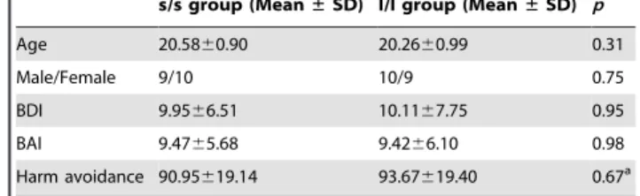
There are 38 participants involved in this study, in which 19 were homozygous for the s allele (s/s group) and 19 were homozygous for the l allele (l/l group) of 5-HTTLPR. The demographic and behavioral data are listed in Table 1. There were no significant differences in age, gender, the score of Beck Depression Inventory II (BDI) [35], the score of Beck Anxiety Inventory (BAI) [36], and the score of harm avoidance of Temperament and Character Inventory-Revised (TCI-R) [37] between the two groups (allps.0.05). Repeated measure analysis of variance (AVOVA) on the accuracy of the emotional task revealed no significant main effects and interaction effect (ps.0.05). ANOVA on the reaction time showed no significant between-group effect, but showed significant within-group effect (F1,36= 11.361, p= 0.002), i.e., slower response to negative
(8926136 ms) stimuli than to neutral (8636131 ms). The reaction time showed a trend of significant interaction (F1,36= 3.903,
p= 0.056). Post hoc pairedt-tests showed that the slower reaction time for negative stimuli existed in l/l group (t18= 3.738,p= 0.002) but not in s/s group (t18= 0.998,p= 0.332) (Figure 1).
Imaging data during resting state
Results of regional homogeneity (ReHo) analysis. One-samplet-tests for the s/s group and the l/l group showed that the default mode network including the posterior cingulate cortex (PCC)/precuneus, medial prefrontal cortex (MPFC), and bilateral angular gyrus, exhibited significantly higher ReHo [38] than the global mean. This pattern was closely consistent with previous studies [38,39].
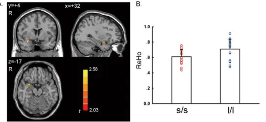
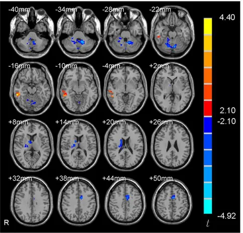
The two-sample t-test showed that the l/l group had signifi-cantly higher ReHo than the s/s group in the right amygdala (Figure 2 and Table 2,p,0.05, corrected) in a region of interest (ROI) analysis. There was no significant correlation between the ReHo value of the right amygdala and the score of behavioral measurements (BAI, BDI, and harm avoidance of TCI) (ps.0.05). In addition, a few other brain regions showed significant between-group difference, including cerebellum, superior temporal gyrus,
Figure 1. Difference in reaction time to negative stimuli and neutral stimuli in l/l group and s/s/ group.**,p,0.01.
and fusiform gyrus in a whole brain analysis (Figure 3 and Table 2,
p,0.05, corrected in the whole brain).
Results of between-group difference in functional connectivity of the amygdala. Some studies have investigated the 5-HTTLPR genotype effects on the functional connectivity of the amygdala during task states [14,15]. However, the results were inconsistent. No resting-state amygdala functional connectivity study has been carried out to investigate the 5-HTTLPR genotype effects. Therefore, as an exploratory study, we compared the functional connectivity with amygdala between s/s and l/l group. Two-sample t-tests showed that no brain region had significant difference in functional connectivity with the right amygdala between the two groups. Whereas, four brain regions including right caudate, middle cingulate gyrus, middle temporal gyrus and cerebellum showed significant difference in functional connectivity with the left amygdala (Figure 4 and Table 3) (p,0.05, corrected). We further performed correlation analysis between the functional connectivity strength of the four brain regions and the behavioral data (the score of BAI, BDI and harm avoidance of TCI) for each group of participants. The right caudate showed significant negative correlation between the functional connectivity strength and the score of harm avoidance in l/l group (r=20.616,
p= 0.007) (Figure 5). No other significant correlation was found.
Imaging data in the emotional processing task
In comparison to the neutral condition, the negative condition showed significantly higher activation in the bilateral amygdala in
both the s/s group and the l/l group (Figure 6,p,0.05, corrected). In addition to the amygdala, there were significantly higher activation to negative stimuli in bilateral posterior fusiform and parahippocampal areas, the anterior cingulate cortex (ACC), and the ventral prefrontal cortex (vPFC). The activation patterns were consistent with previous studies [40].
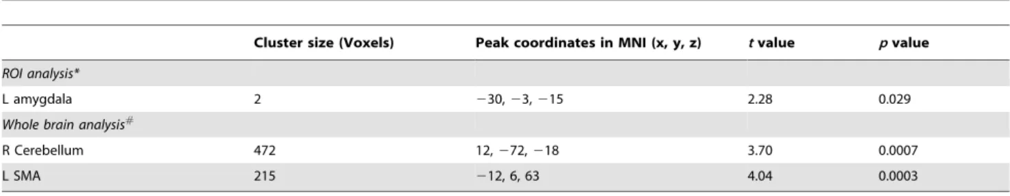
Direct comparison of the two groups didn’t find significant different activation in the bilateral amygdala (p.0.05, corrected in the amygdala). But two voxels showed greater activation (p,0.05, Table 4) in the l/l group than in the s/s group without correction for multiple comparisons. In addition, right cerebellum and left supplementary motor area (SMA) showed significant higher activation in the l/l group than s/s group in an exploratory analysis (Figure 7 and Table 4,p,0.05, corrected within the whole brain).
Discussion
The present study investigated the effects of 5-HTTLPR on amygdala activity both in an emotional processing task and in the resting state in Han Chinese. The result from the resting state demonstrated higher activity in the amygdala of the l/l group compared with that in the s/s group, but no significant difference was found in the emotional task between the two groups.
Studies of 5-HTTLPR genotype effects on emotional task activation have consistently reported higher activation in s/s or s carriers than l/l in Caucasian populations [12–15,32]. The current
Figure 2. Significant higher ReHo in the right amygdala in l/l group than s/s group.A. Voxels with |t|.2.028 and cluster size.297 mm3(11
voxels) corresponded with a correctedp,0.05. R: right side. B. Bars and error bars represent the mean and standard deviation of ReHo value in the right amygdala, which showed significant difference between the two genotype groups in Figure 1A.
doi:10.1371/journal.pone.0036513.g002
Table 2.Significant difference of ReHo in the two genotype groups (s/s vs. l/l).
Cluster size (Voxels) Peak coordinates in MNI (x, y, z) tvalue pvalue
ROI analysis
R amygdala 11 33, 3,218 22.58 0.014
Whole brain analysis
L cerebellum 2959 245,254,242 4.74 0.00003
L STG 253 248, 9,215 24.15 0.0002
L FG 787 269,257,6 25.89 0.000005
study did not find significant difference in task activation in Han Chinese. When we used a liberal threshold (i.e.,p,0.05 without multiple comparison correction), two voxels showed higher activation in l/l group than s/s group. Therefore, this result was not consistent with those from Caucasians. There has been so far only one research article about the genotype effects of 5-HTTLPR on amygdala activity in Asians [18]. In that study, it was found that the l carriers had higher activation in the bilateral amygdala than the s/s group during an angry task. In addition, a review reported a personal communication (SE Taylor, March 22, 2007) and mentioned greater amygdala activation in the l homozygous than in s carriers, most participants being East Asian ancestry [19]. The reaction time of emotional task in the current study provided interesting evidence of 5-HTTLPR genotype effects on emotional processing. The results of non-significant between-group differ-ence was consistent with previous studies [12,13]. However, these studies did not compare the difference between conditions. In the current study, ANOVA showed significant main effect of emotional stimuli on reaction time, i.e., a slower reaction time to negative stimuli than to neutral stimuli. This result was consistent with previous studies [41,42], and suggesting that the emotional task requires more complicated neural processing. Our further analysis showed that such slower reaction time to negative stimuli existed in l/l group but not s/s group. The current result may imply that different genotype of 5-HTTLPR may have different behavior performance during emotional processing. However, such effect is not strong enough (marginal interaction effect of p= 0.056) to draw a conclusion. Several studies had reported that s carriers of 5-HTTLPR exhibited a stronger
attention bias for anxious stimuli than participants with l/l in Caucasians [43–45]. The results from Caucasians and the current result looks contradictory, suggesting that 5-HTTLPR might have ethnic effect. It will be interesting for future studies to further investigate this ethnic effect, especially involving both Caucasians and East Asian populations in a study.
We found higher ReHo in the right amygdala in the l/l group than s/s group. The physiological meaning of the ReHo difference in the right amygdala between l/l and s/s groups remains unclear. ReHo measures the local synchronization of BOLD signal. An electrophysiological study has shown high local synchronization between the local field potentials (LFPs) of multiple cortical electrodes with a physical distance of 2.6–10.6 mm and such local synchronization in resting state could be modulated by stimulation [46]. From this aspect, ReHo measurement of BOLD fMRI was similar with the local synchronization of LFPs of multiple cortical electrodes. ReHo has been used in many studies on brain disorders, of which, the results of two studies on epilepsy were interesting. Increased ReHo was found in the medial temporal lobe of patients with temporal lobe epilepsy (TLE) [47] and also found in the thalamus of patients with generalized tonic-clonic seizures (GTCS) [48]. These results were consistent with the hypothesis that the abnormally increased local synchronization of neural activity may underlie the epileptic discharges [47,48]. Thus the current RS-fMRI result may suggest that the l/l group had higher synchronization of spontaneous neuronal activity in the right amygdala than the s/s group. However, this result of Han Chinese seems to be contrary to those from two RS-fMRI studies in Caucasian population [16,17], which reported that s carriers or
Figure 3. Significant different ReHo in s/s group than l/l group in a whole brain analysis.Voxels with |t|.2.03 and cluster size.6156 mm3 (228 voxels) corresponded with a correctedp,0.05. R: right side.
the s/s homogeneous group had higher CBF in the amygdala than the l/l group. These two studies used arterial spin labeling (ASL) fMRI technique and measured regional CBF while the current study used BOLD fMRI and measured the synchronization (i.e., ReHo) of BOLD signals. It has been reported that resting-state CBF and resting-state ReHo have had a high spatial correlation in the default mode network [49]. Of note, whether such high spatial correlation exists in the amygdala warrants further investigation as the amygdala was not spatially covered in that previous correlation study [49]. Anyway, it is less likely that the reversed results of the current study on Asians compared with that of previous studies on Caucasians were due to the different resting-state imaging techniques (i.e., CBF vs. BOLD). A more reasonable interpretation would be ethnic differences of the effects of the 5-HTTLPR polymorphism.
The right amygdala showed no significant difference between the two groups in functional connectivity. Significant differences in functional connectivity of the left amygdala were found in the right caudate, middle cingulate gyrus, middle temporal gyrus and cerebellum between the two groups. Previous amygdala functional connectivity studies on 5-HTTLPR genotype effects focused on task state and investigated functional connectivity between two predefined regions, e.g., between the amygdala and ventral medial prefrontal cortex [14] and between the amygdala and anterior cingulate cortex [15]. All these four brain regions in the current study had not been reported in previous amygdala functional connectivity studies. An interesting finding in the current study is the higher positive functional connectivity between the right caudate and left amygdala in the l/l group than s/s group. This connectivity strength showed significantly negative correlation
Table 3.Significant difference in functional connectivity with the left amygdala between the two genotype groups (s/s vs. l/l).
Cluster size (Voxels)
Peak coordinates in
MNI (x, y, z) MeanZin s/s MeanZin l/l Tvalue Pvalue
R MTG 129 57,236,212 0.019 20.158* 3.30 0.004
R caudate 147 12, 0, 12 0.163* 0.323* 23.58 0.002
L MCG 119 23, 0, 51 0.016 0.203* 24.01 0.001
L Cere 427 215,251,230 20.090 0.156* 24.92 0.0001
R: right. L: left. MTG: middle temporal gyrus. MCG: middle cingulate gyrus. Cere: cerebellum. *: showing significant functional connectivity by one-samplet-test within group.
doi:10.1371/journal.pone.0036513.t003
with harm avoidance score of TCI in l/l group but not in s/s group. An RS-fMRI study has demonstrated increased connec-tivity between the amygdala and caudate in patients with schizophrenia [50] and a task fMRI study found increased connectivity between the amygdala and the caudate during masked fear condition in borderline personality disorders [51]. However, the existing evidence of the amygdala functional connectivity are not enough to draw a conclusion.
Previous studies showed that 5-HTTLPR can alter amygdala activity in response to salient stimuli but that the direction of this association may be influenced by genetic background (i.e., ethnicity) [19]. The results of both task state and resting state from the current study provide new evidence for this hypothesis. The current study may explain why the Asian population has a higher frequency of the s allele but a lower prevalence of affective disorders compared with Caucasian population [20]. Ethnic differences may be important in determining disease risk factors and optimizing treatment [52].
There were several limitations in the present study. First, the result of no significant difference between the two genotype groups in the emotional processing task may be due to either smaller sample size or ill-designed task. Genetic effects on affective disorder is complex. The impact of one particular gene polymorphism on brain function may be very small and therefore the sample size should be large enough to detect statistical significance [53,54]. The task design could be further optimized. For example, a previous study in Korean women reported higher amygdala activation in l carriers than s/s group, but only for angry emotion and not sad emotion [18]. The negative scene pictures in the current study might have induced complex emotion responses. In addition, we used only negative and neutral stimuli whereas positive stimuli may provide additional information. Therefore, future studies with a well designed task, larger sample size, and even a combination of BOLD and CBF will clarify the genotype difference. Second, we didn’t include a Caucasian population sample and perform the same procedures to elucidate any ethnic
differences of genotype effect. Future studies should be conducted on two population samples, i.e. Caucasians and Asians, about the 5-HTTLPR effect on amygdala activity. Third, our genotyping of the 5-HTTLPR did not differentiate an uncommon alleles (xl) that are longer than the l variant and we did not analyze the recently reported A to G single nucleotide polymorphism (SNP) within the long allele [55,56]. The role of xl is unknown. The lGallele was
found to behave similarly to the lower-expressing s allele [57], and it can increase amygdala activation more than the lAallele [58,59].
Previous studies have reported that the frequency of xl [20] and lG
[60] allele was relatively low in Asians. To exclude these
Figure 6. Activation in the negative condition versus the neutral condition in the two genotype groups.p,0.05, corrected, voxels with |t|.2.10 and cluster size.6156 mm3(228 voxels).
doi:10.1371/journal.pone.0036513.g006
Figure 5. Correlation between harm avoidance score and the strength of functional connectivity between the left amygdala and right caudate in the two genotype groups.FC_Caudate: the strength of functional connectivity between left amygdala and right caudate in each group.
confounding factors would be helpful to reveal the ethnic effect of the genotypes of 5-HTTLPR in the future studies.
In conclusion, our study suggested that 5-HTTLPR can alter the spontaneous activity of the amygdala in Han Chinese and the association between the 5-HTTLPR polymorphism and the activity of the amygdala may be modified by ethnic differences both in response to negative stimuli and during the spontaneous activity of the resting state. Further investigation of both Caucasian and East Asian populations and more detailed genotyping (e.g., A to G SNP within the long allele) will help to clarify this issue.
Methods and Materials
Participants
This study was approved by the Ethics Committee of the Key Laboratory of Cognitive Neuroscience and Learning, Beijing Normal University. There were two stages of informed consent. For the first stage, blood samples were obtained from 663 volunteers who were healthy freshman at Beijing Normal University when they were taking a physical examination. All participants gave written informed consent in which they were told that the genotyping results would not be given to the participants. At the second stage, 42 right-handed participants (mean age 20.4 years, range 18–22 years) were selected for MRI scanning according to genotype (See below) after they gave written informed consent. But they were not aware of their genotypes. None of them had reported any history of psychiatric or neurological illness. Two participants were excluded due to excessive head motion and two participants were excluded due to falling asleep during the resting-state fMRI scanning (See data analysis for details). For the remaining 38 participants, 19 were (9 males) homozygous for the s allele in the s/s group and 19 were (10 males) homozygous for the l allele in the l/l group. Participants in the two genetic groups were similar in age (p= 0.31, two-samplet -test) and gender (p= 0.75, chi-square test).
Mood and personality assessment
The BDI, BAI, and TCI-R were used as behavioral measures of depressive symptoms, anxiety symptoms, and personality dimen-sions, respectively.
RS-fMRI scanning
During the RS-fMRI session, the participants were instructed to keep as still as possible and not to think systematically. Two subjects were excluded due to fall asleep during the scanning. All other subjects reported that they had never fallen asleep during the RS-fMRI scanning.
Emotional task fMRI scanning
During the task-state fMRI scanning, 60 pictures (30 negative and 30 neutral, 30 indoor and 30 outdoor) were chosen from the International Affective Picture System (IAPS) [61]. The negative pictures depict complex unpleasant events including pollution, starving children, and cemeteries [61]. Previous study reported a high correlation (r= 0.913,p,0.01) of the valence scores of IAPS pictures between Han Chinese and Caucasians [62]. Each picture was presented for 1500 ms. The participant was instructed to press the left or right button to judge whether the picture was indoor or outdoor as quickly as possible. Then, a fixation ‘‘+’’ was presented for 500 ms to 6500 ms (randomly jittered between 500, 2500, 4500 and 6500 ms). The order of the categories of pictures (neutral and positive, indoor and outdoor) was randomized. The emotional processing task lasted 5 min 2 s.
MRI data parameters
MR images were collected using a SIEMENS TRIO 3-Tesla in the Brain Imaging Center for brain research, Beijing Normal University. Participants lay supine with head snugly fixed by belt and foam pads to minimize head motion. Each participant underwent an eight-minute RS-fMRI scanning session, an emotional processing task-state fMRI scanning session and a 3D anatomic session. The functional images were obtained with the following parameters: 33 axial slices, thickness/gap = 3/0.6 mm, in-plane resolution = 64664, repetition time (TR) = 2000 ms, echo
time (TE) = 30 ms, flip angle = 90u, field of view (FOV) = 2006200 mm2. The 3D T1-weighted magnetization-prepared rapid gradient echo (MPRAGE) image was acquired
Table 4.Significant difference in activation during the emotional task between the two genotype groups (l/l vs. s/s).
Cluster size (Voxels) Peak coordinates in MNI (x, y, z) tvalue pvalue
ROI analysis*
L amygdala 2 230,23,215 2.28 0.029
Whole brain analysis#
R Cerebellum 472 12,272,218 3.70 0.0007
L SMA 215 212, 6, 63 4.04 0.0003
*: (p,0.05, not corrected).
#
: (p,0.05, corrected).
R: right. L: left. SMA: supplementary motor area. doi:10.1371/journal.pone.0036513.t004
Figure 7. Significant difference in activation in l/l group and s/s group in a whole brain analysis.Voxels with |t|.2.03 and cluster size.6156 mm3(228 voxels) corresponded with a correctedp
,0.05. R: left side.
motion correction using a linear transformation. The transformed 3D images were then segmented into grey matter, white matter and cerebrospinal fluid by the unified segmentation approach. Normalization parameters were applied to the coregistered functional images and resliced to 3 mm isotropic resolution.
ReHo analysis. Resting-State fMRI Data Analysis Toolkit (REST, www.restfmri.net) [64] was then used for linear trend removing, temporally band-pass filtering (0.01,0.08 Hz) [57,65]
and ReHo computation. ReHo used the Kendall coefficient of concordance (KCC) [66] to measure the temporal synchronization of the time series within a functional cluster. This method had been widely used in clinical studies by several different groups [67– 74]. In the current study, 27 nearest neighboring voxels were defined as a cluster and a KCC value (range 0–1) was given to the voxel at the center of this cluster. Then the ReHo maps were spatially smoothed (FWHM = 6 mm) using SPM8. Each individ-ual ReHo map was then standardized by dividing its mean ReHo of the entire brain [38]. One-sample t-tests against one were conducted on the ReHo maps for the two genotype groups separately to show the general patterns of resting-state spontane-ous activity. A two-sample t-test was performed to compare the two genotype groups within the bilateral amygdala. The amygdala ROIs were predefined by the anatomical automatic labeling (AAL) template [75]. Because we had a strong a priori hypothesis regarding differential activity in the amygdala, we used a small volume correction for multiple comparisons within the amygdala ROIs and set corrected p,0.05 as the threshold (individual
p,0.05, cluster size.11 voxels/297 mm3). The statistical result was corrected for multiple comparisons using the ‘‘AlphaSim’’ implemented in REST. This function is based on the Monte Carlo simulation in AFNI (http://afni.nih.gov/afni/docpdf/AlphaSim. pdf). After the ROI analysis, an exploratory investigation was performed in the whole brain to reveal potential between-group difference in other brain regions. Voxels withp,0.05 and cluster size .6156 mm3were considered significant, which corresponds to correctedp,0.05 as determined by AlphaSim in AFNI.
Functional connectivity analysis. A spherical (12 mm in diameter) seed ROI at the center of the right (x, y, z =+24,24,
212) and left (x, y, z =224,24,212) amygdala, respectively, was defined as similarly done in a previous study [14]. For each ROI, a seed reference timecourse was obtained by averaging the time-courses of all voxels in the ROI. Then Pearson’s correlation analysis was performed between the seed reference timecourse and that of each voxel in the brain in a voxel-wise way. The 6 head motion parameters, the global mean timecourse, mean timecourse of the white matter, and mean timecouse of the cerebrospinal fluid were taken as nuisance covariates. Finally, the Fisher’s z transformation was used to improve the normality of the
t-tests was carried out in the left- and right-amygdala mask, respectively. For the left-amygdala mask, voxels withp,0.05 and cluster size.2835 mm3(corresponding to correctedp,0.05) as determined by AlphaSim in AFNI were considered significant. For the right-amygdala mask, voxels with p,0.05 and cluster size
.2754 mm3(corresponding to corrected p,0.05) as determined by AlphaSim in AFNI were considered significant. Then, the Pearson’s correlation analyses were implemented in SPSS 13.0 software (SPSS Inc., Chicago, Illinois, USA) to assess the association between behavioral scores (BAI, BDI, and harm avoidance of TCI, respectively) and the strength of functional connectivity with the amygdala in brain regions showing significant difference between two groups. The strength value of functional connectivity with the amygdala was derived from averaging thezvalue of all voxels in each region which showing significant functional connectivity difference between the two groups.
Task fMRI data analysis
Preprocessing of the task fMRI data, including slice timing, head motion correction, and spatial normalization, were the same as for the RS-fMRI data except for not removing the first 10 time points. In the task-state fMRI scanning, two participants were excluded for further analysis due to head motion of more than 2.0 mm or 2.0u. Predetermined condition effects at each voxel were calculated using at-test, producing a statistical image for the contrast of the negative condition versus the neutral condition for each participant. One-samplet-tests against zero were conducted on the individual contrast maps of the two genotype groups separately to show the general patterns of emotional task activation. Two-sample t-test was conducted on these contrast images to investigate task activation differences between the two genotype groups in the amygdala ROIs defined as in the above ReHo analysis. After the ROI analysis, an exploratory investiga-tion was performed in the whole brain to reveal potential between-group difference in other brain regions. Voxels withp,0.05 and cluster size .6156 mm3 were considered significant, which corresponds to correctedp,0.05 as determined by AlphaSim in AFNI.
Genotyping
previously described [34] on a PE-9700 or a PE-2400 thermal cycle (Perkin Elmer, USA). After initial denaturation at 95uC for 4 min, 35 cycles were carried out at 96uC for 45 sec, 61uC for 90 sec, and 72uC for 90 sec, followed by a final step of elongation at 72uC for 10 min, then the PCR products were separated on a 2% agarose gel supplemented with ethidium bromide for 3 hr. They were then analyzed with Gel Doc 2000 imaging system to detect and record the genotype of each sample. The l variant represented the fragment of 528 bp and the s allele represented the fragment of 488 bp. Genotypes were read by at least two researchers, ambiguous or unidentifiable results were reamplified
and re-scored. Samples that continued to amplify poorly were eliminated from the study.
Acknowledgments
We thank all the research assistants who helped us with data collection.
Author Contributions
Conceived and designed the experiments: YFZ SL Jin Li Jun Li QD. Performed the experiments: SL Jin Li DW. Analyzed the data: SL DW CY. Contributed reagents/materials/analysis tools: QZ DW CY. Wrote the paper: SL YFZ QZ Jun Li.
References
1. Ansorge MS, Zhou M, Lira A, Hen R, Gingrich JA (2004) Early-life blockade of the 5-HT transporter alters emotional behavior in adult mice. Science 306: 879–881.
2. Gaspar P, Cases O, Maroteaux L (2003) The developmental role of serotonin: news from mouse molecular genetics. Nat Rev Neurosci 4: 1002–1012. 3. Lesch KP, Bengel D, Heils A, Sabol SZ, Greenberg BD, et al. (1996) Association
of anxiety-related traits with a polymorphism in the serotonin transporter gene regulatory region. Science 274: 1527–1531.
4. Lotrich FE, Pollock BG (2004) Meta-analysis of serotonin transporter polymorphisms and affective disorders. Psychiatr Genet 14: 121–129. 5. Joo YH, Oh HB, Kim B, Jung SH, Chung JK, et al. (2007) No association
between 5-HTTLPR and harm avoidance in Korean college students. J Korean Med Sci 22: 138–141.
6. Heils A, Teufel A, Petri S, Stober G, Riederer P, et al. (1996) Allelic variation of human serotonin transporter gene expression. J Neurochem 66: 2621–2624. 7. Hauser J, Leszczynska A, Samochowiec J, Czerski PM, Ostapowicz A, et al.
(2003) Association analysis of the insertion/deletion polymorphism in serotonin transporter gene in patients with affective disorder. Eur Psychiatry 18: 129–132. 8. Dorado P, Penas-Lledo EM, Gonzalez AP, Caceres MC, Cobaleda J, et al. (2007) Increased risk for major depression associated with the short allele of the serotonin transporter promoter region (5-HTTLPR-S) and the CYP2C9*3 allele. Fundam Clin Pharmacol 21: 451–453.
9. Hoefgen B, Schulze TG, Ohlraun S, von Widdern O, Hofels S, et al. (2005) The power of sample size and homogenous sampling: association between the 5-HTTLPR serotonin transporter polymorphism and major depressive disorder. Biol Psychiatry 57: 247–251.
10. Kiyohara C, Yoshimasu K (2010) Association between major depressive disorder and a functional polymorphism of the 5-hydroxytryptamine (serotonin) transporter gene: a meta-analysis. Psychiatr Genet 20: 49–58.
11. Egan MF, Goldberg TE, Kolachana BS, Callicott JH, Mazzanti CM, et al. (2001) Effect of COMT Val108/158 Met genotype on frontal lobe function and risk for schizophrenia. Proc Natl Acad Sci U S A 98: 6917–6922.
12. Canli T, Omura K, Haas BW, Fallgatter A, Constable RT, et al. (2005) Beyond affect: a role for genetic variation of the serotonin transporter in neural activation during a cognitive attention task. Proc Natl Acad Sci U S A 102: 12224–12229.
13. Hariri AR, Mattay VS, Tessitore A, Kolachana B, Fera F, et al. (2002) Serotonin transporter genetic variation and the response of the human amygdala. Science 297: 400–403.
14. Heinz A, Braus DF, Smolka MN, Wrase J, Puls I, et al. (2005) Amygdala-prefrontal coupling depends on a genetic variation of the serotonin transporter. Nat Neurosci 8: 20–21.
15. Pezawas L, Meyer-Lindenberg A, Drabant EM, Verchinski BA, Munoz KE, et al. (2005) 5-HTTLPR polymorphism impacts human cingulate-amygdala interactions: a genetic susceptibility mechanism for depression. Nat Neurosci 8: 828–834.
16. Canli T, Qiu M, Omura K, Congdon E, Haas BW, et al. (2006) Neural correlates of epigenesis. Proc Natl Acad Sci U S A 103: 16033–16038. 17. Rao H, Gillihan SJ, Wang J, Korczykowski M, Sankoorikal GM, et al. (2007)
Genetic variation in serotonin transporter alters resting brain function in healthy individuals. Biol Psychiatry 62: 600–606.
18. Lee BT, Ham BJ (2008) Serotonergic genes and amygdala activity in response to negative affective facial stimuli in Korean women. Genes Brain Behav 7: 899–908.
19. Munafo MR, Brown SM, Hariri AR (2008) Serotonin transporter (5-HTTLPR) genotype and amygdala activation: a meta-analysis. Biol Psychiatry 63: 852–857. 20. Goldman N, Glei DA, Lin YH, Weinstein M (2010) The serotonin transporter polymorphism (5-HTTLPR): allelic variation and links with depressive symptoms. Depress Anxiety 27: 260–269.
21. Shi M, Hu J, Dong X, Gao Y, An G, et al. (2008) Association of unipolar depression with gene polymorphisms in the serotonergic pathways in Han Chinese. Acta Neuropsychiatrica 20: 139–144.
22. Kim DK, Lim SW, Lee S, Sohn SE, Kim S, et al. (2000) Serotonin transporter gene polymorphism and antidepressant response. Neuroreport 11: 215–219.
23. Kim JM, Stewart R, Kim SW, Yang SJ, Shin IS, et al. (2007) Interactions between life stressors and susceptibility genes (5-HTTLPR and BDNF) on depression in Korean elders. Biol Psychiatry 62: 423–428.
24. Kunugi H, Hattori M, Kato T, Tatsumi M, Sakai T, et al. (1997) Serotonin transporter gene polymorphisms: ethnic difference and possible association with bipolar affective disorder. Mol Psychiatry 2: 457–462.
25. Ohara K, Nagai M, Tsukamoto T, Tani K, Suzuki Y (1998) Functional polymorphism in the serotonin transporter promoter at the SLC6A4 locus and mood disorders. Biol Psychiatry 44: 550–554.
26. Zhang K, Xu Q, Xu Y, Yang H, Luo J, et al. (2009) The combined effects of the 5-HTTLPR and 5-HTR1A genes modulates the relationship between negative life events and major depressive disorder in a Chinese population. J Affect Disord 114: 224–231.
27. Mrazek DA, Rush AJ, Biernacka JM, O’Kane DJ, Cunningham JM, et al. (2009) SLC6A4 variation and citalopram response. Am J Med Genet B Neuropsychiatr Genet 150B: 341–351.
28. Pollock BG, Ferrell RE, Mulsant BH, Mazumdar S, Miller M, et al. (2000) Allelic variation in the serotonin transporter promoter affects onset of paroxetine treatment response in late-life depression. Neuropsychopharmacology 23: 587–590.
29. Smeraldi E, Zanardi R, Benedetti F, Di Bella D, Perez J, et al. (1998) Polymorphism within the promoter of the serotonin transporter gene and antidepressant efficacy of fluvoxamine. Mol Psychiatry 3: 508–511.
30. Kim H, Lim SW, Kim S, Kim JW, Chang YH, et al. (2006) Monoamine transporter gene polymorphisms and antidepressant response in koreans with late-life depression. JAMA 296: 1609–1618.
31. Yoshida K, Ito K, Sato K, Takahashi H, Kamata M, et al. (2002) Influence of the serotonin transporter gene-linked polymorphic region on the antidepressant response to fluvoxamine in Japanese depressed patients. Prog Neuropsycho-pharmacol Biol Psychiatry 26: 383–386.
32. Heinz A, Smolka MN, Braus DF, Wrase J, Beck A, et al. (2007) Serotonin transporter genotype (5-HTTLPR): effects of neutral and undefined conditions on amygdala activation. Biol Psychiatry 61: 1011–1014.
33. Viviani R, Sim EJ, Lo H, Beschoner P, Osterfeld N, et al. (2010) Baseline brain perfusion and the serotonin transporter promoter polymorphism. Biol Psychiatry 67: 317–322.
34. Collier DA, Stober G, Li T, Heils A, Catalano M, et al. (1996) A novel functional polymorphism within the promoter of the serotonin transporter gene: possible role in susceptibility to affective disorders. Mol Psychiatry 1: 453–460. 35. Beck AT, Steer RA, Ball R, Ranieri W (1996) Comparison of Beck Depression Inventories -IA and -II in psychiatric outpatients. J Pers Assess 67: 588–597. 36. Beck A, Steer R Manual for the Beck Anxiety Inventory: Psychological
Corporation: San Antonio, TX.
37. Cloninger CR, Svrakic DM, Przybeck TR (1993) A psychobiological model of temperament and character. Arch Gen Psychiatry 50: 975–990.
38. Zang Y, Jiang T, Lu Y, He Y, Tian L (2004) Regional homogeneity approach to fMRI data analysis. Neuroimage 22: 394–400.
39. Long XY, Zuo XN, Kiviniemi V, Yang Y, Zou QH, et al. (2008) Default mode network as revealed with multiple methods for resting-state functional MRI analysis. J Neurosci Methods 171: 349–355.
40. Hariri AR, Tessitore A, Mattay VS, Fera F, Weinberger DR (2002) The amygdala response to emotional stimuli: a comparison of faces and scenes. Neuroimage 17: 317–323.
41. Kensinger EA, Corkin S (2003) Effect of negative emotional content on working memory and long-term memory. Emotion 3: 378–393.
42. Yamasaki H, LaBar KS, McCarthy G (2002) Dissociable prefrontal brain systems for attention and emotion. Proc Natl Acad Sci U S A 99: 11447–11451. 43. Beevers CG, Gibb BE, McGeary JE, Miller IW (2007) Serotonin transporter genetic variation and biased attention for emotional word stimuli among psychiatric inpatients. J Abnorm Psychol 116: 208–212.
52. Tian C, Plenge RM, Ransom M, Lee A, Villoslada P, et al. (2008) Analysis and application of European genetic substructure using 300 K SNP information. PLoS Genet 4: e4.
53. Goldberg TE, Weinberger DR (2004) Genes and the parsing of cognitive processes. Trends Cogn Sci 8: 325–335.
54. Lesch KP, Gutknecht L (2005) Pharmacogenetics of the serotonin transporter. Prog Neuropsychopharmacol Biol Psychiatry 29: 1062–1073.
55. Beitchman JH, Baldassarra L, Mik H, De Luca V, King N, et al. (2006) Serotonin transporter polymorphisms and persistent, pervasive childhood aggression. Am J Psychiatry 163: 1103–1105.
56. Hu XZ, Lipsky RH, Zhu G, Akhtar LA, Taubman J, et al. (2006) Serotonin transporter promoter gain-of-function genotypes are linked to obsessive-compulsive disorder. Am J Hum Genet 78: 815–826.
57. Lowe MJ, Mock BJ, Sorenson JA (1998) Functional connectivity in single and multislice echoplanar imaging using resting-state fluctuations. Neuroimage 7: 119–132.
58. Dannlowski U, Ohrmann P, Bauer J, Kugel H, Baune BT, et al. (2007) Serotonergic genes modulate amygdala activity in major depression. Genes Brain Behav 6: 672–676.
59. Smolka MN, Buhler M, Schumann G, Klein S, Hu XZ, et al. (2007) Gene-gene effects on central processing of aversive stimuli. Mol Psychiatry 12: 307–317. 60. Zhang JL, Yang JF, Chan P (2009) No association between polymorphism of
serotonin transporter gene and depression in Parkinson’s disease in Chinese. Neurosci Lett 455: 155–158.
regional homogeneity of resting-state brain activity in autism spectrum disorders. Brain Res 1321: 169–179.
69. Shukla DK, Keehn B, Muller RA (2010) Regional homogeneity of fMRI time series in autism spectrum disorders. Neurosci Lett 476: 46–51.
70. Wu T, Long X, Zang Y, Wang L, Hallett M, et al. (2009) Regional homogeneity changes in patients with Parkinson’s disease. Hum Brain Mapp 30: 1502–1510. 71. Yang T, Cheng Y, Li H, Jiang H, Luo C, et al. (2010) Abnormal regional homogeneity of drug-naive obsessive-compulsive patients. Neuroreport 21: 786–790.
72. Yao Z, Wang L, Lu Q, Liu H, Teng G (2009) Regional homogeneity in depression and its relationship with separate depressive symptom clusters: a resting-state fMRI study. J Affect Disord 115: 430–438.
73. Yuan Y, Zhang Z, Bai F, Yu H, Shi Y, et al. (2008) Abnormal neural activity in the patients with remitted geriatric depression: a resting-state functional magnetic resonance imaging study. J Affect Disord 111: 145–152.
74. Zhu CZ, Zang YF, Cao QJ, Yan CG, He Y, et al. (2008) Fisher discriminative analysis of resting-state brain function for attention-deficit/hyperactivity disorder. Neuroimage 40: 110–120.