Ultraviolet Germicidal Irradiation and Its
Effects on Elemental Distributions in Mouse
Embryonic Fibroblast Cells in X-Ray
Fluorescence Microanalysis
Qiaoling Jin1, Stefan Vogt2, Barry Lai2, Si Chen2, Lydia Finney2, Sophie-Charlotte Gleber2, Jesse Ward2, Junjing Deng1, Rachel Mak1, Nena Moonier3, Chris Jacobsen1,2,4*
1Department of Physics & Astronomy, Northwestern University, Evanston, Illinois, USA,2X-ray Science Division, Advanced Photon Source, Argonne National Laboratory, Argonne, Illinois, USA,3APS Engineering Support Division, Advanced Photon Source, Argonne National Laboratory, Argonne, Illinois, USA,
4Chemistry of Life Processes Institute, Northwestern University, Evanston, Illinois, USA
*[email protected](CJ)
Abstract
Rapidly-frozen hydrated (cryopreserved) specimens combined with cryo-scanning x-ray fluorescence microscopy provide an ideal approach for investigating elemental distributions in biological cells and tissues. However, because cryopreservation does not deactivate po-tentially infectious agents associated with Risk Group 2 biological materials, one must be concerned with contamination of expensive and complicated cryogenic x-ray microscopes when working with such materials. We employed ultraviolet germicidal irradiation to decon-taminate previously cryopreserved cells under liquid nitrogen, and then investigated its ef-fects on elemental distributions under both frozen hydrated and freeze dried states with x-ray fluorescence microscopy. We show that the contents and distributions of most biologi-cally important elements remain nearly unchanged when compared with non-ultraviolet-irradiated counterparts, even after multiple cycles of ultraviolet germicidal irradiation and cryogenic x-ray imaging. This provides a potential pathway for rendering Risk Group 2 bio-logical materials safe for handling in multiuser cryogenic x-ray microscopes without affect-ing the fidelity of the results.
Introduction
X-ray fluorescence microscopy (XFM) and tomography at synchrotron radiation facilities has a long history [1–3] in providing unprecedented sensitivity for low-damage studies of metal and ion distributions in cells and tissues, as discussed in recent reviews [4–9]. While much suc-cessful work has been carried out on room temperature dehydrated specimens, it has long been shown in electron [10,11] and x-ray [12–14] microscopies that rapidly-frozen hydrated speci-men preparations viewed at cryogenic temperatures offer excellent structural preservation.
a11111
OPEN ACCESS
Citation:Jin Q, Vogt S, Lai B, Chen S, Finney L, Gleber S-C, et al. (2015) Ultraviolet Germicidal Irradiation and Its Effects on Elemental Distributions in Mouse Embryonic Fibroblast Cells in X-Ray Fluorescence Microanalysis. PLoS ONE 10(2): e0117437. doi:10.1371/journal.pone.0117437
Academic Editor:James P Brody, Irvine, UNITED STATES
Received:September 22, 2014
Accepted:December 23, 2014
Published:February 23, 2015
Copyright:© 2015 Jin et al. This is an open access article distributed under the terms of theCreative Commons Attribution License, which permits unrestricted use, distribution, and reproduction in any medium, provided the original author and source are credited.
Data Availability Statement:All data are included within the paper and its Supporting Information.
Cryo microscopy methods also provide superior preservation of trace element concentrations, as has been demonstrated in electron microprobe analysis [15–19] and in XFM [20–22].
Many XFM studies employ specimens originated from human organs, tissues and cell lines; since they may contain potentially infectious bloodborne pathogens such as hepatitis B (HBV) and human immunodeficiency viruses (HIV), they are often classified as Risk Group 2 (RG2) materials. Research using RG2 materials has to follow certain biosafety requirements (such as those enforced in the USA by the Occupational Safety & Health Administration), including the use of personal protective equipment for researchers, working with RG2 materials only in des-ignated Biosafety Level 2 (BSL2) areas, and decontaminating all contacted instruments and areas afterwards. These practices are generally easy to follow in a designated BSL2 wet lab.
Consider now the case of a cryo-scanning microscope operating in a vacuum environment, such as the Bionanoprobe (developed by Xradia Inc., which is now Carl Zeiss x-ray Microscopy Inc.). This scanning fluorescence microprobe operates at the Life Sciences Collaborative Access Team (LS-CAT) beamline at the Advanced Photon Source (APS) at Argonne National Labora-tory (ANL) [22]. The Bionanoprobe has a cryo transfer system with samples loaded in car-tridges that are robotically transferred to and from the microscope scanning stage, and the ability to hold four samples in addition to the one under study. While such a failure has not happened yet, imagine the case of having a frozen hydrated RG2 specimen be“lost”inside the vacuum chamber, yet land on a cryo-cooled surface. If the vacuum chamber of the system were to be suddenly vented to air, the cryo-cooled samples would warm up and melt, and one would have hydrated RG2 materials within a complicated microscope system. In this case, decontami-nating the entire system becomes necessary before resuming routine operations. Given the me-chanical complexity involved this would be a daunting task.
While such a scenario might be regarded as unlikely, low probability events must be consid-ered when designing safe protocols. It is therefore important to consider ways of rendering ori-ginaly-RG2 materials into a biologically non-infectious state so that subsequent handling can be according to BSL1 practices. Conventional chemical fixation has been recognized as one such way to deactivate most living organisms [23–25]. Thus one possibility is to employ chemi-cal fixation before biologichemi-cal cells and tissues are rapidly frozen. However, chemichemi-cal fixation is known to cause changes in the chemical makeup of cells [26–30], and cellular structures [31– 35]. Due to the slow penetration of fixatives into cells and tissues as well as subsequent changes in membrane permeability, various cellular ions could be leached, lost or relocated, thus com-plicating investigations aimed at understanding their biological functions. In comparisons of chemically fixed versus cryopreserved mouse fibroblasts, Matsuyamaet al.have shown that chemical fixation leads to significant changes in the distribution of diffusible or weakly bound elements such as Ca, K and Fe, with smaller but noticeable changes observed for tightly bound elements such as Cu and Zn [20]. Thus, to image elemental compositions and distributions as close as possible to the living state in cryo-scanning x-ray fluorescence microscopy, chemical fixations should be avoided.
We have therefore explored a different approach to render frozen hydrated cells into a non-infectious state: the use of ultraviolet germicidal irradiation (UVGI). UV irradiation in the 240–280 nm wavelength range leads to dimerization of thymine molecules on DNA [36]; this alteration in nucleic acids blocks transcription, thereby preventing cell replication. UVGI has been widely acknowledged as an efficient measure to deactivate potentially infectious microor-ganisms [37], including rhinovirus [38],Mycobacterium tuberculosis[39–41],Bacillus cerus
andanthracis[42,43], andSerratia marcescens[41]. It has been frequently applied in various confined and open spaces, such as biological safety cabinet interior surfaces [44], hospital rooms and public buildings [45,46]. It has also been used to sterilize consumable products in food industries [47]. Therefore we explore in the present study whether the employment of
UV Germicidal Irradiation and X-Ray Fluorescence Microanalysis
data collection and analysis, decision to publish, or preparation of the manuscript.
UVGI could be a good approach to decontaminate frozen hydrated specimens, and more im-portantly to investigate whether it will alter elemental content and distribution in biological cells as revealed by XFM imaging.
Results
Because frozen hydrated specimens must be maintained at temperatures below about 135 K to avoid ice crystal formation, we have undertaken tests to see whether UVGI is effective to deac-tivate microorganisms in a liquid nitrogen bath. Compared to UV irradiation conducted in the air, the actual irradiation level received by samples immersed in liquid nitrogen bath might be reduced due to possible UV absorption by liquid nitrogen. Two experiments were designed to evaluate such absorption by using“fastcheck”strips as the indicator of UV irradiation level (as described in Materials and Methods). In one experiment, one set including 3“fastcheck”strips were placed in air, while another set including 3“fastcheck”strips were placed under 3 cm of liquid nitrogen at the same distance from the UV light source; three strips in both sets were then irradiated for 5, 10 or 20 minutes, respectively. Compared to the corresponding strips in the air, the strips immersed in liquid nitrogen bath didn’t develop any color until they were taken out from liquid nitrogen bath and warmed to room temperature, and the indicated UV irradiation level was much reduced. This could be due to a combination of two factors: absorp-tion of UV light in liquid nitrogen, and also reducabsorp-tion in induced chemical change to the“ fas-tcheck”strips when cold (for example, in x-ray experiments we know that while primary bond breaking is not reduced at cryogenic temperatures, secondary effects such as mass loss are greatly reduced [48], so it could be that“fastcheck”color changes are also reduced when UV exposure is done at liquid nitrogen temperature). Nevertheless the indicated irradiation level in the liquid-nitrogen-immersed“fastcheck”strips were still linearly correlated with UV exposure time. Thus, these strips can be still considered to be used as a relative indicator of UV irradia-tion in liquid nitrogen bath. The second experiment was designed to find out whether the“ fas-tcheck”strips placed in the gas phase of liquid nitrogen (0 cm depth) or at different depths of liquid nitrogen bath (1 cm, 3 cm, and 6 cm respectively) developed differently under the same exposure time and the same distance from the UV light source. We observed no difference among these four strips, suggesting that UV absorption by 3 cm liquid nitrogen bath is negligi-ble, which is consistent with previous reports [49,50]. As a result, we conclude that the irradia-tion level received in 3 cm depth of liquid nitrogen bath is roughly equivalent to that received in the air under the same irradiation condition.
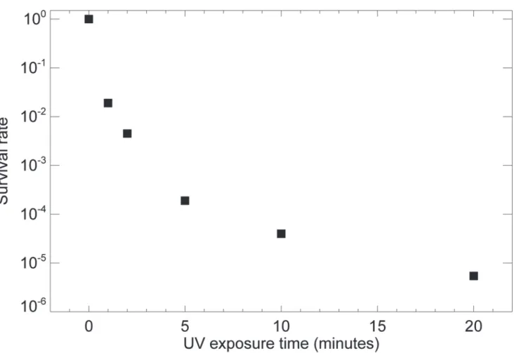
We used a bacterial virus (T7 bacteriophage) to establish the duration of UV exposure re-quired to reduce infectivity. T7 bacteriophage is a double-stranded DNA virus capable of in-fecting susceptible bacterial cells. Although distinct differences exist between T7 bacteriophage and potentially infectious agents such as HIV and HBV associated with RG2 materials, they share essential structures including a protein shell and a DNA/RNA core. As illustrated in Fig. 1, when a droplet of about 6 × 10⁷T7 bacteriophages was plunge frozen but not subject to UV exposure, nearly 100% survived, indicating that plunge freezing itself does not affect infec-tivity. When UV exposures of 5 or 10 minutes were used, all but about 10−4of bacteriophages
exponential relationship of survival rate with UVGI, thus leading to data falling on a straight line on a log (survival rate) versus exposure curve [51], we see in this experiment a slower-than-exponential decrease in survival rate for which we lack a good explanation.
Having demonstrated that UVGI can deactivate T7 bacteriophages even when immersed in liquid nitrogen, we then carried out experiments to examine whether the contents and distribu-tions of key biological elements were affected in eukaryotic cells after ultraviolet exposure. For these studies, we used adherent mouse embryonic fibroblast cells, which though they are RG1 materials, have UV response mechanisms and sensitivity [52] similar to those of human cells such as MCF-7 breast cancer cells [53]. As described in Materials and Methods, these mouse fi-broblasts were grown on silicon nitride windows (Si3N4) which were then plunge-frozen in
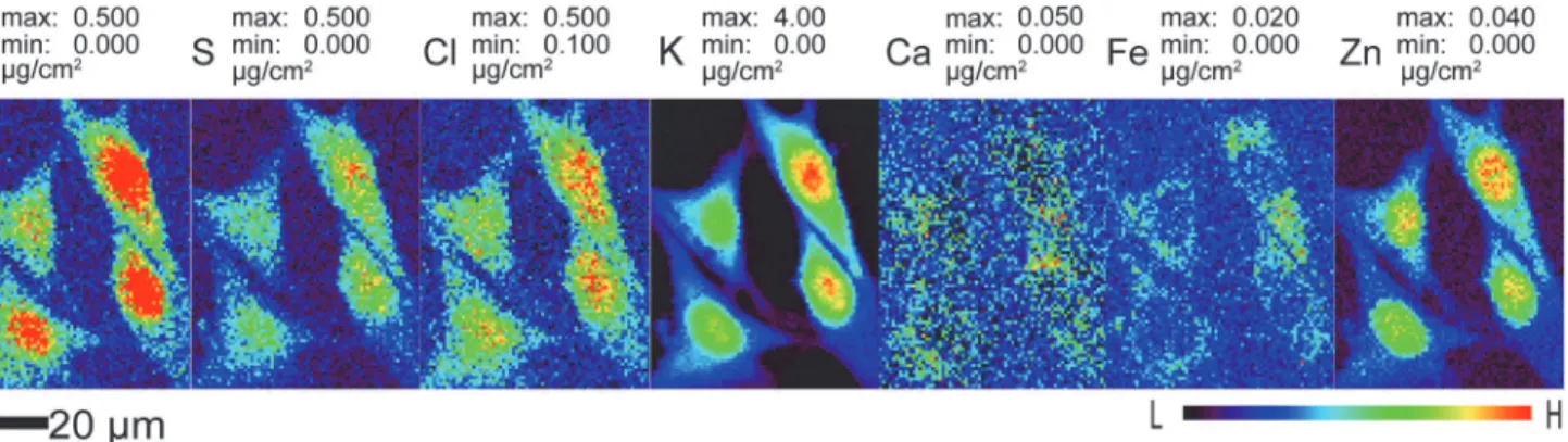
liq-uid ethane and examined at a temperature of approximately 100 K using cold nitrogen gas flow from an Oxford cryojet system. In x-ray fluorescence microscopes, an energy-dispersive detec-tor is used to record the x-ray fluorescence emission spectrum as the cell is scanned through the beam, allowing one to quantitate the distributions of several chemical elements simulta-neously as shown inFig. 2. These images show that diffusible ions (Cl, K, Ca) are nicely con-tained within the cell membrane in cryo preparations.
Our first test was to look for changes in the total contents and distributions of various ele-ments as the same cell preparation was examined with XFM under frozen hydrated state before
Fig 1. The survival rate of T7 bacteriophage under prolonged UV irradiation.About 60 million T7 bacteriophages in 3μl solution were dropped on Si3N4
windows and frozen by liquid-nitrogen-cooled liquid ethane, and then subjected for 0, 1, 2, 5, 10 and 20 minutes of ultraviolet germicidal irradiation while immersed in 3 cm of liquid nitrogen bath. A standard plaque assay was carried out to calculate the survival rate under each exposure, with three independent experiments being averaged to arrive at each point.
doi:10.1371/journal.pone.0117437.g001
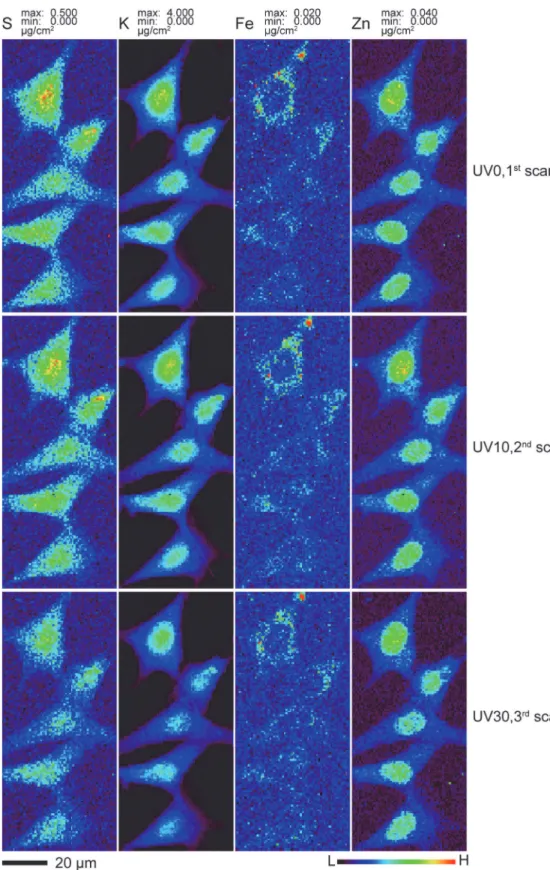
and after different doses of UVGI (Figs. 3). One area containing five cells on Si3N4windows
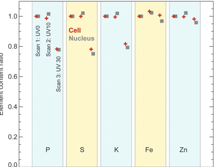
was scanned at 100K for the first time without any UVGI (UV0, first scan). This window was then unloaded for 10 minutes of UVGI under liquid nitrogen and uploaded for the second scan (UV10, second scan). After the second scan was completed, it was unloaded for an addi-tional 20 minutes of UVGI and uploaded for the third scan (UV30, third scan). No apparent difference in cell size and morphology were observed among these three scans; all cells stayed intact without visible breakage and shrinkage. The distribution patterns of various elements in-cluding P, S, Cl, K, Ca, Fe, Cu and Zn remained nearly identical among the three scans under this resolution. Two-dimensional elemental mapping of four representative elements S, K, Fe and Zn was shown inFig. 3. S and K maintained their homogeneous distribution, while Fe was primarily localized in cytoplasm and Zn was more concentrated in the nucleus than in the cy-toplasm, consistent with other observations [20]. To look for any possible changes in total ele-mental content, four out of these five cells and their corresponding nuclei were subjected for ROI (region of interest) analysis. The total elemental contents within these ROIs were calculat-ed and plottcalculat-ed as average fractions to that of UV0. As shown inFig. 4, the average elemental contents between first and second scans remained almost the same for all elements at both the nucleus and the whole cell level. Although the third scan showed a 19–25% decline for lighter elements P, S and K, the decline was no more than 5% for heavier elements such as Fe and Zn. Self-absorption of x-ray fluorescence emission in an 5μm ice layer would account for a signal reduction to 74% of the previous signal for P and 81% for S, which is close to the observed re-ductions to 76% and 80% respectively, so ice accretion between the UV10 and UV30 scans might explain this element-dependent signal change. When the percentage of elemental con-tent in the nucleus versus the whole cell was calculated, the difference between UV0 and UV30 scans was negligible for all measured elements, further supporting this explanation since an ac-creted ice layer would affect nuclear and cytoplasmic regions in the same way.
We have shown above (Figs.3and4) that cellular ions are immobilized in the matrix of a frozen hydrated specimen maintained at cryogenic temperature, so that their distributions and concentrations are unchanged when subject to UVGI. However, most x-ray fluorescence mi-croscopy studies are carried out using dehydrated specimens imaged at room temperature (there are only a few instruments available worldwide with cryo transfer capabilities [20–22]). We therefore attempted to investigate what effects, if any, our proposed sterilization treatment has on cells that are subsequently dehydrated and warmed for room temperature studies. We know for example that polymer films maintained at cryogenic temperatures will exhibit chain
Fig 2. Representative elemental maps of frozen hydrated mouse embryonic fibroblast cells (S1 File).Cells previously cryofixed by plunge freezing were scanned using 10 keV X rays in the cryojet x-ray fluorescence microscope at APS beamline 2-ID-D at 100 K. These scans have 800 nm pixel size and a per-pixel dwell time of 1 second. Data are available in Supporting Information fileS1.zip.
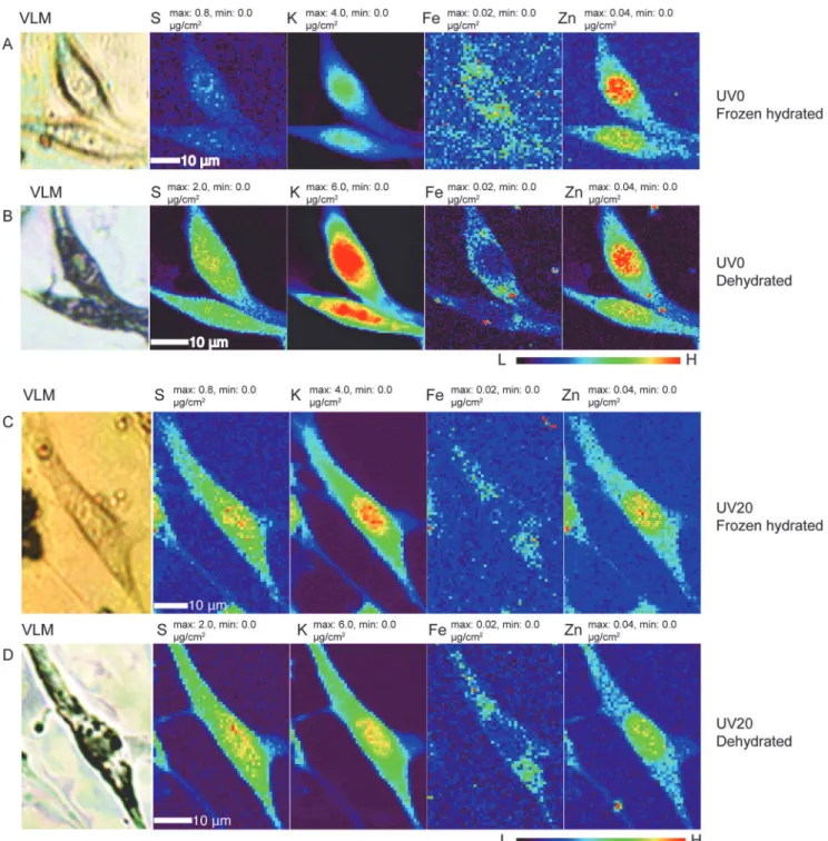
scission at radiation doses of 107Gray, though with little mass loss [48]. We also know that re-gions of cryogenic frozen hydrated cells exposed to doses of about 1010Gray can show signifi-cant mass loss when they are subject to freeze-drying [13]. We therefore made two different frozen hydrated cell preparations, and exposed one to 20 minutes of UVGI while the control sample was not. We then made x-ray fluorescence microscopy images of both preparations in a frozen hydrated state (again involving a radiation dose of about 107Gray) using the cryojet mi-croscope (rows A and C inFig. 5). Afterwards, both preparations were removed from the XFM, UV exposure were used and followed by a third scan (UV30 for 30 minutes accumulative exposure time). The x-ray scan parameters were identical to those used to produce the images ofFig. 2as described in Materials and Methods. The elemental image for the same element was scaled to the same maximum and minimum values among these scans for direct visual comparison. Data are available in Supporting Information filesS2. zip,S3.zip, andS4.zipfor 0, 10, and 30 minutes of UV exposure, respectively.
doi:10.1371/journal.pone.0117437.g003
Fig 4. Elemental content in frozen hydrated mouse embryonic fibroblast cells, based on the images shown inFig. 3.Elemental content within entire cells (red crosses) and their nuclei (gray squares) are plotted as fractions against the content in the first, non-UV-irradiated scan. These measurements were made from four of the five cells shown inFig. 3; the next-to-top cell was excluded due to the difficulty in outlining its area for analysis. The slight decrease observed for the measured content of lighter elements (P, S, K) levels is consistent with a possible explanation of ice or frost accretion on the specimen between the second and third scan, leading to increased self-absorption of these lower energy fluorescent x-rays. This was further supported by the nearly unchanged ratio of elemental contents in the cell’s nucleus versus the entire cell among these three scans.
Fig 5. Comparison of elemental distribution maps of four elements (S, K, Fe, Zn) in frozen hydrated cells (rows A and C) as well as freeze-dried counterparts (panels B and D), with (rows C and D) and without (rows A and B) 20 minutes of ultraviolet germicidal irradiation (UVGI) under liquid nitrogen prior to frozen hydrated imaging.Visible light micrographs (VLM) of frozen hydrated (rows A and C) and freeze dried (rows B and D) cells show that the cells suffered similar degree of shrinkage after x-ray cryo-scanning and freeze drying, whether or not UVGI was used. Please note the maximum and minimum concentration values for the light elements S and K in row A were scaled to be the same as that in row C, but different from that in rows B and D. Scan parameters were the same as used for Figs.2–4. Data are available in Supporting Information as filesS5.zipandS6.zipfor dehydrated cells exposed to UVGI for times of 0 and 20 minutes, respectively, and as filesS7.zipandS8.zipfor frozen hydrated cells exposed to UVGI for times of 0 and 20
minutes, respectively.
doi:10.1371/journal.pone.0117437.g005
subjected to the standard freeze-drying procedure described in Materials and Methods, and x-ray fluorescence micrographs were acquired of the room temperature dehydrated cells (rows B and D inFig. 5). The results show that x-ray irradiation followed by freeze drying leads to the same pattern of elemental distributions but some overall morphological change (approximately 20% shrinkage); some of this change might have been caused by the reactivity of x-ray irradia-tion products in both organic material and in ice, as the warming used in freeze-drying allowed for some chemical reactivity beyond what was possible while the irradiated sample was main-tained at cryogenic temperatures [13]. However, this shrinkage did not appear to depend on whether or not UVGI was used prior to the first frozen-hydrated x-ray image scan. In particu-lar for the lighter elements, the elemental concentrationincreasedin the dehydrated specimens relative to the frozen hydrated specimens; we again attribute this to much reduced fluorescence self-absorption due to the removal of most water/ice upon freeze drying. Most importantly, we did not observe any large-scaledecreasein elemental concentrations that might have otherwise been due to the loss of metal-containing organic functional groups during the freeze-drying process, or the UVGI process.
For all scans shown here, we have made the raw data available as HDF5 files (www. hdfgroup.org/HDF5/) in Supporting Information.
Discussion
As demonstrated extensively in the field of electron microscopy and more recently in x-ray mi-croscopy, cryogenically prepared frozen hydrated cells imaged at cryogenic temperatures show greater fidelity to living cells in morphology, structure, and chemical composition when com-pared to chemically fixed and dehydrated counterparts. Unfortunately, cryopreserved RG2 ma-terials must be considered to be potentially infectious. We have shown here that UVGI in liquid nitrogen bath is sufficient to deactivate T7 bacteriophages (Fig. 1), and more importantly that the application of UVGI has negligible effects on the distributions of metals in frozen hy-drated samples as shown in Figs.2–5. (UV irradiation might lead to changes in metal oxidation states, but we did not carry out x-ray near-edge absorption spectroscopy studies to see whether this is the case). This provides a potential specimen treatment that could be considered to avoid biological contamination of sophisticated cryogenic x-ray microscopes [20–22].
We chose a UVGI procedure of first producing a frozen hydrated cryogenic specimen, and then irradiating that specimen with ultraviolet light while immersed in liquid nitrogen of no more than 3 cm depth. One alternative procedure might be to carry out UVGI on initially-living cells before cryopreservation, which would exclude more instruments and space to come in contact with RG2 materials; however, this might involve complications of specimen heating under ultraviolet irradiation, as well as biochemical reactivity of radiolysis products with possi-ble morphological and elemental distribution changes. In addition, ultraviolet sterilization is easily compromised by any sort of overlying material [43] such as a thicker overlying culture medium. In contrast, cryogenic conditions largely halt the reactions of radiolytical products and thus might prevent additional unintended effects beyond sterilization, and furthermore liquid nitrogen immersion maintains cells at a steady cryogenic temperature during irradiation. Parmegianiet al.have shown that UVGI can be used to sterilize liquid nitrogen used for the preparation and storage of human oocytes [49,50]. Together, these factors led us to attempt UVGI of samples under liquid nitrogen.
phases. To exclude such preexisting variations, we changed to examine the same cells before and after different doses of UVGI under frozen hydrated state, or to examine the same cells be-fore and after freeze drying from samples receiving different doses of UVGI. Our results indi-cate that UVGI has no detectable effects on elemental concentrations and distributions when examined with x-ray fluorescence microscopy under both cryogenic and room temperature conditions (Figs.3–5). This suggests that UVGI immediately after cryofixation could be adopted as a routine practice to render RG2 materials to be handled as RG1 materials, without compromising elemental analysis by x-ray fluorescence microscopy. Finally, while x-ray micro-probes have been used to study both monolayer cell preparations and tissue sections, here we tested cryogenic UVGI on cells only; with tissue sections, one would have to consider issues of UV absorbance in thicker sections.
Materials and Methods
Preparation and UVGI of T7 bacterophages
Approximately 6 × 10⁷plaque forming units (pfu) of T7 bacteriophage in 3μl freshly prepared phage extract were dropped onto the center of Si3N4windows and manually dipped into
liq-uid-nitrogen-cooled liquid ethane using tweezers. After freezing, the windows were transferred from liquid ethane to liquid nitrogen, and laid flat with the bacteriophage facing up within a container and with about 3 cm of liquid nitrogen above. The container was then placed into a Millipore UV Sterilizer equipped with 4 × 8 W germicidal lamps (Sylvania No. G8T5, 253.7 nm wavelength), where it received UV irradiation for 0, 1, 2, 5, 10, or 20 minutes. For treatment duration beyond 10 minutes, UV irradiation was paused every 10 minutes to replenish the liq-uid nitrogen level back to 3 cm. After UVGI, the cryogenic windows were directly dropped into 297μl of Luria-Bertani (LB) broth at room temperature and subjected for a standard plaque assay procedure (T7 select manual from EMD Millipore,www.emdmillipore.com). The surviv-al rate was csurviv-alculated as the number of survivsurviv-al phages in every treatment divided by the num-ber of phages in original 3μl phage solution. The entire experiment was repeated three times with similar results. To convert UV exposure time to an estimated UV irradiation level, UV “fastcheck”strips from UV Process Supply Inc. (catalog number N010-002) were used. By comparing the color changes on strips to the reference chart provided by the manufacturer, ul-traviolet irradiation level can be estimated for any particular UV irradiation settings. Due to the extremely low temperature, the color development of“fastcheck”strips immersed in liquid nitrogen was significantly retarded and reduced compared to strips placed in room tempera-ture air. As a result, the actual irradiation level received under liquid nitrogen was estimated by measuring the level received in the air under the same setting minus any UV absorption by 3 cm of liquid nitrogen as discussed above in Results.
Cell culture and cryogenic sample preparation
Mouse embryonic fibroblast cells NIH/3T3, originally purchased from ATCC (www.atcc.org, Catalog number CRL1658), were cultured at 37°C in a 95% humidified incubator with 5% CO2
in Dulbecco’s Modified Eagle’s Medium ((Life Technologies, Grand Island, NY 14072, USA) supplemented with 10% fetal bovine serum (HyClone, Thermo Fisher Scientific, Inc., Pitts-burgh, USA), 1% non-essential amino acids and 1% L-glutamine (Life Technologies, Grand Is-land, NY 14072, USA), following standard protocols [20,54]. When about 80% confluence was reached, cells were passaged onto a new well of 6-well culture plate with a density of 6 × 10⁴ cells per well in 3 ml of culture medium. For cells to be imaged by XFM, Si3N4windows (Silson
Ltd., catalog number SiRN-5.0-200-1.5-200, 200 nm thick, 1.5×1.5 mm window area) were washed with distilled water followed by 2 mins soaking in 70% ethanol and 2 mins soaking in
100% ethanol, and then dried and taped to the bottom of a well in a 6-well culture plate, with the flat side up. After 20 minutes of UV sterilization, cells were seeded on Si3N4
window-containing wells and grew to 50–70% confluence.
The Si3N4windows with cells grown on them were carefully detached from the well and
rinsed two times by gently dipping into an Eppendorf tube containing 1 ml of Tris-glucose buffer (TG buffer, containing 261 mM glucose and 9 mM acetic acid in 10 mM Tris buffer, pH 7.4) [20]. Excess liquid on the backside of the window (non-cell side) was removed by blotting with filter paper. The windows were then attached to the tweezers supplied with a FEI Vitrobot Mark IV plunge freezer. Plunge freezing was carried out by plunging cells into liquid-nitrogen-cooled liquid ethane following the manufacturer’s instructions with chamber set at a tempera-ture of 30°C, 100% humidity, and with the following blotting parameters: blot time = 2 s, blot force = 0 mm, and blot total = 1. The frozen cells on Si3N4windows were then transferred from
liquid ethane to liquid nitrogen for storage, from which they could be retrieved for ultraviolet irradiation as described above or x-ray imaging experiments.
X-ray fluorescence microscopy
For cells imaged at room temperature, previously plunge-frozen samples were placed into the precooled vacuum chamber of EMS turbo freeze drier K775 and dried with the following cycles: 2 h at -120°C, 1 h from -120°C to -80°C, 3 h at -80°C, 1 h from -80°C to -50°C, 2 h at -50°C, 5 h at 25°C as suggested by the manufacturer. The cryofixation quality of frozen hydrated cells was monitored by observation using a 20x objective in a Nikon 50i light fluorescent microscope equipped with an Instec CLM77K cryo stage, while freeze-dried specimens were examined using a Leica DMXRE microscope. Frozen hydrated cells were scanned at around 100 K by an Oxford cryojet-equipped x-ray fluorescence microscope located at beamline 2-ID-D of APS at Argonne National Laboratory. Freeze-dried cells were scanned by x-ray fluorescence micro-scope located at beamline 2-ID-E at APS. Cells of interest were raster scanned with 0.8μm pixel size and a 10 keV beam with a focus diameter of about 0.5μm. The x-ray induced secondary fluorescence at each scan position was measured using an energy dispersive silicon drift detector (Vortex EM, SII Nanotechnology), and then fitted and quantified by comparison against the fluorescence signals from NBS standards 1832 and 1833 using MAPS software [55,56].
Supporting Information
As Supporting Information, the following HDF5 image files (www.hdfgroup.org/HDF5/) con-tain the elemental concentration maps used to produce the Figures in this paper.
S1 Image.zip. This image contains the representative elemental maps of frozen hydrated mouse embryonic fibroblast cells shown inFig. 2.
(ZIP)
S2 Image.zip. This image contains the fluorescence maps of frozen hydrated cells obtained before UltraViolet Germicidal Irradiation (UVGI), which are shown as the UV0 images in
Fig. 3.
(ZIP)
S3 Image.zip. This image contains the fluorescence maps of frozen hydrated cells obtained after 10 minutes of UVGI, which are shown as the UV10 images inFig. 3.
(ZIP)
S4 Image.zip. This image contains the fluorescence maps of frozen hydrated cells obtained after an additional 20 minutes of UVGI, which are shown as the UV30 images inFig. 3.
S5 Image.zip. This image contains the fluorescence maps of dehydrated cells obtained before UVGI, which are shown as the UV0 Dehydrated images inFig. 5.
(ZIP)
S6 Image.zip. This image contains the fluorescence maps of dehydrated cells obtained after 20 minutes of UVGI, which are shown as the UV20 Dehydrated images inFig. 5.
(ZIP)
S7 Image.zip. This image contains the fluorescence maps of frozen hydrated cells before UVGI, which are shown as the UV0 Frozen hydrated images inFig. 5.
(ZIP)
S8 Image.zip. This image contains the fluorescence maps of frozen hydrated cells after 20 minutes of UVGI, which are shown as the UV20 Frozen hydrated images inFig. 5.
(ZIP)
Acknowledgments
We thank Robert Fischetti and Tatjana Paunesku for helpful discussions.
Author Contributions
Conceived and designed the experiments: QJ SV BL CJ. Performed the experiments: QJ BL SG JW SC. Analyzed the data: QJ SV RM JD CJ. Contributed reagents/materials/analysis tools: QJ LF. Wrote the paper: QJ SV CJ. Biological material procedures: NM.
References
1. Horowitz P, Howell JA (1972) A scanning x-ray microscope using synchrotron radiation. Science 178: 608–611. doi:10.1126/science.178.4061.608
2. Sparks CJ Jr (1980) X-ray fluorescence microprobe for chemical analysis. In: Winick H, Doniach S, edi-tors, Synchrotron Radiation Research. New York: Plenum Press, pp. 459–512.
3. Jones KW, Gordon BM, Hanson AL, Kwiatek WM, Pounds JG (1988) X-ray fluorescence with synchro-tron radiation. Ultramicroscopy 24: 313–328. doi:10.1016/0304-3991(88)90318-XPMID:3281361 4. Paunesku T, Vogt S, Maser J, Lai B, Woloschak G (2006) X-ray fluorescence microprobe imaging in
bi-ology and medicine. Journal of Cellular Biochemistry 99: 1489–1502. doi:10.1002/jcb.21047PMID: 17006954
5. Fahrni CJ (2007) Biological applications of x-ray fluorescence microscopy: exploring the subcellular to-pography and speciation of transition metals. Current Opinion in Chemical Biology 11: 121–127. doi: 10.1016/j.cbpa.2007.02.039PMID:17353139
6. Szczerbowska-Boruchowska M (2008) X-ray fluorescence spectrometry, an analytical tool in neuro-chemical research. X-ray Spectrometry 37: 21–31. doi:10.1002/xrs.1007
7. de Jonge MD, Vogt S (2010) Hard X-ray fluorescence tomography—an emerging tool for structural vi-sualization. Current Opinion in Structural Biology 20: 606–614. doi:10.1016/j.sbi.2010.09.002PMID: 20934872
8. Fittschen UEA, Falkenberg G (2011) Trends in environmental science using microscopic x-ray fluores-cence. Spectrochimica Acta B 66: 567–580. doi:10.1016/j.sab.2011.06.006
9. Majumdar S, Peralta-Videa JR, Castillo-Michel H, Hong J, Rico CM, et al. (2012) Applications of syn-chrotronμ-XRF to study the distribution of biologically important elements in different environmental
matrices: A review. Analytica Chimica Acta 755: 1–16. doi:10.1016/j.aca.2012.09.050PMID: 23146389
10. Taylor K, Glaeser R (1974) Electron diffraction of frozen, hydrated protein crystals. Science 106: 1036–1037. doi:10.1126/science.186.4168.1036
11. Steinbrecht R, Zierold K, editors (1987) Cryotechniques in Biological Electron Microscopy. Berlin: Springer-Verlag.
12. Schneider G (1998) Cryo x-ray microscopy with high spatial resolution in amplitude and phase contrast. Ultramicroscopy 75: 85–104. doi:10.1016/S0304-3991(98)00054-0PMID:9836467
13. Maser J, Osanna A, Wang Y, Jacobsen C, Kirz J, et al. (2000) Soft x-ray microscopy with a cryo STXM: I. Instrumentation, imaging, and spectroscopy. Journal of Microscopy 197: 68–79. doi: 10.1046/j.1365-2818.2000.00630.xPMID:10620150
14. Meyer-Ilse W, Hamamoto D, Nair A, Lelievre S, Denbeaux G, et al. (2001) High resolution protein locali-zation using soft x-ray microscopy. Journal of Microscopy 201: 395–403. doi:10.1046/j.1365-2818. 2001.00845.xPMID:11240856
15. Zierold K (1982) Cryopreparation of mammalian tissue for x-ray microanalysis in STEM. Journal of Mi-croscopy 125: 149–156. doi:10.1111/j.1365-2818.1982.tb00332.xPMID:7086879
16. Saubermann AJ, Echlin P (1975) The preparation, examination and analysis of frozen hydrated tissue sections by scanning transmission electron microscopy and x-ray microanalysis. Journal of Microscopy 105: 155–191. doi:10.1111/j.1365-2818.1975.tb04048.xPMID:765465
17. Saubermann AJ, Heyman RV (1987) Quantitative digital x-ray imaging using frozen hydrated and fro-zen dried tissue sections. Journal of Microscopy 146: 169–182. doi:10.1111/j.1365-2818.1987. tb01338.xPMID:3612771
18. Echlin P (1992) Low-Temperature Microscopy and Analysis. New York: Plenum.
19. Freudenrich CC, Hockett D, Ingram P, LeFurgey A (1994)In situcryofixation of kidney for electron probe x-ray microanalysis. Journal of Structural Biology 112: 173–182. doi:10.1006/jsbi.1994.1018 PMID:7986644
20. Matsuyama S, Shimura M, Fujii M, Maeshima K, Yumoto H, et al. (2010) Elemental mapping of frozen-hydrated cells with cryo-scanning x-ray fluorescence microscopy. X-ray Spectrometry 39: 260–266. doi:10.1002/xrs.1256
21. Bohic S, Cotte M, Salomé M, Fayard B, Kuehbacher M, et al. (2012) Biomedical applications of the ESRF synchrotron-based microspectroscopy platform. Journal of Structural Biology 177: 248–258. doi:10.1016/j.jsb.2011.12.006PMID:22182732
22. Chen S, Deng J, Yuan Y, Flachenecker C, Mak R, et al. (2014) The Bionanoprobe: hard x-ray fluores-cence nanoprobe with cryogenic capabilities. Journal of Synchrotron Radiation 21: 66–75. doi:10. 1107/S1600577513029676PMID:24365918
23. Martin LS, McDougal JS, Loskoski SL (1985) Disinfection and inactivation of the human T lymphotropic virus type III/Lymphadenopathy-associated virus. Journal of Infectious Diseases 152: 400–403. doi: 10.1093/infdis/152.2.400PMID:2993438
24. Martin LS, Loskoski SL, McDougal JS (1987) Inactivation of human T-lymphotropic virus type III/lymph-adenopathy-associated virus by formaldehyde-based reagents. Applied and Environmental Microbiolo-gy 53: 708–709. PMID:3646869
25. Schwebach JR, Jacobs WR, Casadevall A (2001) Sterilization ofMycobacterium tuberculosisErdman samples by antimicrobial fixation in a Biosafety Level 3 Laboratory. Journal of Clinical Microbiology 39: 769–771. doi:10.1128/JCM.39.2.769-771.2001PMID:11158148
26. Cope GH, Williams MA (1968) Quantitative studies on neutral lipid preservation in electron microscopy. Journal of the Royal Microscopical Society 88: 259–277. doi:10.1111/j.1365-2818.1968.tb00612.x PMID:4877254
27. Cope GH, Williams MA (1969) Quantitative studies on the preservation of choline and ethanolamine phosphatides during tissue preparation for electron microscopy. I. Glutaraldehyde, osmium tetroxide, araldite methods. Journal of Microscopy 90: 31–46. doi:10.1111/j.1365-2818.1969.tb00692.xPMID: 4895518
28. Cope GH, Williams MA (1969) Quantitative studies on the preservation of choline and ethanolamine phosphatides during tissue preparation for electron microscopy. II. Other preparative methods. Journal of Microscopy 90: 47–60. doi:10.1111/j.1365-2818.1969.tb00692.xPMID:4895519
29. Kellenberger E, Johansen R, Maeder M, Bohrmann B, Stauffer E, et al. (1992) Artefacts and morpho-logical changes during chemical fixation. Journal of Microscopy 168: 181–201. doi: 10.1111/j.1365-2818.1992.tb03260.xPMID:1464902
30. Coetzee J, van der Merwe CF (1989) Extraction of carbon 14-labeled compounds from plant tissue dur-ing processdur-ing for electron microscopy. Journal of Electron Microscopy Technique 11: 155–160. doi: 10.1002/jemt.1060110210PMID:2709133
31. O’Toole E, Wray G, Kremer J, McIntosh J (1993) High voltage cryomicroscopy of human blood plate-lets. Journal of Structural Biology 110: 55–66. doi:10.1006/jsbi.1993.1004PMID:8494672
32. Vanhecke D, Asano S, Kochovski Z, Fernandez-Busnadiego R, Schrod N, et al. (2011) Cryo-electron tomography: methodology, developments and biological applications. Journal of the Royal Microscopi-cal Society 242: 221–227.
34. LučićV, Rigort A, Baumeister W (2013) Cryo-electron tomography: The challenge of doing structural
bi-ologyin situ. Journal of Cell Biology 202: 407–419. doi:10.1083/jcb.201304193PMID:23918936 35. Carzaniga R, Domart MC, Collinson LM, Duke E (2014) Cryo-soft x-ray tomography: a journey into the
world of the native-state cell. Protoplasma 251: 449–458. doi:10.1007/s00709-013-0583-yPMID: 24264466
36. Russell AD (1982) The destruction of bacterial spores. Academic Press.
37. Blatchley ER III, Peel MM (2001) Disinfection by ultraviolet irradiation. In: Block SS, editor, Disinfection, Sterlization, and Preservation, Lippincott Williams and Wilkins, chapter 41. Fifth edition, pp. 823–851. 38. Myatt TA, Johnston SL, Rudnick S, Milton DK (2003) Airborne rhinovirus detection and effect of
ultravio-let irradiation on detection by a semi-nested RT-PCR assay. BMC Public Health 3: 5. doi:10.1186/ 1471-2458-3-5PMID:12525263
39. Riley RL, Knight M, Middlebrook G (1976) Ultraviolet susceptibility of BCG and virulent tubercle-bacilli. American Review of Respiratory Disease 113: 413–418. PMID:817628
40. Nicas M, Miller SL (1999) A multi-zone model evaluation of the efficacy of upper-room air ultra-violet germicidal irradiation. Applied Occupational and Environmental Hygiene 14: 317–328. doi:10.1080/ 104732299302909PMID:10446484
41. Ko G, First MW, Burge HA (2002) The characterization of upper-room ultraviolet germicidal irradiation in inactivating airborne microorganisms. Environmental health perspectives 110: 95–101. doi:10. 1289/ehp.0211095PMID:11781170
42. Sambol AR, Iwen PC (2006) Biological monitoring of ultraviolet germicidal irradiation in a biosafety level 3 laboratory. Applied Biosafety 11: 81–87.
43. Turnbull PC, Reyes AE, Chute MD, Mateczun AJ (2008) Effectiveness of UV exposure of items con-taminated with anthrax spores in a class 2 biosafety cabinet and a biosafety level 3 laboratory pass-box. Applied Biosafety 13: 164–168.
44. Vesley D, Lauer JL (2000) Decontamination, sterlization, disinfection, and antisepsis. In: Fleming DO, Hunt DL, editors, Biological Safety: Principles and Practices, American Society for Microbiology Press, chapter 25. Third edition.
45. Botzenhart K, Rüden H, Tolon M, von K Scharfenberg M (1976) Clinical uses of ultraviolet light radia-tion. Praktische Anasthesie, Wiederbelebung und Intensivtherapie 11: 320–327. PMID:981142 46. Rudnick SN (2001) Predicting the ultraviolet radiation distribution in a room with multilouvered
germicid-al fixtures. American Industrigermicid-al Hygiene Association Journgermicid-al 62: 434–445. doi:10.1080/ 15298660108984645PMID:11549137
47. Varnam AH, Evans MG (1996) Foodborne pathogens: an illustrated text. London: Manson Publishing Ltd.
48. Beetz T, Jacobsen C (2003) Soft x-ray radiation-damage studies in PMMA using a cryo-STXM. Journal of Synchrotron Radiation 10: 280–283. doi:10.1107/S0909049503003261PMID:12714762
49. Parmegiani L, Cognigni GE, Filicori M (2009) Ultra-violet sterilization of liquid nitrogen prior to vitrifica-tion. Human Reproduction 24: 2969. doi:10.1093/humrep/dep329PMID:19749194
50. Parmegiani L, Accorsi A, Cognigni GE, Bernardi S, Troilo E, et al. (2010) Sterilization of liquid nitrogen with ultraviolet irradiation for safe vitrification of human oocytes or embryos. Fertility and Sterility 94: 1525–1528. doi:10.1016/j.fertnstert.2009.05.089PMID:19591992
51. Qualls RG, Johnson JD (1983) Bioassay and dose measurement in UV disinfection. Applied and envi-ronmental microbiology 45: 872–877. PMID:6405690
52. Deng J, Harding HP, Raught B, Gingras AC, Berlanga JJ, et al. (2002) Activation of GCN2 in UV-irradiated cells inhibits translation. Current Biology 12: 1279–1286. doi:10.1016/S0960-9822(02) 01037-0PMID:12176355
53. Koch-Paiz CA, Amundson SA, Bittner ML, Meltzer PS, Fornace AJ (2004) Functional genomics of UV radiation responses in human cells. Mutation Research 549: 65–78. doi:10.1016/j.mrfmmm.2004.01. 010PMID:15120963
54. Jin Q, Duggan R, Dasa SSK, Li F, Chen L (2010) Random mitotic activities across human embryonic stem cell colonies. Stem Cells and Development 19: 1241–1248. doi:10.1089/scd.2009.0324PMID: 19842916
55. Vogt S (2003) MAPS: A set of software tools for analysis and visualization of 3D x-ray fluorescence data sets. Journal de Physique IV 104: 635–638. doi:10.1051/jp4:20030160
56. Twining BS, Baines SB, Fisher NS, Maser J, Vogt S, et al. (2003) Quantifying trace elements in individ-ual aquatic protist cells with a synchrotron x-ray fluorescence microprobe. Analytical Chemistry 75: 3806–3816. doi:10.1021/ac034227zPMID:14572047