Ge no mics and X-ray micro analysis
indicate that Ca
2+
and thio ls me diate
the aggre gatio n and adhe sio n o f
Xylella fastid iosa
1Departamento de Entomologia, Fitopatologia e Zoologia Agrícola, 2Departamento de Ciências Biológicas,
3Núcleo de Apoio à Pesquisa/Microscopia Eletrônica Aplicada a Pesquisa Agropecuária,
Escola Superior de Agricultura “Luiz de Q ueiroz”, Universidade de São Paulo, Piracicaba, SP, Brasil B. Leite1,
M.L. Ishida1, E. Alves1, H. Carrer2, S.F. Pascholati1 and E.W. Kitajima3
Abstract
The availability of the genome sequence of the bacterial plant patho-gen Xylella fastidiosa, the causal agent of citrus variegated chlorosis, is accelerating important investigations concerning its pathogenicity. Plant vessel occlusion is critical for symptom development. The objective of the present study was to search for information that would help to explain the adhesion of X. fastidiosa cells to the xylem. Scanning electron microscopy revealed that adhesion may occur without the fastidium gum, an exopolysaccharide produced by X. fastidiosa, and X-ray microanalysis demonstrated the presence of elemental sulfur both in cells grown in vitro and in cells found inside plant vessels, indicating that the sulfur signal is generated by the pathogen surface. Calcium and magnesium peaks were detected in association with sulfur in occluded vessels. We propose an explana-tion for the adhesion and aggregaexplana-tion process. Thiol groups, main-tained by the enzyme peptide methionine sulfoxide reductase, could be active on the surface of the bacteria and appear to promote cell-cell aggregation by forming disulfide bonds with thiol groups on the surface of adjacent cells. The enzyme methionine sulfoxide reductase has been shown to be an auxiliary component in the adhesiveness of some human pathogens. The negative charge conferred by the ionized thiol group could of itself constitute a mechanism of adhesion by allowing the formation of divalent cation bridges between the nega-tively charged bacteria and predominantly neganega-tively charged xylem walls.
Co rre spo nde nce
B. Leite
University of Florida 155 Research Road Q uincy, FL 32351 USA
Fax: + 1-850-875-7148 E-mail: bleite@ mail.ifas.ufl.edu
The present address of B. Leite and M.L. Ishida is University of Florida, 155 Research Road, Q uincy, FL 32351, USA.
Research supported by FAPESP.
Received February 26, 2002 Accepted March 19, 2002
Ke y words
•Xylella fastidiosa •Aggregation
•Adhesion
•Methionine sulfoxide reductase
•Biofilm
Xylella fastidiosa is a major problem in Brazilian citrus production areas. The esti-mated annual losses due to citrus variegated chlorosis (CVC) are up 100 million dollars, affecting over 70 million sweet orange trees (1). Some other countries in Latin America
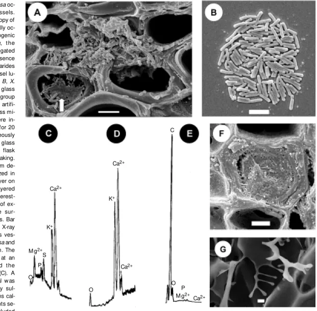
inside the xylem. The fastidium gum (3) released by X. fastidiosa cells has been re-ported to play a central role in the clumping of these bacteria and has been previously assumed to be directly involved in adhesion (4). The role of fastidium gum in adhesion was investigated by examining occluded ves-sels by scanning electron microscopy (SEM). The images generated revealed that most cells are not immersed in the fastidium gum
(Figure 1A). SEM examination of the sur-face of a microscope slide that was immersed in a culture flask and kept there for 20 days under constant shaking showed similar re-sults (Figure 1B), i.e., one cell layer adhered to the glass surface without the fastidium gum. This was considered to be the begin-ning of a biofilm formation.
The bacterial biofilm is a community of microorganisms mobilized on a given
sur-Figure 1. A, Xyllela fastidiosa oc-cluding citrus xylem vessels. Scanning electron microscopy of citrus xylem vessels partially oc-cluded by the plant pathogenic bact erium X. f ast idiosa, t he causal agent of citrus variegated chlorosis. Notice the presence of extracellular polysaccharides (fastidium gum) in the vessel lu-men (arrow ). Bar = 5 µm. B, X. fastidiosa adhered to a glass slide. The figure show s a group of X. fastidiosa cells on an artifi-cial surface, a standard glass mi-croscope slide. Slides w ere in-serted into culture flasks for 20 days. X. fastidosa spontaneously formed colonies on the glass surface, even though the flask w as permanently under shaking. Different degrees of biofilm de-velopment can be visualized in this image, a single cell layer on the border and double-layered cells in the center. It is interest-ing to notice the absence of ex-tracellular polysaccharide sur-rounding the bacterial cells. Bar = 3 µm. Energy dispersive X-ray microanalysis of the citrus ves-sel occluded by X. fastidiosa and the isolated fastidium gum. The X-ray probe w as pointed at an occluded vessel (F) and t he spectrum w as recorded (C). A very strong calcium signal w as detected, accompanied by sul-fur and by the divalent ions cal-cium and magnesium. Points se-lected aw ay from the occluded
vessel (D) did not exhibit the sulfur signal, but calcium w as frequently present. Graphs C and D have the same ordinate scale, 40 counts per second. The fastidium gum (G) show s strong carbon and oxygen signals and in graph E the carbon peak reaches 1300 counts per second. Bar (F) = 2 µm. Bar (G) = 6 µm. M ethodology: X-ray microanalysis w as performed in cross sections of lyophilized leaf petioles coated w ith carbon and observed under a ZEISS 940 A microscope equipped w ith an EDX Oxford system for X-ray detection and spot analysis. The data are representative of repeated preparations performed w ith tissues from distinct infected plants. Uninfected xylem vessels w ere used as control specimens.
Ca2+
C
Ca2+
Ca2+
M g2+
Ca2+
M g2+
K+
K+
S P
O O
face, which improves their chances for sur-vival in the environment. The biofilm begins with a single cell layer followed by the depo-sition of additional layers and by an increase in exopolysaccharide (EPS) production (5). Our findings agree with the predicted stages and suggest that the fastidium gum is not essential in the preliminary steps of X. fastidiosa biofilm formation. In a recent study, Danese and collaborators (6) showed that the adhesion of Escherichia coli K12 was not influenced by the lack of EPS pro-duction. A mutant of E. coli incapable of producing colanic acid, an EPS, was tested for biofilm formation against the wild type. Both the mutant and the wild type E. coli
adhered to a polyvinylchloride surface. How-ever, only the wild type developed a multi-layered biofilm. These investigators con-cluded that the lack of EPS production af-fected the biofilm architecture, but not the cell adhesion. Similarly, our results indicate that X. fastidiosa adhesion is primarily de-pendent on the ability of the bacterium to get in contact with other cells and substrates relying solely on their surface characteris-tics.
Subsequently, SEM coupled with X-ray microanalysis (EDS = energy dispersive
X-ray spectrometry) was performed to com-pare the elemental composition of the fas-tidium gum obtained from cultures of X. fastidiosa and the elemental composition of occluded vessels (Figure 1C-G). EDSallows the detection and identification of the X-rays produced by the impact of the electron beam on the sample, thereby allowing qualitative and quantitative elemental analysis. The anal-ysis is limited to the electron beam penetra-tion, which is usually 1 to 5 micra, and therefore only the surface elemental consti-tution is determined. As expected, most of the fastidium gum (Figure 1G) exhibited carbon and oxygen as major peaks (Figure 1E). Occluded vessels (Figure 1F) consis-tently exhibited calcium, magnesium and sulfur (Figure 1C), contrary to surrounding vessel areas, in which the sulfur signal was absent (Figure 1D). Since the occluded ves-sel contained cells and a previous experi-ment showed that X. fastidiosa cells alone exhibited sulfur (data not shown), we think it is reasonable to conclude that the sulfur signal is generated on the bacterial surface. Two questions were raised by these re-sults: i) If the adhesive material from these two different organisms has the presence of sulfur in common, what is the role of sulfur
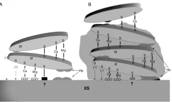
Figure 2. M odel proposed to explain adhesion and aggrega-t ion of Xylella f ast idiosa. In scheme A an X. fastidiosa cell (Xf) is placed in contact w ith the xylem surface (XS) and w ith an-other bacterial cell. All potential points of adhesion are repre-sented. In the interaction be-tw een X. fastidiosa cells and the XS hydrophobic interactions oc-cur betw een hydrophobic (Hp) portions on both sides and addi-tional interactions occur be-tw een the surface sulfur (sulf-hydryl groups) and charges lo-calized on the xylem side (S-? S-+) directly or bridged by cal-cium and m agnesium (S-Ca-COO-, S-M g-COO-). In contrast, cell-to-cell adhesion is believed to be mediated by hydrophobic interactions as just described, by the formation of disulfide bonds (S-S) and by sulfur groups bridged by divalent ions (S-Ca-S, S-M g-S). The situation de-scribed in B involves multilay-ered deposition of cells sup-ported by the architecture pro-vided by exopolysaccharides, w ith the fastidium gum being represented by the shadow ed area. Xf (-)S S S S S S S S S S S S S S S S S S S S S S S S S S S S S S S S Xf Xf Xf (-) .. .. + (-) .. .. . + (-) .. . + +
Ca M g
COO-COO
-Ca M g
Hp ? ? ? ? ? Ca COO -M g COO
-Ca M g
Ca
Ca M g
in adhesion? ii) In addition to giving a nega-tive charge to the bacterial surface, charac-teristic of several microorganisms (7,8), how would sulfur provide adhesive properties to a microorganism surface?
Assays with Sephadex ion-exchange resins, DEAE (positive charges) and car-boxymethyl (negative charges) indicated that
X. fastidiosa cells are more attracted to posi-tively charged DEAE (data not shown). These results are consistent with our hypothesis that the majority of charges on the surface of
X. fastidiosa are negative. Other pathogens, such as E. coli K12 have negatively charged surfaces (8). Cooper and collaborators (9), also using X-ray microanalysis to study ca-cao (Theobroma cacao) vessels occluded by
Verticillium dahlia, observed peak patterns similar to those obtained for X.
fastidiosa-occluded vessels. These investigators con-cluded that sulfur was involved in the resis-tance response of cacao plants to V. dahlia
by assuming that the plant produced and accumulated sulfur in the form of cycloocta-sulfur (S8) to resist the fungal attack.
How-ever, in the case of X. fastidiosa, the sulfur signal is due to the physical presence of the pathogen and is not a localized plant re-sponse. If the sulfur were part of a general metabolic stress response it would also be present in vessels in which the pathogenic organism could not be seen in situ. With proper stimulation, a general response would affect both infected and non-infected areas through signaling and sulfur would be de-tected in the surrounding vessel areas. Re-cently, the adhesive material obtained from
Colletotrichum graminicola conidia was also shown to exhibit calcium and sulfur, even after extensive dialysis (10).
In support of these data, the study of the
X. fastidiosa genome revealed the presence of open reading frames with high homology to several proteins involved in adhesion (4). The enzyme denoted methionine sulfoxide reductase (MsrA; EC 1.8.4.6) may be of particular importance. MsrA is an adhesion
maintenance enzyme that helps maintain the adhesiveness of some human pathogenic bac-terial cells. MsrA is recognized as having a broad substrate specificity and a general mechanism of action controlling a variety of proteins (11). However, the best known sub-strate for MsrA is oxidized methionine (12). The influence of thiol (SH) groups on the adhesiveness of some human pathogenic bacteria has been evaluated. Strainsof Strep-tococcus pneumoniae, Neisseria gonor-rhoeae and E. coli mutants, that lack the capacity to produce MsrA, exhibited reduced ability to adhere when compared to their respective wild type strains (13). Many re-search groups have been working with the adhesive properties of thiols such as the generation of mucoadhesive polymers with thiol groups (14) and adherence of human polymorphonuclear leukocyte (15). The en-hancement of human polymorphonuclear leukocyte adhesion was demonstrated to be dependent on constitutive peripheral SH groups, CD11/C18 integrins (adhesins) and extracellular calcium. The bacterium Thio-bacillus ferrooxidans was also shown to ad-here by means of thiol groups. A 40-kDa surface protein, which strongly binds elemen-tal sulfur, was isolated from the bacterial flagella (16). The X. fastidiosa genome con-tains some surface proteins (Hsf-like) and type 4 fimbriae which could be the targets for MsrA activity.
A model to explain the adhesion of X. fastidiosa to xylem vessels was elaborated and is summarized in Figure 2. The first step of adhesion occurs when the X. fastidiosa
diseased leaves of Vitis vinifera were found to have accumulated Ca2+ and Mg2+ (18),
similar to what was demonstrated for oc-cluded citrus vessels (Figure 1C and F). The high density of negative charges may attract several cations such as Ca2+ and Mg2+, which
are more tightly associated with the negative charges on the cell walls than monovalent cations, such as K+ and Na+ (19). Zinc is
sometimes detected in the elemental profile of X. fastidiosa aggregates (data not shown). Zinc sequestration may possibly explain the symptoms of zinc deficiency in the CVC syndrome. The putative existence of an ad-hesion maintenance enzyme such as MsrA that would maintain the thiol groups with active adhesive properties is another facet of the adhesion model, explaining how cells adhere to a substrate or to other cells, while calcium would form bonds between nega-tive surfaces and neganega-tively charged patho-gen cells (20). In addition, negative sulfur moieties may directly form bonds with posi-tively charged portions of the host tissue. This hypothesis and/or the existence of other forces (such as hydrophobicity) or other charge sources may also be true.
We propose a detailed investigation of the
nutrient status of the xylem fluid, which may be interfering with disease development. This proposition seems to be highly justified and is supported by the fact that xylem cell walls contain fixed negative charges resulting from dissociated polygalacturonic acid, yielding COO- groups (19). The extent and importance of each adhesion/aggregation component has yet to be evaluated.
In summary, our results open an avenue of investigation concerned with the adhe-sion of plant pathogens involving sulfur, calcium, magnesium and MsrA. The study of xylem chemistry may be of significant importance to understand resistance and/or susceptibility to X. fastidiosa. The combina-tion of genomic informacombina-tion and classical research methodologies should uncover the mechanisms of pathogenicity.
Ackno wle gm e nts
We would like to acknowledge Drs. Felipe Rodrigues da Silva and Paulo Arruda, Uni-versidade Estadual de Campinas, for provid-ing a fastidium gum sample for X-ray mi-croanalysis.
Re fe re nce s
1. M onteiro PB, Teixeira DC, Palma RR, Garnier M , Bove JM & Renaudin J (2001). Stable transformation of the Xylella fasti-diosa citrus varigated chlorosis strain w ith
oriC plasmids. Applied and Environmental M icrobiology, 67: 2263-2269.
2. Davis M J, Purcell AH & Thomson SV (1978). Pierce’s disease of grapevines: isolation of the causal bacterium. Science, 199: 75-77.
3. Silva FR, Vettore AL, Kemper EL, Leite A & Arruda P (2001). Fastidium gum: the
Xylella fastidiosa exopolysaccharide pos-sibly involved in bacterial pathogenicity.
FEM S M icrobiology Letters, 203: 165-171.
4. Simpson AJ, Reinach FC, Arruda P et al. (2000). The genome sequence of the
plant pathogen Xylella fastidiosa. Nature, 406: 151-157.
5. Watnick P & Kolter R (2000). Biofilm, city of microbes. Journal of Bacteriology, 182: 2675-2679.
6. Danese PN, Pratt LA & Kolter R (2000). Exopolysaccharide production is required for the development of Escherichia coli K-12 biofilm architecture. Journal of Bacteri-ology, 182: 3593-3596.
7. Buck JW & Andrew s JH (1999). Local-ized, positive charge mediates adhesion of Rhodosporidium toruloides to barley leaves and polystyrene. Applied and Envi-ronmental M icrobiology, 65: 2179-2183. 8. Fletcher JN, Saunders JR, Embaye H,
Obedra RM , Batt RM & Hartr CA (1997). Surf ace propert ies of diarrhoeagenic
Escherichia coli isolates. Journal of M edi-cal M icrobiology, 46: 67-74.
9. Cooper RM , Resende M L, Flood J, Row an M G, Beale M H & Potter U (1996). Detec-tion and cellular localizaDetec-tion of elemental sulphur in disease-resistant genotypes of
Theobroma cacao. Nature, 379: 159-162. 10. Leite B, Ishida M L, Alves E, Pascholati SF & Sugui JA (2000). Detection of calcium in the adhesive material obtained from the plant pathogen Colletotrichum grami-nicola: X ray microanalysis (EDS) evi-dence. Proceedings of M icroscopy and M icroanalysis, 6: 698-699.
12. Low ther WT, Brot N, Weissbach H, Honek JF & M atthew s BW (2000). Thiol-disulfide exchange is involved in the catalytic mechanism of methionine sulfoxide re-ductase. Proceedings of the National Academy of Sciences, USA, 97: 6463-6468.
13. Wizemann TM , M oskovitz J, Pearce BJ, Cundell D, Arvidson CG, So M , Weissbach H, Brot N & M asure HR (1996). Peptide methionine sulfoxide reductase contri-butes to the maintenance of adhesins in three major pathogens. Proceedings of the National Academy of Sciences, USA, 93: 7985-7990.
14. Bernkop-Schnurch A & St eininger S (1999). Synthesis and characterization of mucoadhesive thiolated polymers.
Inter-national Journal of Pharmaceutics, 194: 239-247.
15. Hernandez M & M acia M (1996). Free peripheral sulphydryl groups, CD11/CD18 integrins, and calcium are required in the cadmium and nickel enhancement of hu-man-polymorphonuclear leucocyte adher-ence. Archives of Environmental Contami-nation and Toxicology, 30: 437-443. 16. Ohmura N, Tsugita K, Koizumi J-I & Saiki
H (1996). Sulfur binding protein of flagella of Thiobacillus ferrooxidans. Journal of Bacteriology, 178: 5776-5780.
17. Kristensen S, Tian Y, Klegerman M E & Groves M J (1992). Origins of BCG sur-face charge: effect of ionic strength and chemical modifications of zeta potential of M ycobacterium bovis BCG, Tice
sub-strain, cells. M icrobios, 70: 284-285. 18. Goodw in PH, De Vay JE & M eredith CP
(1988). Physiological responses of Vitis vinifera cv “ Chardonnay” to infection by Pierce’s disease bacterium. Physiological and M olecular Plant Pathology, 32: 17-32. 19. van Ieperer W, van M eeteren U & van Gelder H (2000). Fluid ionic composition influences hydraulic conductance of xy-lem conduits. Journal of Experimental Botany, 51: 769-776.

