Nitric oxide at the CVLM is involved in the attenuation of the reflex bradycardia
in renovascular hypertensive rats
Uberdan Guilherme Mendes de Castro
a,b, Graziele Galdino de Sousa
a, Raquel do Pilar Machado
a,b,
Mauro César Isoldi
a,b, Marcelo Eustáquio Silva
b, Ana Paula Nadu
c, Luiz Eduardo Sousa
a,b,
Robson Augusto Souza dos Santos
c, Maria José Campagnole-Santos
c, Andréia Carvalho Alzamora
a,b,⇑ aDepartamento de Ciências Biológicas, Instituto de Ciências Exatas e Biológicas, Universidade Federal de Ouro Preto, Ouro Preto, MG, BrazilbNúcleo de Pesquisa em Ciências Biológicas, Universidade Federal de Ouro Preto, Ouro Preto, MG, Brazil
cDepartamento de Fisiologia e Biofísica, Instituto de Ciências Biológicas, Universidade Federal de Minas Gerais, Belo Horizonte, MG, Brazil
a r t i c l e
i n f o
Article history:
Received 22 February 2011 Revised 6 December 2011 Available online 14 January 2012
Keywords:
Caudal ventrolateral medulla Reflex bradycardia Nitric oxide Superoxide anion
Renovascular hypertension-Goldblatt-2K1C
a b s t r a c t
Hypertension is associated to an increase in central oxidative stress and an attenuation of the baroreflex control of arterial pressure. The present study evaluated the effect of alterations in the levels of nitric oxide (NO) and superoxide anion in the caudal ventrolateral medulla (CVLM), a key area of the brainstem for the baroreflex control of arterial pressure, in renovascular hypertensive rats (2K1C). Baseline mean arterial pressure (MAP), heart rate (HR), and reflex bradycardia were evaluated 30 days after renal artery occlusion in anesthetized (urethane, 1.2 g/kg, i.p.) 2K1C or normotensive (SHAM) rats. The MAP, HR, and baroreflex control of HR were evaluated before and after CVLM microinjections of the non-selective NOS inhibitorL-NAME (10 nmol), the NO precursorL-ARG (50 nmol), or the antioxidant ascorbic acid, Vit C (10 nmol). In both 2K1C and SHAM animals, CVLM microinjection ofL-NAME produced a decrease in MAP, whereasL-ARG induced a significant increase in MAP. However, microinjection of Vit C into the CVLM produced a decrease in MAP and HR only in 2K1C and not in SHAM rats. Cardiovascular effects pro-duced by microinjection ofL-ARG into the CVLM were abolished by prior microinjection ofL-NAME in the CVLM of 2K1C and SHAM rats. Microinjection ofL-NAME into the CVLM increased the sensitivity of reflex bradycardia in 2K1C animals. In contrast, the CVLM microinjection ofL-ARG reduced reflex bradycardia only in SHAM rats. Vit C in the CVLM did not change reflex bradycardia in either 2K1C or in SHAM rats. These results suggest that increased oxidative stress in the CVLM during hypertension contributes to the reduced baroreflex sensitivity and to maintain hypertension in the 2K1C model.
Ó2012 Elsevier Inc.
Introduction
Evidence supports the hypothesis that the increase in oxidative stress plays a role in the pathophysiology of arterial hypertension [1,2]. It has been shown that increase in reactive oxygen species
(ROS), nitric oxide (NO) and superoxide anion (O2 ) may be altered in central areas related to cardiovascular control in hypertensive animals[3,4].
NO plays an important role in cardiovascular regulation, in both physiological and pathological conditions [5,6]. It has been described as a neurotransmitter in the brain, involved in the mod-ulation of excitatory and inhibitory neurons, and acting through interneurons or directly stimulating the release of neurotransmit-ters [7,8]. Increasing evidence indicates that NO modulates responses mediated by the sympathetic nervous system (SNS) and parasympathetic nervous system (PNS) through its action in the main areas in the brain involved in the control of cardiovascu-lar functions, such as the paraventricucardiovascu-lar nucleus of the hypothal-amus (PVN)[9–11], the nucleus of the solitary tract (NTS)[12,13] and the areas of the ventrolateral medulla (VLM)[12,14,15]. Previ-ous studies have shown that mRNA expression of NO synthase (NOS) isoforms and the NO production in the brain may vary in hypertensive rats, according to the time course of the hypertension
1089-8603Ó2012 Elsevier Inc. doi:10.1016/j.niox.2012.01.002
Abbreviations:2K1C, Goldblatt renovascular hypertension 2-kidney, 1-clip; BP, blood pressure; CNS, central nervous system; CVLM, caudal ventrolateral medulla; HR, heart rate;L-ARG,L-arginine; MAP, mean arterial pressure; ms, millisecond;
NO, nitric oxide; NOS, nitric oxide synthase; NTS, nucleus of the solitary tract; PI, pulse interval; PNS, parasympathetic nervous system; PVN, paraventricular nucleus of the hypothalamus; ROS, reactive oxygen species; RVLM, rostral ventrolateral medulla; SHAM, normotensive rats; SHR, spontaneous hypertensive rats; SNS, sympathetic nervous system; Vit C, ascorbic acid – vitamin C; VLM, ventrolateral medulla.
⇑Corresponding author. Address: Departamento de Ciências Biológicas, Instituto de Ciências Exatas e Biológicas, Universidade Federal de Ouro Preto, Morro do Cruzeiro, 35 400-000 Ouro Preto, MG, Brazil. Fax: +55 31 3559 1633.
E-mail addresses:[email protected], [email protected]
(A.C. Alzamora).
Contents lists available atSciVerse ScienceDirect
Nitric Oxide
j o u r n a l h o m e p a g e : w w w . e l s e v i e r . c o m / l o c a t e / y n i o x
Open access under the Elsevier OA license.
or the CNS region[12,16–20]. Additionally, an elevation of super-oxide in brain areas related to cardiovascular control has been de-scribed as one of the factors responsible for increased SNS activity in hypertensive models[1,21].
The CVLM is a depressor region, and stimulation of this area causes hypotension with consequent vasodilatation of several peripheral vascular beds[22–24]. The tonic activity of the CVLM is important for the maintenance of normal blood pressure (BP), and involves a combination of excitatory (glutamatergic) and inhibitory (GABAergic) synapses, although the source of this activ-ity is not fully understood[25,26]. It is well established that CVLM is rich in GABAergic neurons, and many of these GABAergic neu-rons tonically inhibit sympatho-excitatory neuneu-rons in the rostral VLM (RVLM), leading to a decrease in SNS activity and a conse-quent reduction in BP[27–31]. Therefore, inhibition of CVLM neu-ronal activity significantly increases the neuneu-ronal activity of RVLM, SNS activity, and BP. Further, the CVLM is essential for most sym-patoinhibitory reflexes produced by activation of NTS, such as the baroreflex control of BP[31,32].
The contribution of the CVLM to the genesis and maintenance of hypertension has not been completely elucidated[33–35]. Sev-eral studies have shown that hypertension occurs during hyperac-tivity of neurons in the RVLM[36–38]. Moreover, in spontaneous hypertensive rats (SHR) and in renovascular hypertensive rats [39,40], the inhibition caused by GABAergic CVLM on the RVLM excitatory neurons seems to be attenuated, which could explain the increase in SNS activity observed in these models of hyperten-sion[39–41].
In the present study, we evaluated the possibility that NO and the superoxide anion (O2 ) could not only contribute to the base-line control of BP, but could also modulate baroreflex bradycardia at the CVLM in hypertensive rats. For this purpose, we evaluated baseline BP and baroreflex control of HR after CVLM microinjection of the non-selective NOS inhibitorL-NAME, microinjection of the NO precursorL-arginine (L-ARG), or microinjection of a potential scavenger of superoxide anions (ascorbic acid – vitamin C; Vit C) in 2K1C renovascular hypertensive rats.
Materials and methods
Animals
Experiments were performed in male Fisher rats from ENUT, Universidade Federal de Ouro Preto, Brazil. The animals were housed in separate cages in groups of four rats according to their group (2K1C or SHAM), with free access to rat chow and tap water in a temperature- and light-controlled room. All animal procedures were in accordance with the Guidelines for Ethical Care of Experi-mental Animals, and were performed as approved by the Institu-tional Ethics Committee of the Federal University of Ouro Preto, Minas Gerais, Brazil (Protocol # 022/2007).
Induction of renovascular hypertension
Goldblatt renovascular hypertension was induced as described by Goldblatt et al.[42]. Briefly, the rats (weighing 150–180 g) were anesthetized with a mixture of ketamine and xylazine (50 and 10 mg/kg, respectively, i.p.), and a silver clip (0.20 mm ID) was placed around the left renal artery through a midline incision (Goldblatt renovascular hypertension, 2-kidney, 1-clip model; 2K1C). Other rats were submitted to similar procedures but with-out the renal-artery clip placement (SHAM group or normotensive rats). BP recording and CVLM microinjections were carried out 30 days after the surgery. At this time the animals weighed 260–300 g.
Arterial pressure measurements
Pulsatile arterial pressure was monitored by a Gould pressure transducer (PM-1000, CWE) coupled to a blood pressure signal amplifier (UIM100A, Powerlab System). Mean arterial pressure (MAP) and heart rate (HR) were determined from the arterial pres-sure wave. All variables were continuously recorded with a Power-Lab digital acquisition system (Powerlab – 4/20, AD Instruments) with an 800 Hz sampling rate.
CVLM microinjections
2K1C and SHAM rats were anesthetized with urethane (1.2 g/kg, i.p.) and underwent a tracheostomy. Next, a polyethylene catheter was inserted into the abdominal aorta through the femoral artery, for arterial pressure measurement, and another catheter was in-serted into the inferior cava vein through the femoral vein, for drug injection. The animals were placed in a stereotaxic frame (David Kopf Instruments, CA) as described by Rodrigues et al.[43].
Unilateral microinjections ofL-NAME (10 nmol/100 nl),L-ARG (50 nmol/100 nl), Vit C (10 nmol/100 nl) or sterile saline (vehicle – NaCl 0.9%) in a volume of 100 nl were made over a 20–30 s per-iod into the CVLM (0.7 mm anterior, 1.8 mm lateral to the obex, and just above the pia mater on the ventral surface), as described by Alzamora et al.[44]. The doses ofL-NAME,L-ARG, and Vit C were based on previous studies[1,15,45,46].
Evaluation of the sensitivity of the baroreflex bradycardia
The baroreflex bradycardia was tested in different groups of animals (2K1C and SHAM), before and 5–10 min after CVLM micr-oinjections ofL-NAME (n= 8 in each group),L-ARG (n= 6–7), and Vit C (n= 10–11). Baroreflex control of HR was determined by recording reflex heart rate changes (baroreflex bradycardia) in re-sponse to transient increases in MAP produced by repeated bolus injections of graded doses of phenylephrine (0.5–50
lg, i.v.) in
ure-thane-anesthetized rats, according to previous studies[47]. The HR was converted to pulse interval (PI, ms) by the formula: 60,000/HR. A best-fit regression line was drawn from MAP and HR changes ob-tained with the different doses of phenylephrine for each animal. The slope of the regression line was used as an index of baroreflex sensitivity (baroreflex gain).Experimental procedures
The arterial pressure and HR of urethane-anesthetized 2K1C and SHAM rats were continuously recorded. In three different groups of rats, after a 10-min stabilization period, the micropipette was positioned in the CVLM, andL-NAME (10 nmol,n= 6–7) or sal-ine (NaCl, 0.9% – 100 nL); L-ARG (50 nmol, n= 6–11) or saline (NaCl, 0.9% – 100 nL); and Vit C (10 nmol,n= 13–14) or saline were microinjected, in a random order. In some animals, phosphate-buffered saline (pH 7.4; 100 nl) was microinjected into the CVLM as a control for the Vit C vehicle. In order to test the ability of L-ARG to induce a rise in NO level, approximately 50 min after the CVLM microinjection of theL-ARG,L-NAME was microinjected, and after 5 min L-ARG was repeated in 2K1C (n= 6) or SHAM (n= 11) rats.
Finally, the baroreflex test was evaluated in three additional groups of 2K1C and SHAM animals, before and 5–10 min after CVLM microinjection of L-NAME (n= 8),L-ARG (n= 6–7), or Vit C (n= 10–11).
In another group of rats SHAM and 2K1C rats (n= 6–12) was evaluated the mRNA expression of nNOS and eNOS by qRT-PCR analysis.
Histological verification of microinjection sites
At the end of each experiment, the animals were then killed with an excess of anesthetic, and the brain was carefully removed and fixed in 10% phosphate-buffered formalin. Serial coronal sec-tions (40–50
lm) of the medulla oblongata were made and stained
with neutral red for histological examination. Microinjection sites were identified by tissue rupture produced by the microinjections under light microscopy, and was referred to standard anatomical structures of the brain stem according to the atlas of Paxinos and Watson[48].Fig. 1shows, on the left side, an image of a histolog-ical section of the medulla, illustrating the microinjection site in a representative animal. On the right side, diagrams of frontal sec-tions of the medulla from the atlas of Paxinos and Watson[48] show the location (black area) of the center of the microinjections in all animals of this study. The microinjections into the CVLM were located in the ventral portion of the lateral reticular nucleus, at the level of 13.3 to 13.8 mm, posterior to the bregma (Fig. 1).Reverse transcription and real-time PCR
Normotensive – SHAM and 2K1C rats (n= 6–12) were decapi-tated, their brains were removed, the CVLM region was dissected, quickly frozen on liquid nitrogen and stored at 70°C until
pro-cessed. A pool of CVLM region from two animals was homogenized, and the total RNA was isolated with TRIzol reagent (Invitrogen Life Technologies) according to the manufacturer’s protocol. RNA sam-ples, approximately 2
lg, were treated with DNAse (Invitrogen Life
Technologies) to eliminate genomic DNA present in the samples. Approximately 1lg of DNased mRNA was used as template for
M-MLV reverse transcriptase (Promega) using the reverse specifics oligonucleotides. Real-time PCR was carried out following genera-tion of first strand cDNA. Endothelial (eNOS), neuronal NOS (nNOS) and the endogenous S26 ribosomal cDNA were amplified using specific primers (Table 1) and SYBR Green PCR Master Mix (Applied Biosystems) in an ABI Prism 7000 Sequence Detection Systems.Drugs
The drugs (urethane, phenylephrine,L-NAME, andL-ARG; Sigma Chemical Company, St. Louis, MO, USA) were all dissolved in saline. Vit C (Sigma Chemical Company, St. Louis, MO, USA) was diluted in phosphate-buffered saline (pH 7.4).
Statistical analysis
The results are expressed as means ± SEM. Comparisons be-tween two groups were assessed by Student’sttest. Comparisons of three or more groups were made by one-way ANOVA followed by Newman–Keuls. Statistical analyses were performed with the
softwareGraphpad Prism(version 4.00). The criterion for statistical significance was set atp< 0.05.
Results
Baseline values of MAP and HR
The baseline values of MAP of anesthetized 2K1C (140 ± 2 mm Hg,n= 45) were significantly higher than the baseline values of MAP of anesthetized SHAM rats (105 ± 1 mm Hg,n= 44). The base-line values of HR were not significantly different between the 2K1C group (370 ± 6 beats/min,n= 45) and the SHAM group (372 ± 4 beats/min,n= 44).
BP effect of CVLM microinjections
Unilateral microinjection ofL-NAME into the CVLM produced a significant decrease in MAP in 2K1C rats ( 17 ± 3 vs 5 ± 1 mm Hg,n= 7; saline;Fig. 2A), which was similar to that produced by microinjection ofL-NAME into the CVLM in SHAM rats ( 17 ± 3 vs 4 ± 0.4 mm Hg, saline;n= 6;Fig. 2A). No significant changes were observed in the duration of the hypotensive effect induced by L-NAME in 2K1C rats (6 ± 1 min, n= 6) or in SHAM rats (4 ± 0.5 min,n= 6). In addition,L-NAME also induced a significant fall in HR in 2K1C rats ( 29 ± 7 beats/min vs 1 ± 1 beats/min, n= 7; saline; Fig. 2D), similar to that observed in SHAM rats ( 39 ± 15 beats/min vs 1 ± 1 beats/min,n= 6; saline;Fig. 2D).
Microinjection ofL-ARG into the CVLM produced a significant increase in MAP in 2K1C rats (11 ± 3 vs 0.3 ± 0.6 mm Hg,n= 6; saline;Fig. 2B), which was similar to the pressor effect produced by microinjection of L-ARG into the CVLM in SHAM rats (11 ± 1 mm Hg,n= 11; Fig. 2B). However, L-ARG lengthened the hypertensive effect in 2K1C rats (26 ± 7 min,n= 6) compared to the SHAM group (10 ± 2 min,n= 11). The pressor effect of L-ARG was not accompanied by a significant effect on the HR in 2K1C rats ( 12 ± 6 vs 3 ± 1 beats/min,n= 6) or in SHAM rats (8 ± 8 beats/ min vs 1 ± 1 beats/min,n= 11; saline;Fig. 2E).
Microinjection of Vit C into the CVLM produced a significant de-crease in MAP in 2K1C rats ( 17 ± 2 vs 3 ± 0.5 mm Hg,n= 14; sal-ine;Fig. 2C). In contrast, microinjection of Vit C into the CVLM produced no significant decrease in MAP in SHAM rats ( 5 ± 1 vs 3 ± 0.6 mm Hg,n= 13; saline;Fig. 2C). Vit C also induced a signif-icant decrease in HR in 2K1C rats ( 26 ± 6 beats/min vs 0.5 ± 0.5 beats/min,n= 14 saline;Fig. 2F) and had no significant effect on HR in SHAM rats ( 13 ± 4 beats/min vs 3 ± 2 beats/min,n= 13; saline;Fig. 2F).
As shown inFig. 3, microinjection ofL-L-NAME into the CVLM abolished the pressor effect induced by L-ARG on the CVLM in 2K1C rats (11 ± 3 mm Hg, beforeL-NAME vs 3 ± 2 mm Hg, after L-NAME;n= 6;Fig. 3A) and in SHAM rats (11 ± 1 mm Hg, before
L-NAME vs 3 ± 2 mm Hg, afterL-NAME;n= 11;Fig. 3A). Microinjec-tion ofL-ARG afterL-NAME had no significant effect on HR in both groups (data not shown).
Evaluation of the sensitivity of the baroreflex bradycardia
As expected, the reflex bradycardia of 2K1C rats (0.12 ± 0.02 ms/ mmHg,n= 25) was significantly lower compared to that of SHAM
Fig. 1.Left, image of a histological section of the medulla illustrating disruption of the tissue caused by CVLM microinjection. Right, diagram of frontal sections of the medulla (13.80 mm caudal to the bregma) showing the center of the microinjection into the CVLM (black area). The diagram is from the atlas of Paxinos and Watson, 1986. AP = area postrema; Amb = nucleus ambiguus; LR = lateral reticular nucleus; Py = pyramidal tract; Sol = nucleus of solitary tract; nXII = hypoglossal nucleus.
Table 1
Primers sequence used to perform real-time PCR.
Gene Sequence (50–30) forward Sequence (50–30) reverse
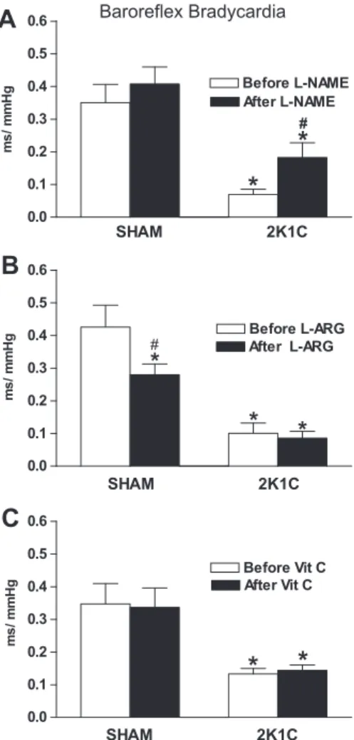
rats (0.37 ± 0.07 ms/mmHg,n= 25).Fig. 4A illustrates the barore-flex bradycardia induced before and after the CVLM microinjection of L-NAME. As shown in Figs. 4A and 5A, L-NAME significantly increased baroreflex bradycardia in 2K1C rats (0.18 ± 0.04 vs 0.07 ± 0.01 ms/mmHg,n= 8; beforeL-NAME). In contrast, microin-jection ofL-NAME into the CVLM did not change the baroreflex bra-dycardia of the SHAM rats (0.41 ± 0.05 vs 0.35 ± 0.06 ms/mmHg, n= 8, beforeL-NAME;Fig. 5A).
On the other hand,L-ARG significantly decreased baroreflex bra-dycardia in SHAM rats (0.28 ± 0.03 vs 0.42 ± 0.07 ms/mmHg,n= 6; beforeL-ARG; Figs.4B and5B). In contrast, microinjection ofL-ARG into the CVLM did not change the baroreflex bradycardia in 2K1C rats (0.08 ± 0.02 vs 0.10 ± 0.03 ms/mmHg, n= 7, beforeL-NAME; Fig. 5B).
In addition, microinjection of Vit C into the CVLM did not change the baroreflex bradycardia in 2K1C rats (0.14 ± 0.02 vs 0.13 ± 0.02 ms/mmHg,n= 10, before Vit C;Fig. 5C) and in SHAM rats (0.34 ± 0.06 vs 0.35 ± 0.06 ms/mmHg, n= 11, before Vit C; Fig. 5C).
Expression of nNOS mRNA and eNOS mRNA in the CVLM
As shown inTable 2the mRNA expression of nNOS and eNOS, obtained by qRT-PCR analysis, were lower in the CVLM of 2K1C rats (values and values, respectively) as compared to SHAM rats (values and values, respectively).
Discussion
The main finding of the present study was that CVLM inhibition of NO synthase withL-NAME increased the sensitivity of the baro-reflex bradycardia in 2K1C hypertensive rats, suggesting the exis-tence of an inhibitory nitrergic influence on CVLM neurons that participates in baroreflex pathway. Interestingly, the response in-duced in baseline arterial pressure was not different in 2K1C rats in comparison to SHAM. On the contrary, the administration of an antioxidant agent, Vit C, did not alter baroreflex modulation at the CVLM, but induced a higher hypotensive response in 2K1C rats.
This result is in keeping with those of other studies showing that NO plays an inhibitory role in the brain [49], especially in areas related to cardiovascular control such as the PVN[9,10,49], NTS[50–52], and RVLM[53–55]. Overall, these studies have shown that an increase in NO level in the PVN or in the RVLM leads to ef-fects that include marked reductions in BP and in sympathetic activity, through an interaction between NO and GABAergic neu-rons. These studies are consistent with the observations reported here, and reinforce the hypothesis that NO also has an inhibitory role in neurons of the CVLM.
Several studies using a variety of methods have identified NOS in different nuclei associated with baroreflex control, including the PVN, NTS, CVLM, and RVLM[20,56–58]. The role of NO in different areas of the brain related to baroreflex control is not completely -30
-25 -20 -15 -10 -5
0 SHAM 2K1C
*
*
L-NAME Saline
mmH
g
-30 -20 -10 0 10 20
30 SHAM 2K1C
*
*
L-ARG Saline
mmH
g
-70 -60 -50 -40 -30 -20 -10
0 SHAM 2K1C
*
Vit C Salina
*
be
at
s/
m
in
-70 -50 -30 -10 10 30 50
70 SHAM 2K1C
L-ARG Saline
bea
ts
/m
in
Changes in HR
Changes in MAP
A
B
C
E
D
F
-30 -25 -20 -15 -10 -5 0
Saline Vit C
SHAM 2K1C
*
mmH
g
-70 -60 -50 -40 -30 -20 -10 0
SHAM 2K1C
*
*
L-NAME Saline
be
at
s
/m
in
Fig. 2.Mean arterial pressure (DMAP, mmHg) and heart rate (HR, beats/min) changes produced by CVLM microinjections ofL-NAME (10 nmol/100 nl; panels A and D) orL -ARG (50 nmol/100 nl; panels B and E) or Vit C (10 nmol/100 nl, panels C and F) or saline (100 nl) in normotensive (SHAM,n= 6–13) or hypertensive (2K1C,n= 6–14) rats.
⁄p< 0.05 in comparison to saline (ANOVA followed by Newman–Keuls test).
Fig. 3.Mean arterial pressure changes (DMAP, mmHg) produced by CVLM microinjections ofL-ARG (50 nmol/100 nl) before and 5 min after microinjection ofL-NAME (10 nmol) in normotensive (SHAM,n= 11) or hypertensive (2K1C,n= 6) rats.⁄p< 0.05 compared to saline (ANOVA followed by Newman–Keuls test) and #p< 0.05 compared toL-ARG before (ANOVA followed by Newman–Keuls test).
understood. In the NTS, a major site for baroreflex modulation, some investigators have suggested that NO increases baroreflex sensitivity[59], whereas others have found either that NO medi-ated an inhibitory effect [17,60,61] or that it had no effect [17,62]. Conversely, studies have shown that the overexpression
of eNOS in RVLM improved baroreflex function in stroke-prone SHR[55], whereas nNOS gene transfer into the RVLM improves baroreflex function in rats with heart failure[63]. These effects may have resulted from a cardiac sympathoinhibitory effect of NO in RVLM neurons.
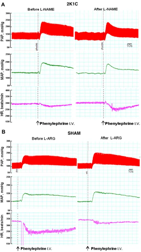
Fig. 4.Pulsatile (PAP, mmHg), mean arterial pressure (MAP, mmHg) and heart rate (HR, beats/min) recordings illustrating the typical effect produced by injection of phenylephrine (25lg, i.v.) before and after CVLM microinjection ofL-NAME (10 nmol) in 2K1C rats (panel A) orL-ARG (50 nmol) in SHAM rats (panel B). Arrows indicate
Although it has been shown that NO acts as a modulator of activity of the SNS, studies showed different changes in the mRNA expression of nNOS and eNOS in different areas of the CNS and at different stages or models of hypertension, such as SHR[17,18,67] and in 2K1C renovascular hypertensive rats[19]. Our present re-sults showed that the mRNA expression of nNOS and eNOS in the CVLM were lower in 2K1C rats (4 weeks of renal artery occlusion) compared to SHAM rats. In fact, Krukoff et al.[19]showed that nNOS gene expression in CVLM was increased at 3 weeks and de-creased at 6 weeks after renal artery occlusion. Plochocka-Zulinska and Krukoff[18]demonstrated an increase in nNOS expression in the CVLM of adult SHR, but not in young pre-hypertensive rats [18]. However, despite of the reduced mRNA levels of NOS found
in our study, our data showed an improvement in baroreflex sen-sitivity afterL-NAME microinjection into the CVLM in hypertensive rats, suggesting that NO is playing a modulatory role on baroreflex control at this site. Whether this fact can be attributed to a different source of NO or to an increased activity of the enzymes involved in NO production at this site remains to be investi-gated. In addition, the relative contribution of each NOS isoform(s) for the production of NO in the CVLM at different stages of hyper-tension awaits future investigation. In the present study, the use of a non-specific inhibitor of the NOS,L-NAME, as a first approach to understand the involvement of NO in the CVLM, showed that NO acting at the CVLM is contributing to the lower baroreflex sensitiv-ity in renovascular hypertensive rats, at least at a time point where the disease is dependent on the activation of the circulating renin-angiotensin system.
Our data showed that microinjection ofL-ARG, a precursor for NO synthesis, into the CVLM induced a significant attenuation in baro-reflex bradycardia in normotensive rats, which reinforces the hypothesis that increased NO in the CVLM leads to a decrease in bar-oreflex sensitivity. On the other hand, the present study showed that CVLM microinjection ofL-ARG in the 2 K1C rats did not alter the already low sensitivity of the baroreflex, which may be related to the very low value for baroreflex sensitivity that these animals showed at the baseline. Interestingly, CVLM microinjection of L-NAME induced a significant improvement in the baroreflex brady-cardia in hypertensive rats, in contrast to the effect in normotensive rats. This finding further suggests that endogenous NO may partic-ipate in the modulation of the baroreflex control of HR, at least in the CVLM of 2 K1C rats.
The inhibitory role of NO in baroreflex modulation at the CVLM could be due to one or more of the following mechanisms: (1) the well-known inhibitory GABAergic pathway from CVLM to RVLM [29,31,39,64], (2) the projection from the NTS to the CVLM [27,65], and (3) the projections from the CVLM to the nucleus ambiguus[66]. Future studies will be necessary to evaluate which pathways are being modulated by NO at the CVLM, especially con-sidering that activation of the parasympathetic system is the major mechanism involved in baroreflex bradycardia.
In the present study, we have shown that CVLM microinjection ofL-NAME induced similar decreases in MAP and HR in 2K1C and in normotensive rats. Accordingly, CVLM microinjection ofL-ARG induced similar increases in BP in 2K1C and in normotensive rats. This agrees with previous studies by us[15]and others[14], which showed that CVLM microinjection ofL-ARG induced a significant increase in MAP, whereasL-NAME induced a significant hypoten-sion. Shapoval et al.[14]showed that unilateral CVLM microinjec-tions of sodium nitroprusside (an NO donor) and L-ARG in anesthetized cats produced an increase in MAP and RSNA activity, whereas microinjection of L-NMMA, a non-selective inhibitor of NOS isoforms, produced opposite effects. Similarly, Lage et al. [15], observed that microinjection ofL-NAME in the CVLM of nor-motensive rats produced hypotension, whereas microinjection of L-ARG promoted an increase in BP in normotensive rats.
Kishi et al.[54]showed in stroke-prone SHR that overexpres-sion of an eNOS-related gene in the RVLM increases the release of both glutamate and GABA. However, the predominant effect was on GABAergic activity, which resulted in a reduction in the SNS activity in these animals. It has been reported that hyperten-sion is accompanied by a decrease in the activity of CVLM gabaer-gic neurons that project to sympathetic neurons in the RVLM [39,40,68]. Taken together, the data from these studies suggest that NO has an inhibitory effect on neurons involved in the tonic control of arterial pressure, possibly by suppressing the CVLM-RVLM GAB-Aergic pathway.
As mentioned before, our data showed that there was an in-creased baroreflex response to CVLM L-NAME microinjection in 0.0
0.1 0.2 0.3 0.4 0.5 0.6
SHAM 2K1C
SHAM 2K1C
SHAM 2K1C
Before L-NAME After L-NAME
*
#*
ms
/ mmH
g
Baroreflex Bradycardia
B
C
A
0.0 0.1 0.2 0.3 0.4 0.5 0.6
Before L-ARG After L-ARG
*
*
*
#
ms
/ mmH
g
0.0 0.1 0.2 0.3 0.4 0.5 0.6
Before Vit C After Vit C
*
*
ms
/ mmH
g
Fig. 5.Sensitivity of bradycardia (ms/mmHg) represented by mean of individual slope of the regression line for the reflex changes in HR and changes in MAP obtained by graded doses of phenylephrine in normotensive (SHAM,n= 6–11) or hypertensive (2K1C,n= 7–10) rats.⁄p< 0.05 compared to the respective SHAM
group (ANOVA followed by Newman–Keuls test). #p< 0.05 compared to 2K1C
before (ANOVA followed by Newman–Keuls test).
Table 2
mRNA expression of nNOS and eNOS (arbitrary unit) in the caudal ventrolateral medulla (CVLM) of renovascular hypertensive rats 2K1C or normotensive SHAM rats.
nNOS mRNA (a.u.) eNOS mRNA (a.u.) SHAM 1.28 ± 0.15 1.01 ± 0.10 2K1C 0.40 ± 0.07* 0.20 ± 0.06*
n 3–4 3–6
Values are mean ± SEM;n= number of samples. Each sample represents a pool of the CVLM of 2 animals.
*p< 0.05 compared to the respective SHAM group (Student’sttest).
hypertensive rats. However, 2K1C rats showed a similar BP effect after CVLM L-NAME microinjection compared with SHAM rats. These data suggest that sympathetic and cardiac baroreflex are independently regulated by distinct signaling system in the CVLM [34]Further, NO is specifically affecting the activity of neurons in-volved in baroreflex.
On the other hand, literature data have shown that during hypertension, oxidative stress also may increase the production of other free radicals such as the superoxide anion (O2 ), which is also considered a modulator of SNS activity [1]. Furthermore, the reaction between the superoxide anion and NO leads to pro-duction of another free radical, peroxynitrite, which also contrib-utes to cardiac sympathovagal imbalance in hypertensive states [3].
Our present data showed that CVLM microinjection of Vit C, which has the property of scavenging superoxide anions, did not alter baroreflex bradycardia in 2K1C or in SHAM rats. This suggests that endogenous superoxide anions are not involved in baroreflex modulation, at least at this brain site. Our data also showed that CVLM microinjection of Vit C caused significant hypotension and bradycardia only in the 2K1C rats. No cardiovascular effect was ob-served in normotensive rats after Vit C microinjection. Similarly, Oliveira-Sales et al.[1]reported that microinjection of Vit C into the RVLM lowered BP and HR only in 2K1C rats and not in SHAM rats, showing that oxidative stress is an important additional mechanism for the high BP of 2K1C rats. As mentioned above, it is interesting that Vit C microinjection did not alter baroreflex bra-dycardia in 2K1C or normotensive rats, which is in line with the concept that different pathways are involved in baseline MAP and reflex control of HR[34], and superoxide may only affect one of them.
Furthermore, microinjection ofL-NAME and Vit C in the CVLM induced similar effects on MAP and HR in 2K1C rats by inhibiting the formation of NO and the superoxide anion, respectively. Con-sidering also that CVLM microinjection of Vit C did not alter the MAP of SHAM rats, our data suggest the involvement of superoxide anions in the CVLM in maintaining the high levels of MAP in 2K1C hypertensive rats.
In summary, our data emphasize the inhibitory role of NO in the CNS, by showing that NO in the CVLM may be an additional mech-anism involved in the attenuated baroreflex bradycardia that is ob-served in renovascular hypertensive 2K1C rats. The data also showed that, during hypertension, there is an increased production of superoxide in the CVLM, which in turn would participate in maintaining high blood pressure in the 2K1C model. Overall, the data from the present study showed that increased oxidative stress in the CVLM contributes importantly to reducing the sensitivity of reflex bradycardia and to the high levels of MAP that occur in reno-vascular hypertension.
Acknowledgments
This study was supported by FAPEMIG-RedeToxifar (Fundação de Amparo à Pesquisa do Estado de Minas Gerais), CNPq (Conselho Nacional de Desenvolvimento Científico e Tecnológico), INCT-Nanobiofar-FAPEMIG-CNPq, Pronex Project Grant (FAPEMIG/CNPq) and CAPES (Coordenação de Aperfeiçoamento de Pessoal de Nível Superior). Uberdan Guilherme Mendes de Castro received a CAPES fellowship (Master’s Degree) in the ‘‘Programa de Pós-graduação Ciências Biológicas’’, NUPEB, UFOP.
References
[1] E.B. Oliveira-Sales, E.E. Nishi, B.A. Carillo, M.A. Boim, M.S. Dolnikoff, C.T. Bergamaschi, R.R. Campos, Oxidative stress in the sympathetic premotor
neurons contributes to sympathetic activation in renovascular hypertension, Am. J. Hypertens. 22 (5) (2009) 484–492.
[2] E.B. De Oliveira-Sales, E.E. Nishi, M.A. Boim, M.S. Dolnikoff, C.T. Bergamaschi, R.R. Campos, Upregulation of AT1R and iNOS in the rostral ventrolateral medulla (RVLM) is essential for the sympathetic hyperactivity and hypertension in the 2K–1C Wistar rat model, Am. J. Hypertens. 23 (7) (2010) 708–715.
[3] E.J. Danson, D.J. Paterson, Reactive oxygen species and autonomic regulation of cardiac excitability, J. Cardiovasc. Electrophysiol. 17 (Suppl 1) (2006) S104–S112. [4] V.M. Campese, R.K. Sindhu, S. Ye, Y. Bai, N.D. Vaziri, B. Jabbari, Regional expression of NO synthase, NAD(P)H oxidase and superoxide dismutase in the rat brain, Brain Res. 1134 (1) (2007) 27–32.
[5] R.M. Touyz, Reactive oxygen species, vascular oxidative stress, and redox signaling in hypertension: What is the clinical significance? Hypertension 44 (3) (2004) 248–252.
[6] F.X. Guix, I. Uribesalgo, M. Coma, F.J. Munoz, The physiology and pathophysiology of nitric oxide in the brain, Prog. Neurobiol. 76 (2) (2005) 126–152.
[7] S. Wang, A.G. Teschemacher, J.F. Paton, S. Kasparov, Mechanism of nitric oxide action on inhibitory GABAergic signaling within the nucleus tractus solitarii, FASEB J. 20 (9) (2006) 1537–1539.
[8] M. Engelmann, G. Wolf, T.F. Horn, Release patterns of excitatory and inhibitory amino acids within the hypothalamic supraoptic nucleus in response to direct nitric oxide administration during forced swimming in rats, Neurosci. Lett. 324 (3) (2002) 252–254.
[9] T. Horn, P.M. Smith, B.E. Mclaughlin, L. Bauce, G.S. Marks, Q.J. Pittman, A.V. Ferguson, Nitric oxide actions in paraventricular nucleus: Cardiovascular and neurochemical implications, Am. J. Physiol. 266 (1 Pt 2) (1994) R306–R313. [10] K. Zhang, K.P. Patel, Effect of nitric oxide within the paraventricular nucleus on
renal sympathetic nerve discharge: Role of GABA, Am. J. Physiol. 275 (3 Pt 2) (1998) R728–R734.
[11] K. Powers-Martin, J.K. Phillips, V.C. Biancardi, J.E. Stern, Heterogeneous distribution of basal cyclic guanosine monophosphate within distinct neuronal populations in the hypothalamic paraventricular nucleus, Am. J. Physiol. Regul. Integr. Comp. Physiol. 295 (4) (2008) R1341–R1350. [12] C.J. Tseng, H.Y. Liu, H.C. Lin, L.P. Ger, C.S. Tung, M.H. Yen, Cardiovascular effects of
nitric oxide in the brain stem nuclei of rats, Hypertension 27 (1) (1996) 36–42. [13] G.P. Pajolla, D. Accorsi-Mendonca, G.J. Rodrigues, L.M. Bendhack, B.H. Machado, C.N. Lunardi, Fluorescent indication that nitric oxide formation in NTS neurons is modulated by glutamate and GABA, Nitric Oxide 20 (3) (2009) 207–216. [14] L.N. Shapoval, V.F. Sagach, L.S. Pobegailo, Nitric oxide influences ventrolateral
medullary mechanisms of vasomotor control in the cat, Neurosci. Lett. 132 (1) (1991) 47–50.
[15] R.C. Lage, M.J. Campagnole-Santos, M.A. Fontes, R.A. Santos, Cardiovascular effects produced by nitric oxide-related drugs in the caudal ventrolateral medulla, NeuroReport 10 (4) (1999) 731–735.
[16] Y. Hirooka, J.W. Polson, R.A. Dampney, Pressor and sympathoexcitatory effects of nitric oxide in the rostral ventrolateral medulla, J. Hypertens. 14 (11) (1996) 1317–1324.
[17] V. Pontieri, M.K. Venezuela, C. Scavone, L.C. Michelini, Role of endogenous nitric oxide in the nucleus tratus solitarii on baroreflex control of heart rate in spontaneously hypertensive rats, J. Hypertens. 16 (12 Pt 2) (1998) 1993–1999. [18] D. Plochocka-Zulinska, T.L. Krukoff, Increased gene expression of neuronal nitric oxide synthase in brain of adult spontaneously hypertensive rats, Brain Res. Mol. Brain Res. 48 (2) (1997) 291–297.
[19] T.L. Krukoff, F. Gehlen, D. Ganten, J. Wagner, Gene expression of brain nitric oxide synthase and soluble guanylyl cyclase in hypothalamus and medulla of two-kidney, one clip hypertensive rats, Hypertension 26 (1) (1995) 171–176. [20] A. Kantzides, E. Badoer, NNOS-containing neurons in the hypothalamus and
medulla project to the RVLM, Brain Res. 1037 (1–2) (2005) 25–34. [21] M.H. Tai, L.L. Wang, K.L. Wu, J.Y. Chan, Increased superoxide anion in rostral
ventrolateral medulla contributes to hypertension in spontaneously hypertensive rats via interactions with nitric oxide, Free Radic. Biol. Med. 38 (4) (2005) 450–462.
[22] P.G. Guertzenstein, O.U. Lopes, Cardiovascular responses evoked from the nicotine-sensitive area on the ventral surface of the medulla oblongata in the cat, J. Physiol. 347 (1984) 345–360.
[23] R.N. Willette, S. Punnen-Grandy, A.J. Krieger, H.N. Sapru, Differential regulation of regional vascular resistance by the rostral and caudal ventrolateral medulla in the rat, J. Auton. Nerv. Syst. 18 (2) (1987) 143–151.
[24] S.L. Cravo, S.F. Morrison, D.J. Reis, Differentiation of two cardiovascular regions within caudal ventrolateral medulla, Am. J. Physiol. 261 (4 Pt 2) (1991) R985– R994.
[25] W.W. Blessing, D.J. Reis, Evidence that GABA and glycine-like inputs inhibit vasodepressor neurons in the caudal ventrolateral medulla of the rabbit, Neurosci. Lett. 37 (1) (1983) 57–62.
[26] P.G. Guyenet, T.M. Filtz, S.R. Donaldson, Role of excitatory amino acids in rat vagal and sympathetic baroreflexes, Brain Res. 407 (2) (1987) 272–284. [27] S.K. Agarwal, A.J. Gelsema, F.R. Calaresu, Inhibition of rostral VLM by
baroreceptor activation is relayed through caudal VLM, Am. J. Physiol. 258 (5 Pt 2) (1990) R1271–R1278.
[28] I. Jeske, S.F. Morrison, S.L. Cravo, D.J. Reis, Identification of baroreceptor reflex interneurons in the caudal ventrolateral medulla, Am. J. Physiol. 264 (1 Pt 2) (1993) R169–R178.
[30] J.B. Minson, I.J. Llewellyn-Smith, J.P. Chalmers, P.M. Pilowsky, L.F. Arnolda, C-fos identifies GABA-synthesizing barosensitive neurons in caudal ventrolateral medulla, NeuroReport 8 (14) (1997) 3015–3021.
[31] A.M. Schreihofer, P.G. Guyenet, The baroreflex and beyond: Control of sympathetic vasomotor tone by GABAergic neurons in the ventrolateral medulla, Clin. Exp. Pharmacol. Physiol. 29 (5–6) (2002) 514–521.
[32] Z.J. Gieroba, W.W. Blessing, Effect of nucleus tractus solitarius lesions on cardiovascular responses elicited from the caudal ventrolateral medulla, J. Auton. Nerv. Syst. 39 (2) (1992) 97–104.
[33] D.B. Averill, D.I. Diz, Angiotensin peptides and baroreflex control of sympathetic outflow: Pathways and mechanisms of the medulla oblongata, Brain Res. Bull. 51 (2) (2000) 119–128.
[34] A.F. Sved, S. Ito, C.J. Madden, Baroreflex dependent and independent roles of the caudal ventrolateral medulla in cardiovascular regulation, Brain Res. Bull. 51 (2) (2000) 129–133.
[35] L.M. Cangussu, U.G. De Castro, R. Do Pilar Machado, M.E. Silva, P.M. Ferreira, R.A. Dos Santos, M.J. Campagnole-Santos, A.C. Alzamora, Angiotensin-(1–7) antagonist, A-779, microinjection into the caudal ventrolateral medulla of renovascular hypertensive rats restores baroreflex bradycardia, Peptides 30 (10) (2009) 1921–1927.
[36] R.K. Chan, Y.S. Chan, T.M. Wong, Electrophysiological properties of neurons in the rostral ventrolateral medulla of normotensive and spontaneously hypertensive rats, Brain Res. 549 (1) (1991) 118–126.
[37] T. Suzuki, K. Takayama, M. Miura, Distribution and projection of the medullary cardiovascular control neurons containing glutamate, glutamic acid decarboxylase, tyrosine hydroxylase and phenylethanolamine N-methyltransferase in rats, Neurosci. Res. 27 (1) (1997) 9–19.
[38] J. Minson, L. Arnolda, I. Llewellyn-Smith, P. Pilowsky, J. Chalmers, Altered c-fos in rostral medulla and spinal cord of spontaneously hypertensive rats, Hypertension 27 (3 Pt 1) (1996) 433–441.
[39] J.K. Smith, K.W. Barron, The rostral and caudal ventrolateral medulla in young spontaneously hypertensive rats, Brain Res. 506 (1) (1990) 153–158. [40] E. Colombari, M.A. Sato, S.L. Cravo, C.T. Bergamaschi, R.R. Campos Jr., O.U.
Lopes, Role of the medulla oblongata in hypertension, Hypertension 38 (3 Pt 2) (2001) 549–554.
[41] D.A. Mandel, A.M. Schreihofer, Glutamatergic inputs to the CVLM independent of the NTS promote tonic inhibition of sympathetic vasomotor tone in rats, Am. J. Physiol. Heart Circ. Physiol. 295 (4) (2008) H1772–H1779.
[42] H. Goldblatt, J. Lynch, R.F. Hanzal, W.W. Summerville, Studies on experimental hypertension: I. The production of persistent elevation of systolic blood pressure by means of renal ischemia, J. Exp. Med. 59 (3) (1934) 347–379.
[43] M.C. Rodrigues, M.J. Campagnole-Santos, R.P. Machado, M.E. Silva, J.L. Rocha, P.M. Ferreira, R.A. Santos, A.C. Alzamora, Evidence for a role of AT(2) receptors at the CVLM in the cardiovascular changes induced by low-intensity physical activity in renovascular hypertensive rats, Peptides 28 (7) (2007) 1375–1382. [44] A.C. Alzamora, R.A. Santos, M.J. Campagnole-Santos, Hypotensive effect of ANG II and ANG-(1–7) at the caudal ventrolateral medulla involves different mechanisms, Am. J. Physiol. Regul. Integr. Comp. Physiol. 283 (5) (2002) R1187–R1195.
[45] S. Kagiyama, T. Tsuchihashi, I. Abe, M. Fujishima, Cardiovascular effects of nitric oxide in the rostral ventrolateral medulla of rats, Brain Res. 757 (1) (1997) 155–158.
[46] D.N. Mayorov, Nitric oxide synthase inhibition in rostral ventrolateral medulla attenuates pressor response to psychological stress in rabbits, Neurosci. Lett. 424 (2) (2007) 89–93.
[47] A.C. Alzamora, R.A. Santos, M.J. Campagnole-Santos, Baroreflex modulation by angiotensins at the rat rostral and caudal ventrolateral medulla, Am. J. Physiol. Regul. Integr. Comp. Physiol. 290 (4) (2006) R1027–R1034.
[48] G. Paxinos, C. Watson, The Rat Brain in Stereotaxic Coordinates, Academic Press, New York, 1986.
[49] K.P. Patel, Y.F. Li, Y. Hirooka, Role of nitric oxide in central sympathetic outflow, Exp. Biol. Med. (Maywood) 226 (9) (2001) 814–824.
[50] T. Tagawa, T. Imaizumi, S. Harada, T. Endo, M. Shiramoto, Y. Hirooka, A. Takeshita, Nitric oxide influences neuronal activity in the nucleus tractus solitarius of rat brainstem slices, Circ. Res. 75 (1) (1994) 70–76.
[51] H.C. Lin, F.J. Wan, B.H. Kang, C.C. Wu, C.J. Tseng, Systemic administration of lipopolysaccharide induces release of nitric oxide and glutamate and c-fos expression in the nucleus tractus solitarii of rats, Hypertension 33 (5) (1999) 1218–1224.
[52] S. Wang, J.F. Paton, S. Kasparov, Differential sensitivity of excitatory and inhibitory synaptic transmission to modulation by nitric oxide in rat nucleus tractus solitarii, Exp. Physiol. 92 (2) (2007) 371–382.
[53] J. Zanzinger, J. Czachurski, H. Seller, Inhibition of basal and reflex-mediated sympathetic activity in the RVLM by nitric oxide, Am. J. Physiol. 268 (4 Pt 2) (1995) R958–R962.
[54] T. Kishi, Y. Hirooka, K. Sakai, H. Shigematsu, H. Shimokawa, A. Takeshita, Overexpression of eNOS in the RVLM causes hypotension and bradycardia via GABA release, Hypertension 38 (4) (2001) 896–901.
[55] T. Kishi, Y. Hirooka, Y. Kimura, K. Sakai, K. Ito, H. Shimokawa, A. Takeshita, Overexpression of eNOS in RVLM improves impaired baroreflex control of heart rate in SHRSP. Rostral ventrolateral medulla. Stroke-prone spontaneously hypertensive rats, Hypertension 41 (2) (2003) 255–260. [56] S.R. Vincent, H. Kimura, Histochemical mapping of nitric oxide synthase in the
rat brain, Neuroscience 46 (4) (1992) 755–784.
[57] J. Zanzinger, Role of nitric oxide in the neural control of cardiovascular function, Cardiovasc. Res. 43 (3) (1999) 639–649.
[58] A.Y. Chang, J.Y. Chan, S.H. Chan, Differential distribution of nitric oxide synthase isoforms in the rostral ventrolateral medulla of the rat, J. Biomed. Sci. 10 (3) (2003) 285–291.
[59] Y. Hirooka, T. Kishi, K. Sakai, H. Shimokawa, A. Takeshita, Effect of overproduction of nitric oxide in the brain stem on the cardiovascular response in conscious rats, J. Cardiovasc. Pharmacol. 41 (Suppl 1) (2003) S119–S126.
[60] J.F. Paton, J. Deuchars, Z. Ahmad, L.F. Wong, D. Murphy, S. Kasparov, Adenoviral vector demonstrates that angiotensin II-induced depression of the cardiac baroreflex is mediated by endothelial nitric oxide synthase in the nucleus tractus solitarii of the rat, J. Physiol. 531 (Pt 2) (2001) 445–458.
[61] H. Waki, S. Kasparov, L.F. Wong, D. Murphy, T. Shimizu, J.F. Paton, Chronic inhibition of endothelial nitric oxide synthase activity in nucleus tractus solitarii enhances baroreceptor reflex in conscious rats, J. Physiol. 546 (Pt 1) (2003) 233–242.
[62] J. Zanzinger, J. Czachurski, H. Seller, Effects of nitric oxide on sympathetic baroreflex transmission in the nucleus tractus solitarii and caudal ventrolateral medulla in cats, Neurosci. Lett. 197 (3) (1995) 199–202. [63] Y. Wang, K.P. Patel, K.G. Cornish, K.M. Channon, I.H. Zucker, NNOS gene
transfer to RVLM improves baroreflex function in rats with chronic heart failure, Am. J. Physiol. Heart. Circ. Physiol. 285 (4) (2003) H1660–H1667. [64] P.J. Gatti, P.J. Homby, A.K. Mandal, W.P. Norman, A.M. Dasilva, R.A. Gillis,
Cardiovascular neurons in cat caudal ventrolateral medulla: Location and characterization of GABAergic input, Brain Res. 693 (1–2) (1995) 80–87. [65] S.A. Aicher, O.S. Kurucz, D.J. Reis, T.A. Milner, Nucleus tractus solitarius efferent
terminals synapse on neurons in the caudal ventrolateral medulla that project to the rostral ventrolateral medulla, Brain Res. 693 (1–2) (1995) 51–63. [66] D.J. Mckitrick, F.R. Calaresu, Reciprocal connection between nucleus ambiguus
and caudal ventrolateral medulla, Brain Res. 770 (1–2) (1997) 213–220. [67] N. Clavier, J.R. Tobin, J.R. Kirsch, M. Izuta, R.J. Traystman, Brain nitric oxide
synthase activity in normal, hypertensive, and stroke-prone rats, Stroke 25 (8) (1994) 1674–1677 (discussion 77–78).
[68] J.K. Smith, K.W. Barron, Cardiovascular effects ofL-glutamate and tetrodotoxin microinjected into the rostral and caudal ventrolateral medulla in normotensive and spontaneously hypertensive rats, Brain Res. 506 (1) (1990) 1–8.