Tomás Monteiro Fernandes
Licenciado em Bioquímica
Characterization of extracellular electron
transfer components of
Geobacter
bacteria
Dissertação para obtenção do Grau de Mestre em Bioquímica
Orientador: Doutora Leonor Morgado, Investigadora de Pós-
Doutoramento, UCIBIO, Faculdade de Ciências e Tecnologia,
Universidade Nova de Lisboa
Co-orientador: Prof. Doutor Carlos A. Salgueiro, Professor
Associado com Agregação, Faculdade de Ciências e
Tecnologia, Universidade Nova de Lisboa
Júri:
Presidente: Prof. Doutor José Ricardo Ramos Franco Tavares Arguente: Prof. Doutor Eurico José da Silva Cabrita
i
Tomás Monteiro Fernandes
Licenciado em Bioquímica
Characterization of extracellular electron
transfer components of
Geobacter
bacteria
Dissertação para obtenção do Grau de Mestre em Bioquímica
Orientador: Doutora Leonor Morgado, Investigadora de Pós-
Doutoramento, UCIBIO, Faculdade de Ciências e Tecnologia,
Universidade Nova de Lisboa
Co-orientador: Prof. Doutor Carlos A. Salgueiro, Professor
Associado com Agregação, Faculdade de Ciências e
Tecnologia, Universidade Nova de Lisboa
Júri:
Presidente: Prof. Doutor José Ricardo Ramos Franco Tavares Arguente: Prof. Doutor Eurico José da Silva Cabrita
v
-- -- -
ix
-x
-xi
-
xiii
-
--
xv
--
xvii
1 I ntroduction ... 1
1.1 Geobacter bacteria ... 4
1.2 Geobacter sulfurreducens electron transfer pathways ... 5
1.3 Gene knockout and proteomic studies in Geobacter sulfurreducens ... 10
1.4 Geobacter metallireducens ... 11
1.5 Geobacter sulfurreducens versus Geobacter metallireducens ... 13
1.6 Functional diversity of cytochromes ... 14
1.7 Multiheme cytochromes ... 19
1.8 Structural and thermodynamic characterization of cytochromes c... 20
1.9 Objectives and thesis outline ... 26
1.10 References ... 27
2 Thermodynamic characterization of PpcA from G. metallireducens ... 39
2.1 Materials and methods ... 44
2.1.1 Nuclear magnetic resonance fundamentals ... 44
2.1.2 Exchange spectroscopy and its importance for cytochromes characterization ... 54
2.1.3 Expression and purification of PpcA from G. metallireducens ... 56
2.1.4 NMR studies ... 57
2.1.4.1 Sample preparation ... 57
2.1.4.2 NMR experiments... 58
2.1.5 Assignment of the heme substituents signals in the reduced state ... 59
2.1.6 Assignment of the heme substituents signals in the oxidized state ... 61
2.1.7 Thermodynamic model ... 63
2.2 Results and discussion ... 68
2.2.1 Order of oxidation of the heme groups ... 68
2.2.2 Thermodynamic properties of PpcA from G. metallireducens... 71
2.2.3 The effect of pH on the heme oxidation profiles ... 75
2.2.4 Functional mechanism of PpcA at physiological pH ... 76
2.2.5 Functional comparison with the homologous PpcA from G. sulfurreducens ... 78
2.3 Conclusions ... 81
xviii
3 Expression, purification and biochemical characterization of GSU0105 ... 91
3.1 Materials and methods ... 95
3.1.1 Expression and purification ... 95
3.1.2 NMR studies ... 96
3.1.2.1 Sample preparation ... 96
3.1.2.2 NMR experiments... 96
3.1.3 Electrochemistry ... 97
3.1.3.1 Fundamentals ... 97
3.1.3.2 Protein electrochemistry ... 99
3.1.3.3 Electrochemical studies ... 101
3.2 Results and discussion ... 102
3.2.1 Optimization of the expression and purification of GSU0105 ... 102
3.2.1.1 Optimization of the strains and protein expression induction ... 102
3.2.1.2 Optimization of the purification ... 108
3.2.1.3 Final conclusions ... 109
3.2.2 Preliminary spectroscopic characterization of GSU0105 ... 109
3.2.2.1 UV-visible features of GSU0105 ... 109
3.2.2.2 NMR features of GSU0105 ... 112
3.2.3 Electrochemical characterization of GSU0105 ... 115
3.3 Conclusions ... 120
3.4 References ... 122
4 Exploring membrane proteins of Geobacter sulfurreducens ... 127
4.1 Materials and methods ... 134
4.1.1 Insertion of His-tag on the pGSU2643 (OmaW) plasmid ... 134
4.1.2 Expression of OmaW and OmaV of Geobacter sulfurreducens ... 138
4.1.3 Purification of OmaW and OmaV of Geobacter sulfurreducens ... 138
4.1.4 Purification of His-tagged OmaW ... 140
4.2 Results and discussion ... 141
4.2.1 Optimization of the expression and purification protocols ... 141
4.2.1.1 Purification of OmaW and OmaV using mild techniques ... 141
4.2.1.2 Purification of OmaW and OmaV using detergents ... 147
4.2.1.3 Purification of His-tagged OmaW ... 151
4.3 Conclusions ... 156
xix
5 Future perspectives ... 163
5.1 References ... 168
6 Appendix ... 169
6.1 Reagents ... 171
6.2 SDS-PAGE electrophoresis ... 173
6.2.1 Heme staining of SDS-PAGE electrophoresis gels ... 174
6.2.2 BlueSafe staining of SDS-PAGE electrophoresis gels ... 174
6.3 Agarose gel electrophoresis ... 175
6.4 NMR signal assignments ... 176
6.5 NMR pH titration of PpcA from G. metallireducens ... 181
6.6 NMR redox titrations of PpcA from G. metallireducens ... 184
6.7 Preparation of sodium dithionite solutions... 186
6.8 Electrochemistry data ... 187
6.9 Redox and pH dependence of paramagnetic chemical shifts ... 190
xxi
Figure 1.1 In situ test plot for U(VI) bioremediation station. ... 4 Figure 1.2 Initial model suggested for EET in G. sulfurreducens, with iron oxides serving as final electron
acceptors. ... 8
Figure 1.3 Current proposed model for the extracellular electron transfer pathways in G. sulfurreducens... 9
Figure 1.4 Current extracellular electron transfer pathways model in G. metallireducens. ... 13
Figure 1.5 Different types of hemes found in cytochromes. ... 15 Figure 1.6 Spin-states of octahedral Fe(III) and Fe(II). ... 17 Figure 1.7 Schematic representation of a c-type heme and the correspondent polypeptide binding motif. 18
Figure 1.8 Crystal structure of the dodecaheme cytochrome GSU1996 from G. sulfurreducens, obtained in
the oxidized state. ... 19
Figure 1.9 Structures of several cytochromes from G. sulfurreducens, obtained in the oxidized state. ... 22
Figure 1.10 Electronic distribution scheme for a triheme cytochrome with a proton-linked equilibrium,
showing the 16 possible microstates. ... 23
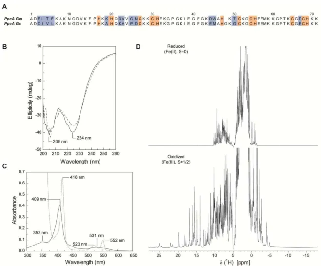
Figure 2.1 Amino acid sequence and spectroscopic properties of PpcA from G. metallireducens. ... 42
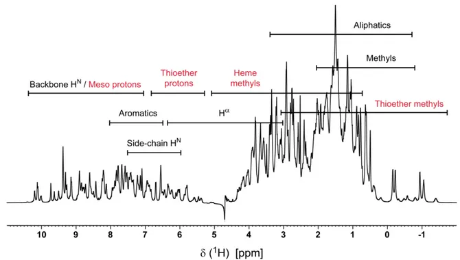
Figure 2.2 Energy diagram of a nucleus with increasing magnetic field strength. ... 45 Figure 2.3 Nuclei precess in the presence of a magnetic field. ... 46 Figure 2.4 Simplest 1D pulse sequence. ... 48 Figure 2.5 Expansion of the 1D 1H-NMR spectrum of methyl acrylate, in acetone solution. ... 48 Figure 2.6 1D 1H-NMR spectrum of the triheme cytochrome PpcA from G. metallireducens in the
reduced state. ... 49
Figure 2.7 Pulse sequence of the 2D 1H-Correlation Spectroscopy (COSY) experiment. ... 49 Figure 2.8 Pulse sequence of the 2D 1H-TOCSY experiment. ... 50 Figure 2.9 Pulse sequence of the 2D 1H-NOESY experiment. ... 51 Figure 2.10 Pulse sequence of the 2D 1H,13C-HMQC experiment. ... 52 Figure 2.11 Size limitation of the NMR technique. ... 53 Figure 2.12 Plot of relaxation time versus correlation time. ... 53
Figure 2.13 Exchange spectroscopy basics. ... 55 Figure 2.14 Diagram of heme c, numbered according to the IUPAC-IUB nomenclature. ... 60
xxii
Figure 2.16 2D 1H,13C-HMQC spectra of PpcA from G. sulfurreducens at pH 5.5 and 298 K. ... 62
Figure 2.17 Thermodynamic model of a triheme cytochrome. ... 64 Figure 2.18 Energy interactions in a triheme cytochrome with one redox-Bohr center. ... 65 Figure 2.19 Illustration of the heme oxidation profiles for PpcA from G. metallireducens (pH 5.8, 288 K).
... 69
Figure 2.20 2D 1H,13C-HMQC spectrum of PpcA from G. metallireducens at pH 5.8, 288 K. ... 70
Figure 2.21 Fitting of the thermodynamic model to the experimental data for PpcA from G.
metallireducens. ... 72
Figure 2.22 pH dependence of the heme methyl proton chemical shifts of PpcA from G. metallireducens
in the oxidized state.. ... 75
Figure 2.23 Redox dependence of the heme oxidation fractions of PpcA from G. metallireducens at
different pH values. ... 76
Figure 2.24 Electron/proton transfer pathways of PpcA from G. metallireducens. ... 77
Figure 2.25 Preferential electron/proton coupled transfer pathways in the homologous PpcA cytochromes
from G. metallireducens and G. sulfurreducens at physiological pH. ... 79
Figure 3.1 Amino acid alignment of the PpcA-family cytochromes (PpcA, PpcB, PpcC, PpcD and PpcE)
with the periplasmic cytochrome GSU0105. ... 93
Figure 3.2 Potential-time excitation signal in a cyclic voltammetric experiment. ... 98 Figure 3.3 Typical cyclic voltammogram for a reversible redox couple, during a single potential cycle. .... 99 Figure 3.4 Purification of GSU0105. ... 103 Figure 3.5 Size exclusion chromatography elution profile of GSU0105. ... 105 Figure 3.6 SDS-PAGE analysis of the GSU0105 protein expression induction temperature dependence. 106 Figure 3.7 Cation exchange chromatography elution profiles of the periplasmic fractions of E.coli JM109
and E. coli SF110 cells. ... 107
Figure 3.8 Protein expression of JM109 and SF110 E. coli strains. ... 108
Figure 3.9 Cation exchange chromatography elution profile of GSU0105. ... 109 Figure 3.10 - UV-visible spectra features of cytochrome GSU0105 in the oxidized and reduced states. ... 110 Figure 3.11 Expansion of the 1D 1H-NMR spectra of GSU0105, in 32 mM sodium phosphate pH 8 (100
mM ionic strength), at 298 K. ... 113
Figure 3.12 CV assays of 200 μM GSU0105 in phosphate buffer with NaCl (170 mM final ionic strength),
at pH 7. ... 115
Figure 3.13 CV assays of 200 μM GSU0105 in phosphate buffer with NaCl (170 mM final ionic strength),
at pH 7. ... 116
xxiii
Figure 4.1 Schematic diagram of typical membrane proteins in a biological membrane. ... 129 Figure 4.2 The proposed Mtr and Pcc extracellular electron transfer pathways. ... 131 Figure 4.3 Scheme for electron transfer from the inner cytoplasmic membrane to the extracellular
environment in S. oneidensis. ... 132
Figure 4.4 Amino acid sequence alignment of OmaW and OmaV from G. sulfurreducens. ... 133
Figure 4.5 Overview of the Q5® Site-Directed Mutagenesis kit from New England Biolabs. ... 135 Figure 4.6 SDS-PAGE of OmaV and OmaW supernatants and pellets after periplasmic fraction isolation
and mechanical lysis with the French-press. ... 142
Figure 4.7 SDS-PAGE analysis of the different mild solubilization techniques (Part 1). ... 144 Figure 4.8 SDS-PAGE analysis of the different mild solubilization techniques (Part 2). ... 145 Figure 4.9 Cation exchange chromatography of OmaV in the denaturated state. ... 147 Figure 4.10 SDS-PAGE analysis of the different detergent or glycerol based solubilization techniques. .. 150 Figure 4.11 Gel electrophoresis of PCR products in 1% agarose gel, 1x TAE buffer. ... 152 Figure 4.12 Final sequence of the His-tagged OmaW... 152 Figure 4.13 Affinity chromatography of OmaW. ... 153 Figure 4.14 UV-visible spectrum of the fraction containing OmaW in the oxidized state... 154 Figure 4.15 SDS-PAGE analysis of the affinity chromatographies performed in Ni2+ beads. ... 155
Figure 6.1 Protein molecular weight marker Precision Plus ProteinTM Dual Xtra Standards (Bio-Rad). .. 173 Figure 6.2 1 kb DNA ladder from New England Biolabs. ... 175 Figure 6.3 1H,13C-HMQC spectrum of PpcA from G. metallireducens, at pH 8.1, 288 K. ... 176
Figure 6.4 1H,13C-HMQC spectrum of PpcA from G. metallireducens, at pH 8.1, 298 K. ... 178
Figure 6.5 1D 1H-NMR pH titration of PpcA from G. metallireducens in the oxidized state, at 288 K (pH
5.3 7.2). ... 181 Figure 6.6 1D 1H-NMR pH titration of PpcA from G. metallireducens in the oxidized state, at 288 K (pH
7.3 8.2). ... 182
Figure 6.7 1D 1H-NMR pH titration of PpcA from G. metallireducens in the oxidized state, at 288 K (pH
8.3 9.5). ... 183
Figure 6.8 UV-visible spectrum of sodium dithionite... 186 Figure 6.9 Cyclic voltammetry control assays with 200 μM BSA in phosphate buffer with NaCl (170 mM
final ionic strength), at pH 7. ... 187
xxv
Table 1.1 Heme reduction potentials and pairwise interactions of the fully reduced and protonated forms
of several priplasmic cytochromes from G. sulfurreducens. ... 24
Table 1.2 Macroscopic pKa values of the redox-Bohr center of several periplasmic cytochromes from G.
sulfurreducens at each stage of oxidation. ... 25
Table 1.3 Data set of G. sulfurreducens and G. metallireducensc-type cytochromes participating in EET
pathways. ... 25
Table 2.1 Amino acid sequence identity percentages within and between the PpcA-families from G. metallireducens and G. sulfurreducens. ... 41
Table 2.2 Properties of some NMR active nuclei. ... 44 Table 2.3 Redox-dependent heme methyl chemical shifts of PpcA from G. metallireducens at pH 5.8, 288
K. ... 71
Table 2.4 Thermodynamic parameters of the fully reduced and protonated form of PpcA, obtained at 288
K and 250 mM ionic strength. ... 73
Table 2.5 Heme reduction potentials of triheme cytochromes from G. metallireducens, G. sulfurreducens
and Desulfuromonas acetoxidans in the fully reduced and protonated state. ... 74
Table 2.6 Thermodynamic parameters of the fully reduced and protonated forms of PpcA from G. metallireducens and PpcA from G. sulfurreducens, obtained at 288 K and 250 mM ionic strength. ... 78
Table 3.1 Biochemical characteristics of periplasmic cytochromes from G. sulfurreducens. ... 94
Table 3.2 Heme reduction potentials of triheme cytochromes from G. metallireducens, G. sulfurreducens
and Desulfuromonas acetoxidans at pH 7 and 293 K. ... 119
Table 4.1 Biochemical characteristics of OM cytochromes from G. sulfurreducens. ... 133
Table 4.2 Sequences of the DNA insert and primers used to produce the pGSU2643H plasmid, encoding
for the N-terminal His-tagged OmaW cytochrome. ... 134
xxvi
Table 4.7 Solutions used for total membrane disruption and/or solubilization of the OmaV and OmaW
cytochromes. ... 139
Table 4.8 Treatments for the extraction of peripheral membrane proteins. ... 143 Table 4.9 Classification of detergents, according to Helenius and Simons. ... 148 Table 4.10 Critical micelle concentration (CMC) for detergents commonly used in integral membrane
proteins extraction. ... 149
Table 5.1 Features of the recently cloned OM proteins. ... 167
Table 6.1 List of the reagents used in this Thesis. ... 171 Table 6.2 SDS-PAGE gel recipe for 5% stacking gel + 15% running gel. ... 173 Table 6.3 Solutions for heme staining. ... 174 Table 6.4 Heme methyls and propionates assignment (1H and 13C) of PpcA from G. metallireducens in the
oxidized state, at 250 mM ionic strength, pH 8.1, 288 K. ... 177
Table 6.5 Heme methyls and propionates assignment (1H and 13C) of PpcA from G. metallireducens in the
oxidized state, at 250 mM ionic strength, pH 8.1, 298 K. ... 179
Table 6.6 Heme methyls and propionates assignment (1H and 13C) of PpcA from G. metallireducens in the
oxidized state, at 250 mM ionic strength, pH 5.8, 288 K. ... 180
Table 6.7 Chemical shifts of the heme methyl protons of PpcA from G. metallireducens in the reduced
and oxidized states, at pH 5.8 and 288 K. ... 180
Table 6.8 Heme methyl chemical shifts of PpcA from G. metallireducens at different stages of oxidation
(pH 5.8 8.9). ... 184
xxvii
--
-- --
-- - - -
-- - -
-xxviii
-β
--
-xxix
--
-γ
ω
oτ
cδ
-ε
𝜇⃗
--
-- -
-xxx
Amino acid abbreviations
Alanine Ala A
Arginine Arg R
Asparagine Asn N
Aspartate Asp D
Cysteine Cys C
Glutamate Glu E
Glutamine Gln Q
Glycine Gly G
H istidine His H
I soleucine Ile I
Leucine Leu L
Lysine Lys K
M ethionine Met M
Phenylalanine Phe F
Proline Pro P
Serine Ser S
Threonine Thr T
Tryptophan Trp W
Tyrosine Tyr Y
1
Introduction
2
1 I ntroduction ... 3
1.1 Geobacter bacteria ... 4
1.2 Geobacter sulfurreducens electron transfer pathways ... 5
1.3 Gene knockout and proteomic studies in Geobacter sulfurreducens ... 10
1.4 Geobacter metallireducens ... 11
1.5 Geobacter sulfurreducens versus Geobacter metallireducens ... 13
1.6 Functional diversity of cytochromes ... 14
1.7 Multiheme cytochromes ... 19
1.8 Structural and thermodynamic characterization of cytochromes c ... 20
1.9 Objectives and thesis outline ... 26
3
1 Introduction
Throughout history, humans have both affected and been affected by the natural world. Environmental problems are becoming more complex, especially as issues arise on a more global level, such as that of aquatic and atmospheric pollution, global warming or equitable access to safe and affordable drinking water. The interactions between human society and the environment are constantly changing. The environment is used and altered by a wide variety of people with many different interests and the challenge is to find approaches to environmental management that give people the quality of life they seek, while protecting the environmental systems that are also the foundations of their well-being. An adequate mindset, implemented by schools and families, is a strong first step towards this goal. A multidisciplinary learning approach can be established by the contributions of a wide range of fields, providing a deeper understanding of the technological, political, and social options, as well as strategies for studying and managing the relationship between our society and the environment.
In 2015, the United Nations set the Sustainable Development Goals, which comprise 17 global goals that cover a broad range of environmental, social and economic development issues [1]. These include poverty, hunger, health, education, climate change, gender equality, water sanitation, energy, environment and social justice. Among these goals, the insurance of access to affordable, reliable, sustainable and modern energy for all, as well as the need to guarantee availability and sustainable management of clean water, stand out as the ones that can be achieved with biotechnological applications based on Geobacter bacteria.
The first report that bacteria can generate electricity appeared almost a hundred years ago, by Potter [2]. Some microorganisms, such as Geobacter, can convert chemical energy from a
wide range of organic and inorganic substances into electric current. On the other hand,
Geobacter species have been shown to play important roles in the bioremediation of
groundwater contaminated with petroleum and landfill leachate [3-7]. Since Geobacter
discovery, many studies have been made in order to understand the mechanisms underlying these unique characteristics which make Geobacter an excellent target to develop Microbial
4
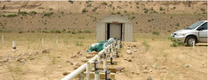
Figure 1.1 I n situ test plot for U(V I ) bioremediation station This site, which was part of the Uranium Mill Tailings Remedial Action program from the U.S Department of Energy, was constructed on the grounds of a former uranium ore processing facility in Rifle, Colorado, USA. Geobacter sulfurreducens performs microbial reduction of soluble U(VI) existing in contaminated groundwaters to insoluble U(IV), facilitating the immobilization of the heavy metal. This strategy is then coupled with a soil washing technique, allowing the concentration of uranium from contaminated soils. Anderson and co-workers [8], responsible for the implementation of this station, discovered that Geobacter species can be simply and effectively stimulated for U(VI) reduction with the addition of an acetate solution to the contaminated groundwaters. They also verified that U(VI) concentrations decreased in as little as 9 days after acetate injection and that within 50 days, uranium concentration had declined below the prescribed treatment level of 0.18
μ
M [8] (photo taken from http://www.geobacter.org/bioremediation).
The work developed and presented in this Thesis focuses on the functional, thermodynamic, and biochemical characterization of electron transfer components from Geobacter bacteria.
1.1
Geobacter
bacteria
The family Geobacteraceae is part of the order Desulfuromonadales in the
-subclass of theProteobacteria. The order branches phylogenetically between the orders Desulfovibrionales and
Desulfobacterales and consists of the genus Geobacter and the sole species Pelobacter
propionicus [9].
Geobacter species are Gram-negative bacteria who play an important biogeochemical role in
a diversity of natural environments. Geobacter species are mostly known for their capability of
making electrical contacts with extracellular electron acceptors and other microorganisms [10]. This remarkable versatility permits Geobacter species to fill important niches in a variety of
5 electrodes, in current consuming biofilms, a process that is currently explored in microbial electrosynthesis systems.
Geobacter species show an impressive respiratory adaptability, since they are capable of
sustaining their growth by using extracellular compounds, such as Fe(III), U(VI) or Mn(IV) oxides, as terminal electron acceptors, in addition to the more frequent respiratory processes, which use soluble electron donors (e.g. acetate) and acceptors (e.g. fumarate) [12]. Some of these compounds are toxic or radioactive, making this organism a potential target for bioremediation and biotechnological applications [12, 13].
Geobacter bacteria were firstly classified as strict anaerobes that are naturally found in a
variety of soils and sediments. However, data obtained for Geobacter sulfurreducens (G.
sulfurreducens) indicates that Geobacter species can also grow at low levels of molecular
oxygen, providing an explanation for their abundance in oxic subsurface environments [14] and for the existence of genes predicted to code for proteins involved in response to oxidative stress, such as rubredoxin, catalase, superoxide dismutase and superoxide reductase, as well as for a terminal cytochrome c oxidase [15].
These bacteria can be easily cultured and genetically manipulated for physiological studies.
G. sulfurreducens was the first Geobacter for which methods for genetic manipulation were
developed and, therefore, it has served as model for functional genomic studies designed to regulation and extracellular electron transfer (EET) mechanisms [16-22].
1.2
Geobacter
sulfurreducens
electron transfer pathways
The bacterium G. sulfurreducens was described for the first time in 1994, after being isolated
from surface sediments of a hydrocarbon-contaminated ditch near Norman, Oklahoma, USA [23]. G. sulfurreducens has a versatile approach to capturing energy and carbon, having three
enzyme systems capable of converting pyruvate to acetyl-CoA. These systems include a pyruvate-ferredoxin oxidoreductase and a pyruvate-formate lyase, both used by anaerobes, and a putative pyruvate dehydrogenase complex, found largely in aerobic organisms [24].
The genome of G. sulfurreducens has an unprecedented number of putative c-type
cytochromes, with 111 coding sequences containing at least one match to the c-type
6
highlights the importance of electron transport to this microorganism and suggests that this bacterium possesses electron transfer networks with high flexibility and redundancy, allowing the reduction of diverse metal ions in natural environments [15]. In fact, several studies revealed a complex transcriptional response by G. sulfurreducens to different electron
acceptors. Currently, no single gene deletion on the G. sulfurreducens genome was found to
eliminate electron transfer to all electron acceptors, thus confirming the complexity of the electron transfer networks in G. sulfurreducens [21, 25-34].
The cytochromes from G. sulfurreducens are strategically localized at the bacterial inner
membrane (IM), periplasm or outer membrane (OM), allowing the transfer of electrons from intracellular carriers, such as NADH, to extracellular acceptors.
The main IM electron transfer components studied in recent years have been the IM-associated peroxidase MacA (GSU0466 the nomenclature of the genome of G. sulfurreducens
is in agreement with http://www.genome.jp/kegg/genome.html), CbcL (GSU0274) and ImcH
(GSU3259) cytochromes. MacA is a dihemic, 35 kDa protein, thought to be involved in the
electron transfer from the IM to the periplasmic components of the EET network [35, 36]. This cytochrome was identified as a peroxidase and is also capable of exchanging electrons with the periplasmic c-type cytochrome PpcA [37]. The CbcL protein contains a HydC/FdnI
diheme b-type cytochrome linked to a 9-heme periplasmic cytochrome c domain [38]. On the
other hand, the cytochrome ImcH was predicted to contain up to three transmembrane helices (depending on processing of a putative signal anchor), a region of NapC/NirT homology, and up to 7 heme c-type heme binding motifs [39].
Amongst all the proteins involved in the EET processes of G. sulfurreducens, the most
studied is the periplasmic cytochrome PpcA (GSU0612), that belongs to a family composed by
five low molecular weight (10 kDa) triheme cytochromes with approximately 70 residues each. The other four cytochromes of this family are designated PpcB (GSU0364), PpcC (GSU0365),
PpcD (GSU0124), PpcE (GSU1760) and share 77% (PpcB), 62% (PpcC), 57% (PpcD) and 65%
(PpcE) amino acid sequence identity with PpcA. Due to the cellular location of these five cytochromes, it was proposed that they are likely reservoirs of electrons, destined for the cell outer surface, bridging the electron transfer between the cytoplasm and the cell exterior [19, 27].
Also located in the periplasm of G. sulfurreducens, there are other relevant cytochromes,
7 four similar triheme domains, designated A, B, C, D and the cytochrome is thought to act as a periplasmic electron capacitor [40, 41]. PccH was unequivocally identified as crucial for G.
sulfurreducens cells to be able to accept electrons from electrodes [42]. GSU0105, together with
two other uncharacterized cytochromes (GSU0701 and GSU2515), is highly expressed in Fe(III) reducing conditions but is not expressed at all in cultures grown on fumarate [27].
G. sulfurreducens is said to have a similar OM architecture compared to Shewanella
oneidensis (S. oneidensis), meaning that the electrons reach the cellular exterior through
porin-cytochrome trans-outer membrane complexes [43]. The OM complexes OmaB(GSU2738
)-OmbB(GSU2739)-OmcB(GSU2737)/OmaC(GSU2732)-OmbC(GSU2733)-OmcC(GSU2731) in
G. sulfurreducens may have similar functions to the ones described for the MtrA-MtrB-MtrC
complex in S. oneidensis [44]. These complexes consist of a porin-like OM protein (OmbB or
OmbC), a periplasmic octaheme cytochrome c (OmaB or OmaC) and an OM dodecaheme
cytochrome c (OmcB or OmcC). Recently, other OM complexes, presenting a similar complex
organization, OmaW(GSU2643)-OmbW(GSU2644)-OmcW(GSU2642) and OmaV(GSU2725
)-OmbV(GSU2726)-OmcV(GSU2724), were shown to be involved in EET to Fe(III) and Mn(IV)
oxides [45]. Shi and co-workers [46] suggested that the OmaW-OmbW-OmcW complex has an extra periplasmic component, the GSU2645 cytochrome. Otero and co-workers [47] identified a five-component OM complex and named it ExtHIJKL (ExtH(GSU2940
)-ExtI(GSU2939)-ExtJ(GSU2938)-ExtK(GSU2937)-ExtL(GSU2936)). This complex was found to
be important in Fe(III) citrate reduction [47].
Other OM cytochromes have been studied in G. sulfurreducens, namely OmcE, OmcF,
OmcS and OmcZ. The 26 kDa tetraheme c-type cytochrome OmcE (GSU0618) is located in
the exterior of the OM of the bacterium and was shown to have an important role in Fe(III) oxide reduction [30]. OmcF (GSU2432) is a monoheme cytochrome c predicted to be localized
at the OM of G. sulfurreducens, having a crucial role in Fe(III) citrate reduction [29]. Finally,
OmcS (GSU2504, 47 kDa) and OmcZ (GSU2076, 30 kDa) are extracellular c-type cytochromes,
containing six and eight heme groups, respectively [48, 49]. OmcS is usually associated with conductive pili [49], whereas OmcZ is responsible for promoting electron transfer in current-producing G. sulfurreducens biofilms [50].
As mentioned above, G. sulfurreducens also possesses electrically conductive pili that enable
8
the inhibition of the electron transport to Fe(III) oxides, interspecies electron exchange and the development of thick electrically conductive biofilms in strains with deletion of the PilA (GSU1496), the pilus monomer [31, 51, 53, 55]; (ii) the inhibition of Fe(III) oxide reduction
and reduced biofilm conductivity, in strains with genetically modified pilA, specially designed
to yield poor conductivity [58]; (iii) infective Fe(III) oxide reduction and current production, in a strain of G. sulfurreducens expressing the poorly conductive pili of Pseudomonas
aeruginosa [59]; (iv) the individual pilin filaments are electrically conductive [51, 55, 60]; and
(v) the pili propagate charge similarly to carbon nanotubes [61].
Although many EET components of G. sulfurreducens are known, these mechanisms and
the proteins implicated in each electron transfer pathway are still under investigation. The initial model presented by Lovley in 2006 [36] suggested that the electrons coming from the menaquinone pool were transferred to the periplasmic components of the bacterium through the IM associated cytochrome MacA (Figure 1.2).
9 Electrons were then shuttled via PpcA and related cytochromes to the outside of the cell
through the OmcB multiheme cytochrome, followed by electron transfer reactions with other OM components and pili. Although the idea for the routes of EET remained the same, in 2014, a slightly different model was presented, since OmcB was found to actually be part of a three-component OM complex (OmaB-OmbB-OmcB) [11, 44]. More recently, a series of findings about important EET components led to a redesign of the EET routes of electron transfer in G. sulfurreducens (Figure 1.3).
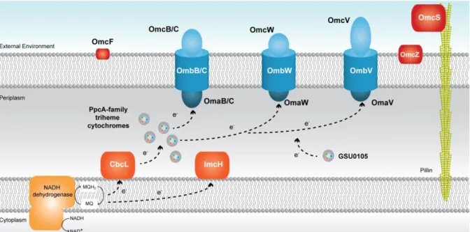
Figure 1.3 Current proposed model for the extracellular electron transfer pathways in G.
sulfurreducens The black arrows represent the proposed electron transfer pathway. Either CbcL or
ImcH (both in dark orange) receive electrons from the menaquinol (MQH2)/ menaquinone (MQ) pool, at the IM, depending on the final electron acceptor redox potential. The periplasmic components, namely the PpcA-family of triheme cytochromes (PpcA-E) are reduced by the electrons received from either CbcL or ImcH. These cytochromes mediate the electron transfer from the periplasm to the OM complexes (in blue), that are likely directly involved in insoluble Fe(III) oxides reduction, which are not represented in the figure. Other components that participate in electron transfer mechanisms in G. sulfurreducens are also represented (GSU0105 and other OM cytochromes
OmcF, OmcS and OmcZ which are represented in red).
The current model for EET maintains the theory that electrons are transferred to the menaquinone pool via the NADH dehydrogenase located in the IM [62]. Then, depending on
the redox potential of the final electron acceptor, different proteins are involved in the quinone regeneration: the CbcL-dependent pathway operates with acceptors at or below redox potentials of 100 mV (versus the normal hydrogen electrode, NHE), whereas the ImcH
10
putatively supplied from these two IM associated proteins to periplasmic triheme cytochromes. In order to reduce extracellular compounds, the electrons are transferred from the periplasmic cytochromes to OM associated cytochromes, through porin-cytochrome trans
-outer membrane complexes [63].
1.3 Gene knockout and proteomic studies in
Geobacter
sulfurreducens
Several studies have been made in order to identify the proteins involved in the different reduction pathways used by G. sulfurreducens. Proteomic analysis under different growth
conditions and gene deletion studies were performed, with special focus on the c-type
cytochromes who may play important roles in electron transfer to extracellular electron acceptors. In this section, the main conclusions taken from those studies are summarized.
The IM associated cytochrome MacA is more abundant during growth with Fe(III) oxides
versus Fe(III) citrate [26] and the deletion of its gene was shown to result in the inhibition of
the expression of the omcB gene [64]. However, the expression of the OM cytochrome OmcB
in the macA deficient mutant restored the capacity for Fe(III) reduction [64]. In addition to
this, a high similarity in the expression patterns and mutant phenotypes between MacA and OmcB, suggests that these two cytochromes may function in the same or similar routes of electron transfer [45]. The deletion of the genes encoding for the IM cytochromes CbcL and ImcH inhibited reduction of Fe(III) oxides and Fe(III) citrate , respectively [38, 39].
Regarding the periplasmic cytochromes, it was shown that the deletion of the gene coding for PpcA affects the ability of G. sulfurreducens to reduce Fe(III) oxides and U(VI) [19, 33].
Also, PpcA was detected in G. sulfurreducens cultures that grow in presence of Fe(III) citrate
and Fe(III) oxide [26]. Similarly, PpcB and PpcC were also detected in Fe(III) citrate and Fe(III) oxide cultures [26]. The deletion of the genes encoding for these two cytochromes, which are part of the same locus, was shown to affect U(VI) reduction [33]. The deletion of the
gene encoding for PpcD affects the reduction of U(VI) and the cytochrome was shown to be more abundant during growth with Fe(III) oxide versus Fe(III) citrate [27, 33]. PpcE was only
detected in cultures with Fe(III) citrate and its gene deletion also affected U(VI) reduction [27, 33]. Finally, PccH is crucial for fumarate reduction in current-consuming biofilms [42].
Several OM cytochromes have been proven to play relevant roles in EET pathways and on the overall regulation of energy transduction in G. sulfurreducens. OmcE, for example, is more
abundant in Geobacter cells during growth with Fe(III) oxides than in cells grown in Fe(III)
11 reduce Fe(III) oxides, suggesting that other EET components can partially compensate for the absence of OmcE [30]. The deletion of the omcF gene affects the expression of other OM
cytochromes, namely OmcB, OmcC and OmcS [18]. Specifically, the deletion of omcF resulted
in a loss of expression of omcB and omcC, as well as an overexpression of omcS, during growth
on Fe(III) citrate [29]. Recently, different studies revealed that a omcF-deficient strain was
unable to grow in presence of Fe(III) oxide and presented a significant decrease in current production [29, 45]. On the other hand, the deletion of omcS affects Geobacters growth in the
presence of Fe(III) oxides [30, 45]. The OmcS cytochrome was shown to be more abundant during growth with Fe(III) oxides versus Fe(III) citrate [30, 31], whereas OmcZ is more
abundant in the opposite conditions [26].
Other studies performed in vitro demonstrated that OmcZ transfers electrons to a diversity
of potential extracellular electron acceptors, such as Fe(III) citrate, U(VI), Cr(VI), Au(III), Mn(IV) oxide and AQDS [26, 48]. These studies also demonstrated that OmcZ does not transfer electrons to Fe(III) oxides, justifying its higher abundance during growth with Fe(III) citrate versus Fe(III) oxides. The OM complexes OmaB-OmbB-OmcB and
OmaC-OmbC-OmcC were found to be important in Mn(IV) oxide reduction [25].
Finally, the OmaW-OmbW-OmcW and OmaV-OmbV-OmcV complexes were found to be overexpressed during growth with Mn(IV) oxide versus Fe(III) citrate and with Fe(III) oxide
versus Fe(III) citrate [25, 47].
1.4
Geobacter metallireducens
G. metallireducens is a rod shaped, Gram-negative, anaerobic bacterium and was first
isolated from freshwater sediments in 1987. It was the first Geobacter ever to be isolated [65].
This bacterium was also the first organism found to completely oxidize organic compounds to carbon dioxide with Fe(III) oxide serving as the electron acceptor [65-67].
Initially, G. metallireducens was reported to be a non-motile bacterium [68]. However, in
2002, motility was observed for G. metallireducens cells grown in insoluble Fe(III) by
phase-contrast microscopy [69]. This bacterium is capable of storing energy through dissimilatory reduction of iron, manganese, uranium and other metals [65].
12
aromatic compounds [71-74]. Furthermore, G. metallireducens can also use nitrate as electron
acceptor, having a functional enzyme complex (which includes a multiheme cytochrome c)
that exhibits both nitrate and nitrite reductase activities [68, 75].
In 2006, studies revealed that G. metallireducens can reduce even very stable iron complexes,
such as Prussian Blue (cyanide-metal complex, Fe4[Fe(CN)6]3, typically found in soils and
aquifers of industrial sites, usually released or spread by outgassing or transport with the groundwater), using it as a primary electron acceptor [76].
G. metallireducens is also capable of reducing ionic mercury (Hg2+) to elemental mercury
(Hg) without having to use a mercury reductase [77]. This feature of G. metallireducens can be
explored in order to mobilize mercury from contaminated groundwaters and produce methylmercury in anoxic environments [77]. Also, G. metallireducens is able to use vanadium
as a final electron acceptor, as proven by Aklujkar and co-workers [78].
In 2008, Tremblay and co-workers [79] developed a genetic system for G. metallireducens
and identified a very important role for pili in Fe(III) reduction and electron transfer to electrodes. G. metallireducens unique motility is one of the reasons why this bacterium is more
efficient in the reduction of Fe(III) oxides, compared to G. sulfurreducens [79]. As mentioned,
in G. sulfurreducens it has been proven that in addition to pili, OM c-type cytochromes are
important for extracellular electron exchange with Fe(III) oxide [30, 80], U(VI) [33], humic substances [81], electrodes [28, 31] and other cells [53]. However, there is poor conservation of OM cytochromes between G. sulfurreducens and G. metallireducens. Further studies of the
functional homologues in G. metallireducens are likely to provide important insights into the
features that c-type cytochromes may share to permit similar function in the absence of
sequence homology [79].
In the recent years, a few proteomic and gene knockout studies have been published for G.
metallireducens [73, 74, 82, 83]. These studies revealed that there are some EET components
that are conserved in G. metallireducens when compared with G. sulfurreducens, namely the
PpcA-family of cytochromes and some IM and OM cytochromes. However, there is not much information about the functional properties of these components, in contrast with G.
sulfurreducens (as demonstrated in the previous section).
For the family of periplasmic cytochromes existing in G. metallireducens (composed by
PpcA (Gmet_2902), PpcB (Gmet_3166), PpcC (Gmet_3165), PpcE (Gmet_1846) and PpcF
13 http://www.genome.jp/kegg/genome.html), one can infer that their functions will be very similar to the ones of the PpcA-family from G. sulfurreducens, due to the high homology
between both families. This means that in G. metallireducens, this family of cytochromes is
probably responsible for controlling the electron flow from the IM to the OM electron transfer components (Figure 1.4). This subject will be addressed in more detail in Chapter 2.
Figure 1.4 Current extracellular electron transfer pathways model in G. metallireducens The
black arrows represent the proposed electron transfer pathway. The periplasmic PpcA-family triheme cytochromes (PpcA, PpcB, PpcC, PpcE and PpcF) mediate the electron transfer between the IM associated cytochromes (in orange) to the porin-cytochrome complexes in the OM (in blue).
1.5
Geobacter sulfurreducens versus Geobacter metallireducens
Globally, G. sulfurreducens and G. metallireducens share several metabolic features,
including the ability to reduce U(VI) [84], high current production [85], as well as the capability to transfer electrons to different species without the involvement of electron carrier molecules, a process designated direct interspecies electron transfer (DIET) [54, 86, 87].
Although there are several metabolic characteristics shared between the two bacteria, a marked difference relates to their ability to oxidize organic compounds. While G.
sulfurreducens can only oxidize acetate, formate, lactate and pyruvate, G. metallireducens can
oxidize all the above mentioned, as well as benzaldehyde, butanol, ethanol, phenol, propionate and propanol [78]. Furthermore, G. metallireducens is able to use more inorganic compounds
as final electron acceptors, such as vanadium, in contrast with G. sulfurreducens [10, 78]. G.
14
application in several systems, such as: (i) bioremediation of waters contaminated with organic wastes, particularly important for the removal of azo compounds produced by textile industries [88]; (ii) bioremediation of contaminated waters with vanadium [89] and (iii) full degradation of several compounds to methane via DIET mechanisms with methanogenic
bacteria [87]. This feature can be applied to the treatment of wastewaters where methanogenic microorganisms tend to degrade organic matter, with concomitant methane production [86].
Considering all the above mentioned, to optimize these processes, it is first necessary to understand the mechanisms underlying EET in these bacteria. The recent development of a genetic system for G. metallireducens is likely to refocus attention on this organism and it will
allow the unravelling of the above mentioned physiological mechanisms, as well as other relevant novel properties, such as anaerobic benzene degradation, Fe(III) reduction or the use of cytochromes as capacitors to permit respiration in the absence of exogenous electron acceptors, among other remarkable features [78, 79].
1.6 Functional diversity of cytochromes
Heme-containing proteins display a diversity of biological functions, including (i) simple electron transfer reactions, such as those catalyzed by b- and c-type cytochromes [90]; (ii)
oxygen transport and storage via hemoglobin and myoglobin [91]; (iii) oxygen reduction to
the level of water by cytochrome oxidase [92]; (iv) oxygenation of organic substrates, as facilitated by the cytochromes P-450 [93] and (v) the reduction of peroxides by catalases and peroxidases [94]. By combining heme groups with other cofactors, such as flavins and/or metal ions (molybdenum or copper), these proteins can have an even higher range of functions, extending them to a large range of enzymatic processes, which include dehydrogenations [95] and the reduction of numerous small molecules [96].
15
Figure 1.5 D ifferent types of hemes found in cytochromes The classification of the heme groups is based on the differences between the porphyrin molecules and types of polypeptide covalent attachments. a) b-type protoheme IX; b) c-type substituted protoheme IX; c) a-type, d) d-type; e) and f) d1-type. This figure was taken from [97].
16
ligands to iron; (iii) the heme solvent exposure; (iv) the heme accessibility to exogenous ligands; (v) the distribution of polar and charged groups in the heme neighborhood and (vi) the specific properties of the heme-binding site in the protein, stand out as the most crucial ones [95, 98].
scribe a group of heme proteins undergoing oxidation/reduction reactions, characterized in the reduced form by intense absorption bands in the 510-615 nm range [99]. Cytochromes are named accordingly to their heme type letter (in italic) and a number in subscript, depending on some intrinsic characteristics related to the protein axial ligands coordination, number of heme groups, optical or functional properties [100].
Cytochromes c, specifically, are heme proteins containing a c-type heme and often function
as electron carriers in biological systems. The polypeptide chain of these proteins is covalently bound to one or several c-type heme groups through thioether linkages established with the
sulfhydryl groups of two cysteine residues in a conserved binding motif sequence CXXCH [98, 101]. The heme group displays a central role in the functional modulation of these proteins. It is constituted by four pyrrole subunits connected by methane bridges (protoporphyrin IX) and in the center, the iron ion is equatorially coordinated by four nitrogen atoms (Figure 1.5b).
Iron (0) has an electronic structure 1s22s22p63s23p63d64s2 (where the first numbers
corresponds to the principal quantum number; the letters correspond to the orbital quantum number; and the superscripts correspond to the electron occupancies). The electronic structure of iron is sometimes described as [Ar]3d64s2 to emphasize that the electron
occupancies of the higher energy orbitals (3d and 4s), superimposed on an argon core, are responsible for the electronic properties and, ultimately, for the chemical behavior of iron [101]. In heme proteins, iron usually exists in two more common oxidation states: the ferrous state (Fe(II): [Ar]3d64s0) and the ferric state (Fe(III): [Ar]3d54s0).
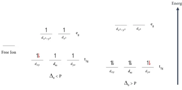
In an iron atom there are five 3d-orbitals two orbitals of higher energy (dx2-y2 and dz2, also
known as the eg set of orbitals) and three of lower energy (dxy, dxz and dyz, also known as the t2g
17 electrostatic field created by the ligands and the energy required to pair electrons in the same orbital (pairing energy - P) [101].
The pairing energy is always unfavorable, but what is relevant is whether it requires less energy to pair electrons in the t2g orbitals than it does to keep them unpaired in the eg orbitals.
In a weak crystal field, the d-orbital splitting is smaller than the pairing energy (
0
< P) and the electrons remain unpaired in separate orbitals (Figure 1.6). This electron distribution is the high-spin configuration. On the other hand, in a strong crystal field, the electrons enter the t2gorbitals and pair to produce the low-spin configuration (Figure 1.6).
Figure 1.6 Spin-states of octahedral Fe(I I I ) and Fe(I I ) The free iron ion has five 3d-orbitals of the same energy. In the figure, these orbitals are represented as a single one, for simplicity. When placed in an octahedral field, the d-orbitals of the iron atom are rearranged in two sets the t2g set (of lower energy, composed by the dxy, dxz and dyz orbitals) and the eg set (composed by the dx2-y2 and dz2orbitals). For Fe(III), whose electrons are represented by black arrows, a weak crystal field (
0
< P) leads to a total spin of 5/2 (high-spin configuration), whereas a strong crystal field (0
> P) results in a total spin of 1/2 (low-spin configuration). On the other hand, for Fe(II), whose extra electron is represented by a red arrow, a weak crystal field (0
< P) leads to a total spin of 2 (high-spin configuration), whereas a strong crystal field (0
> P) results in a total spin of 0 (low-spin configuration).18
spin state can be determined by a variety of spectroscopic techniques, namely Nuclear Magnetic Resonance (NMR), UV-visible and Electron Paramagnetic Resonance (EPR) [103].
In cytochromes c, one of the two axial coordination positions is occupied by the side chain
of a histidine (designated as proximal ligand, see Figure 1.7). On the other hand, the distal ligand is more variable and can be the side chain of a (i) methionine, which predominates in monoheme cytochromes c, (ii) histidine, particularly predominant in multiheme cytochromes
c or (iii) asparagine, lysine or tyrosine, although with less frequency. In certain conditions, the
distal position of the heme can also be transiently vacant, as observed for various cytochromes with enzymatic activity [62, 100, 104].
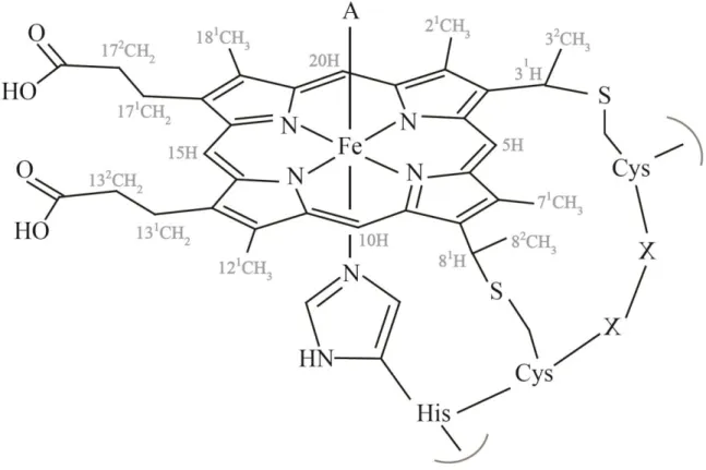
Figure 1.7 Schematic representation of a c-type heme and the correspondent polypeptide
binding motif (CXXCH ) The distal coordination position, labeled with A, can be free or occupied by different amino acids side chains. The IUPAC-IUB nomenclature for tetrapyrroles is illustrated in gray [105]. This figure was taken from [62].
Therefore, in hemes c with histidine or methionine as distal ligands, the d-orbital splitting is
19 reactions usually have both axial positions occupied and hold histidine or methionine residues as distal ligands [62, 101].
Furthermore, the heme redox potential is also strongly dependent on the nature of the axial ligands. The side chain methionine sulfur is a good electron acceptor and favors the electron-rich reduced state, resulting in a more positive redox potential compared to bis-histidinyl coordinated heme groups [97, 106].
1.7 Multiheme cytochromes
Multiheme cytochromes are crucial components of several biological processes and are involved in electron transfer [107, 108], enzymatic reactions [108-110] and on signal transduction events [111]. They were also shown to work as electron biocapacitors (e.g.
GSU1996, Figure 1.8), thus contributing for the enhancement of the electron-storage capacity of several bacteria [40, 41, 112].
Figure 1.8 Crystal structure of the dodecaheme cytochrome GSU1996 from G. sulfurreducens,
obtained in the oxidized state The cytochrome (PDB ID: 3OV0 [41]) is organized in four similar domains (A-D), each containing three hemes, connected by a flexible linker. Each domain has structural homology to the triheme cytochromes of the PpcA-family from G. sulfurreducens, except for the heme IV, which contains His-Met axial coordination. In the figure, the backbone is represented in blue, whereas the hemes are represented in red. Roman numerals indicate the hemes in their order of attachment to the CXXCH motif in the polypeptide chain. The structure was drawn using the PyMOL molecular graphics system [113].
Multiheme cytochromes generally have a set of the same type of heme groups, as seen for the PpcA-family from G. sulfurreducens (in which all the hemes are from the c-type).
20
namely the cytochrome cd1-nitrite reductase (hemes c and d1) [108, 114] and the IM CbcL
cytochrome from Geobacter sulfurreducens, which contains a HydC/FdnI diheme b-type
cytochrome linked to a 9-heme periplasmic cytochrome c domain [38].
These proteins are able to receive or donate multiple electrons in a cooperative way, depending on the intrinsic properties of the neighboring hemes (redox interactions) or heme surrounding protonable centers (redox-Bohr effect) [115-117]. The several heme groups ranges, as a consequence of the contribution of each individual heme redox potential. Typically, the heme iron-iron distances between adjacent hemes do not exceed 15 Å, allowing a fast electronic exchange between the redox centers and assuring efficient redox reactions [104, 118, 119].
The redox potential of hemes in multiheme cytochromes is affected by several factors, namely (i) the differences in the free energy between the oxidized and reduced sates, resulting from molecular interactions; (ii) the modulation of the electrostatic interactions within the protein or with the solvent; (iii) the heme solvent accessibility; (iv) the extent to which the heme group is distorted from planarity; (v) the protonation state of the heme propionate groups and (vi) the type of axial ligands and heme iron coordination, as stated previously [101, 120-126]. Furthermore, multiheme cytochromes have a lower amino acid residue to heme ratio compared to monoheme cytochromes, meaning that the heme groups are more solvent exposed. The high heme solvent exposure and the typical His-His heme coordination strongly contributes to the lower redox potential values generally observed in multiheme cytochromes.
1.8 Structural and thermodynamic characterization of cytochromes
c
The determination of multiheme cytochromes structures is crucial to understand their functional mechanisms. Three-dimensional structures of biological macromolecules can be determined by X-ray crystallography and NMR at atomic resolution. These techniques are well established complementary high-resolution methods to analyze protein structure-function relationships.
NMR spectroscopy enables the determination of protein structures in conditions similar to
21 Advances in protein expression protocols have contributed to increase the expression yields for mature multiheme cytochromes. Higher yields of protein expression allowed the overcome of traditional difficulties associated with the determination of solution structures using natural abundance samples [130-134]. This rendered the isotopic labelling of multiheme cytochromes much more cost-effective, facilitated the NMR signal assignment procedure and provided the foundations for the identification of redox partners and mapping of their interacting regions [135-137].
The isotopic labeling protocol developed by Fernandes and co-workers [138] was used to produce 15N and 13C-15N labeled cytochromes and allowed to obtain proteins uniformly
labeled, with the correct folding and post-translational incorporation of heme groups [139-141]. Taking advantage of these methodologies, the NMR solution structures of two different cytochromes from Geobacter sulfurreducens have been determined. Currently, there are
solution structures available for the periplasmic triheme cytochrome PpcA, in both oxidized [142] and reduced states [143], and for the OM monoheme cytochrome OmcF, in the reduced state [144]. Crystal structures of Geobacter sulfurreducens cytochromes are available in the
oxidized state, namely those of PpcA [145], PpcB [146], PpcC [147], PpcD [147], PpcE [147], OmcF [148, 149], GSU1996 [41], PccH [150] and MacA [37]. The structures of some of the referred cytochromes are presented in Figure 1.9. In the GSU1996 cytochrome and in the PpcA-family cytochromes (Figures 1.8 and 1.9, respectively), the hemes are numbered I, III and IV. This designation derives from the superimposition of the hemes in cytochromes c7
with those of the structurally homologous tetraheme cytochromes c3 [151]. On the other hand,
22
23 In monoheme cytochromes only the reduced and oxidized states may coexist in solution, whereas for multiheme cytochromes, several one-electron reversible transfer steps convert the fully reduced state (stage 0, S0) into the fully oxidized state (stage X, Sx). In addition, in each
stage, the group responsible for the redox-Bohr effect may be protonated or deprotonated, which further increases the number of microstates in solution. For example, in a triheme cytochrome there are 16 different microstates in solution (Figure 1.10).
Figure 1.10 Electronic distribution scheme for a triheme cytochrome with a proton-linked
equilibrium, showing the 16 possible microstates The light gray and dark gray circles correspond to the protonated and deprotonated microstates, respectively. The protonated microstates are also -Bohr center. Inner hexagons represent heme groups. The reduced hemes I, III, and IV are colored green, orange and blue, respectively. The oxidized hemes are colored white. The microstates are grouped, according to the number of oxidized hemes, in four oxidation stages connected by three one-electron redox steps. P0H and P0 represent the
reduced protonated and deprotonated microstates, respectively. PijkH and Pijk indicate, respectively,
the protonated and deprotonated microstates, where i, j and k represent the heme(s) that are oxidized in that particular microstate.
24
titrations describe the whole-protein macroscopic redox behavior (macroscopic redox potentials), without deconvoluting the reduction potentials of the individual redox centers (microscopic redox potentials) [50, 149, 152, 153]. For multiheme cytochromes, the detailed thermodynamic characterization involves the determination of several parameters, namely the redox potentials, redox interactions, redox-Bohr interactions and pKa value(s) of the
redox-Bohr center(s). To calculate these thermodynamic parameters, it is necessary to monitor the stepwise oxidation of each heme oxidation at several pH values. This information can be achieved by NMR spectroscopy, which allows the determination of the individual heme oxidation profiles, due to the different spectral signatures of the NMR spectra of low-spin multihemes, in the reduced and oxidized states. This topic is explained in more detail in Chapter 2, where a full thermodynamic characterization of the triheme cytochrome PpcA from Geobacter metallireducens is presented.
In the last few years, several thermodynamic parameters of G. sulfurreducens and G.
metallireducens cytochromes have been published, namely the (i) heme redox potentials, redox
interactions, redox-Bohr interactions (Table 1.1) and the macroscopic pKa values of the
redox-Bohr center of several periplasmic cytochromes (Table 1.2), as well as the (ii) Eapp values of
several periplasmic, inner- and outer-membrane cytochromes (Table 1.3).
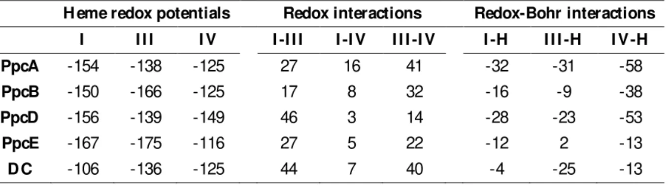
Table 1.1 H eme reduction potentials and pairwise interactions (mV ) of the fully reduced and protonated forms of several priplasmic cytochromes from G. sulfurreducens [129, 154] The values were determined at 288 K . DC stands for Domain C of the GSU1996 cytochrome.
H eme redox potentials Redox interactions Redox-Bohr interactions I I I I I V I -I I I I -I V I I I -I V I -H I I I -H I V -H
PpcA -154 -138 -125 27 16 41 -32 -31 -58
PpcB -150 -166 -125 17 8 32 -16 -9 -38
PpcD -156 -139 -149 46 3 14 -28 -23 -53
PpcE -167 -175 -116 27 5 22 -12 2 -13
25
Table 1.2 M acroscopic pKa values of the redox-Bohr center of several periplasmic cytochromes from G. sulfurreducens at each stage of oxidation [129, 154] The values were determined at 288 K. DC stands for Domain C of the GSU1996 cytochrome.
pKa
Oxidation stage PpcA PpcB PpcD PpcE D C
0 8.6 7.4 8.7 7.7 6.0
1 8.0 7.1 8.1 7.6 5.6
2 7.2 6.8 7.4 7.5 5.4
3 6.5 6.3 6.9 7.4 5.2
pKa 2.1 1.1 1.8 0.3 0.8
Table 1.3 D ata set of G. sulfurreducens (Gs) and G. metallireducens (Gmet) c-type cytochromes
participating in EET pathways The redox potential values were determined at pH 7.
Protein H eme axial ligands Eapp (mV ) vs NH E
M acA Gs His-Met -188 [37]
GSU1996 His-His; His-Met -124 [11]
PpcA Gs His-His -117 [146]
PpcB Gs His-His -137 [146]
PpcC Gs His-His -143 [11]
PpcD Gs His-His -132 [129]
PpcE Gs His-His -134 [129]
PpcA Gmet His-His -78 [155]
PpcF Gmet His-His -56 [156]
OmcF Gs His-Met +180 [149]
OmcS Gs His-His -212 [152]
26
1.9 Objectives and thesis outline
The structural and functional characterization of individual electron transfer components is crucial to understand the EET mechanisms of Geobacter. This knowledge can be further used
to design optimized Geobacter mutant strains, with increased bacterial respiratory rates.
The goal of this Thesis was to study different electron transfer components from Geobacter
bacteria, namely the periplasmic cytochrome PpcA from G. metallireducens, the periplasmic
cytochrome GSU0105 from G. sulfurreducens and the OM cytochromes OmaV and OmaW
from G. sulfurreducens. The Thesis is divided in five Chapters. A general introduction to
Geobacter and cytochromes is presented in Chapter 1. In Chapter 2, the functional and
thermodynamic characterization of the properties of the triheme cytochrome PpcA from G.
metallireducens and their comparison with those of PpcA from G. sulfurreducens are
presented. Chapter 3 describes the optimization of the expression and purification protocols of the periplasmic cytochrome GSU0105 from G. sulfurreducens, as well as preliminary insights
![Figure 1.2 I nitial model suggested for EET in G. sulfurreducens by Lovley [36], with iron oxides serving as final electron acceptors The electrons are transferred from the menaquinone pool to the IM associated cytochrome MacA, who then shutt](https://thumb-eu.123doks.com/thumbv2/123dok_br/16502938.734114/40.892.195.698.524.947/suggested-sulfurreducens-acceptors-electrons-transferred-menaquinone-associated-cytochrome.webp)
![Figure 1.8 Crystal structure of the dodecaheme cytochrome GSU1996 from G. sulfurreducens , obtained in the oxidized state The cytochrome (PDB ID: 3OV0 [41]) is organized in four similar domains (A-D), each containing three hemes, co](https://thumb-eu.123doks.com/thumbv2/123dok_br/16502938.734114/51.892.119.784.548.793/crystal-structure-dodecaheme-cytochrome-sulfurreducens-cytochrome-organized-containing.webp)
![Figure 1.9 Structures of several cytochromes from G. sulfurreducens , obtained in the oxidized state The solution structure of PpcA (lowest energy, PDB ID: 2MZ9 [142]) and the crystal structures of PpcB (chain A, PDB ID: 3B](https://thumb-eu.123doks.com/thumbv2/123dok_br/16502938.734114/54.892.121.771.97.893/structures-cytochromes-sulfurreducens-obtained-oxidized-solution-structure-structures.webp)