Proprioceptive deicits in Parkinson’s disease: from clinical
data to animal experimentation
Letícia Ribeiro
1, Tadeu Mello e Souza
1, Lisiane Bizarro
1and Alcyr Oliveira
21 - Universidade Federal do Rio Grande do Sul, Porto Alegre, RS, Brazil.
2 - Universidade Federal de Ciências da Saúde de Porto Alegre, Porto Alegre, RS, Brazil
Abstract
Parkinson’s disease (PD) is characterized by the manifestation of akinesia, slowness to initiate movement, muscle rigidity, and tremors. However, recent evidence indicates that this pathology also causes alterations in proprioception. Disturbances in proprioceptive mechanisms directly affect postural control and the ability to calculate the velocity and amplitude of movement, suggesting that these alterations are related to the motor symptoms of PD. This article reviews the clinical data on these symptoms and presents evidence of a connection between proprioceptive deicits and the physiology of PD. The identiication of proprioceptive impairments in different forms of Parkinsonism can provide valuable clues on the physiopathology of proprioception in idiopathic PD. Keywords: proprioception, Parkinson’s disease, animal models of parkinsonism.
Received 11 November 2010; received in revised form 26 April 2011; accepted 13 May 2011. Available on line 10 October 2011
Letícia Ribeiro and Lisiane Bizarro, Programa de Pós-Graduação em Psicologia, Departamento de Psicologia do Desenvolvimento e da Personalidade, Universidade Federal do Rio Grande do Sul, Brasil; Tadeu Mello e Souza, Departamento de Bioquímica, Universidade Federal do Rio Grande do Sul; Alcyr Oliveira, Departamento de Psicologia, Programa de Pós-Graduação em Ciências da Reabilitação, Universidade Federal de Ciências da Saúde de Porto Alegre, RS, Brasil. Correspondence regarding this article should be directed to Alcyr Oliveira, PhD. Phone: ++55-51-33038826. E-mail: [email protected]
Introduction
Parkinson’s disease (PD) is a progressive neurodegenerative disorder that affects dopaminergic neurons in the substantia nigra pars compacta (SNpc; Snider, Fahn, Isgreen, & Cote, 1976). Since the irst report of PD in 1817, it has been characterized by clinical manifestations of akinesia, slowing of physical movement, muscle rigidity, tremors (Alexi et al., 2000) and proprioceptive impairment (Jacobs & Horak, 2006; Vaugoyeau, Viel, Assaiante, Amblard, & Azulay, 2007).
Proprioception is responsible for the perception of body position in space and whether members remain static or are moving (Jacobs & Horak, 2006). A disruption in proprioceptive mechanisms may cause impairments in postural control, leading to limitations in the estimation of the amplitude and speed of movement. The involvement of proprioception in the maintenance of motor skills in normal subjects suggests that proprioceptive deicits may be linked to
the motor symptoms of PD (Vaugoyeau et al., 2007). Parkinson’s disease patients show dificulties performing movements that rely on proprioceptive integration, such as requiring upper-limb movements or estimating position without vision (Klockgether, Borutta, Rapp, Spieker, & Dichgans, 1995; Seiss, Praamstra, Hesse, & Rickards, 2003). Performance in tasks that require reaching a target without visual perception of the foreinger or walking to a location without leg visibility is impaired in the initial stages of this disorder (Adamovich, Berkinblit, Hening, Sage, & Poizner, 2001; Keijsers, Admiraal, Cools, Bloem, & Gielen, 2005). These patients often present dificulty keeping the body still and an inability to use the legs to recover balance when having gait disturbances (Rocchi, Chiari, Cappello, Gross, & Horak, 2004). These symptoms suggest the impaired capacity to integrate proprioceptive information to adjust movement and restore balance (Contreras-Vidal & Gold, 2004; Jacobs & Horak, 2006).
present article explores the implication of deicits in the proprioceptive system by examining evidence that suggests a physiological basis for several symptoms of PD.
Parkinson’s disease characteristics
Parkinson’s disease affects 2.5% of the world’s population and is the second most frequent neurodegenerative disorder that affects the elderly (Hattori, Kobayashi, Sasaki-Hatano, Sato, & Mizuno, 2003). However, it also affects young individuals (Alexi et al., 2000). One of the hallmarks of PD is the progressive death of dopaminergic neurons in the SNpc, the cause of which is yet unknown. Studies have indicated a multifactorial origin for the disease that involves both genetic and environmental factors (Hattori et al., 2003). Mitochondrial dysfunction, oxidative stress, genetic alterations of parkin and a-synuclein
protein expression, and the consequent presence of Lewy bodies in SNpc neurons are some of the molecular mechanisms that promote PD (Hattori et al., 2003).
Neuronal loss of approximately 50% in the SNpc leads to dopaminergic depletion of approximately 80% in the striatum, one of the target structures of nigral projections. When neuron deterioration reaches such levels, compensation mechanisms are insuficient to contain deicits, leading to the manifestation of initial PD motor symptoms (Simola, Morelli, & Carta, 2007).
Motor-skill disturbances in PD largely affect the patient’s quality of life. These motor deicits appear during ordinary daily tasks such as walking, sitting, and moving members to reach objects. Adequate performance in all of these tasks requires proper integration between sensory inputs and motor responses, which essentially depend on proprioception (Varraine, Bonnard, & Pailhous, 2002).
Proprioception: deinition and concept
The term “proprioception” derives from the Latin word
proprius, meaning “own.” Sherrington initially used it to
deine sensorial processes involved in the consciousness of the spatial location of an individual, posture, and movement (Voight & Cook, 2001). This mechanism is essential for establishing an appropriate relationship between space and the ability to move accordingly.
The control of posture and movement ultimately depends on the orchestrated feedback of muscle contraction, muscle stretching, and the corresponding response relected in the spinal cord. Movement thus depends on the speed and precision with which mechanoreceptors located in muscle ibers signal these alterations (Voight & Cook, 2001). Since the 1970s, however, researchers have suggested that the contribution of sensorial afferents cannot be excluded from proprioception (Goodwin, McCloskey, &
Matthews, 1972; O’Suilleabhain, 2004; Vaugoyeau et al., 2007). Stimuli from the skin and articulation receptors may also contribute to the conscious judgment of kinetic and static activity (Goodwin et al., 1972; Vaugoyeau et al., 2007). The adequate functioning of these receptors as contributors to the proprioceptive system may be important for the sensorial perception of alterations found in PD patients (O’Suilleabhain, 2004; Snider et al., 1976).
Sensorial alterations in PD and their
implications for proprioception
Parkinson’s disease patients may manifest itching, tingling, and burning sensations or clumsiness of the arms and legs, symptoms collectively known as paresthesia (O’Suilleabhain, 2004). Few studies have investigated the presence of paresthesia in PD or systematically quantiied the appearance, course, and magnitude of these symptoms (O’Suilleabhain, 2004). Quantitative sensorial studies in PD patients have shown that they have increased thresholds for tactile and thermal stimuli and a signiicant reduction in mechanical pain perception (Nolano et al., 2008; Tinazzi et al., 2008; Table 1). Some studies have suggested that sensorial alterations follow the course of the disease and tend to manifest along with other motor symptoms. Snider and colleagues (1976), however, stated that paresthesia emerges as an initial symptom of the disorder in approximately 20% of cases. A clinical survey of 101 patients diagnosed with PD revealed that 42% of the patients suffered from paresthesia (Snider et al., 1976). In a similar study involving 99 patients, 63% complained of these sensations, and the authors related their manifestation to the course of the disease (Shulman, Singer, Bean, & Weiner, 1996). Koller (1984) studied 50 PD patients and reported that 20% suffered from a continuous form of paresthesia, whereas 18% complained of sporadic symptoms. Another report with a sample of 85 PD patients found that 70% complained of paresthesia, many of them speciically during the off periods of treatment with dopaminergic agonists (Gunal, Nurichalichi, Tuncer, Bekiroglu, & Aktan, 2002). These results suggest that paresthesia is not a rare symptom. One study found that of 50 patients interviewed during the off period of dopamine agonist treatment, approximately 40% presented these symptoms. In this study, ive subjects complained that the intensity of these symptoms in the off state was more disabling than the typical motor symptoms (Witjas et al., 2002).
exposure to L-3,4-dihydroxyphenylalanine (L-DOPA) may be related to the presence and intensity of these manifestations (O’Suilleabhain, 2004).
Little is known about the neurocircuitry involved in the physiopathology of paresthesia in PD. Studies conducted to track detectable electrophysiological damage to the skin and nerve endings in PD patients revealed an absence of observable peripheral alterations (Koller, 1984; O’Suilleabhain, 2004). A study conducted in 26 elderly patients, however, concluded that 53% of the subjects presented electrophysiological abnormalities in nerve conduction velocity in the cervical spine, and 73% presented the same impairments in the lumbar spine (Lee & Trojanowski, 2006). The absence of evidence of radiculopathy in other studies of PD patients, however, suggests that paresthesia may have other origins (O’Suilleabhain, 2004). Nerve conduction velocity studies, however, have failed to track large sensory endings and small ibers, which appears to be necessary to elucidate peripheral sensory alterations.
Based on morphological analyses of skin biopsies (Nolano et al., 2008), one study suggested peripheral deafferentation in PD, showing a signiicant loss of epidermal nerve ibers and Meissner corpuscles. Additional indings found increased branching, nerve sprouting, and vascular bed enlargement, which may be attributable to compensatory mechanisms (Duffau & Capelle, 2001; Nolano et al., 2008). What remains unknown is the connection between this peripheral loss and dopamine denervation in the basal ganglia. Impairments in basal ganglia and thalamocortical-basal ganglia circuitry might be the main cause of the sensorial problems observed in PD, the mechanisms for which are yet unknown (Tinazzi et al., 2008).
Stimuli applied to the skin generate responses in the basal ganglia, especially in the SNpc, which plays a crucial role in the modulation of sensorial processing in the thalamus (Bendrups & McKenzie, 1974). Data from proprioception-related evoked potential studies revealed that sensorial potential triggered in the parietal cortex in PD patients is usually normal (Nakashima,
Reference/Year Subjects Methods Results
Snider et al.,
1976 101 patients Self-report and clinical evaluation 43% suffered from paresthesia.
Shulman et al.,
1996 100 patients + 50 age-matched controls Questionnaire 73% of PD patients had sensory symptoms. 44% had internal tremor.
Koller, 1984 50 patients Self-report 38% had sensory complaints and no objective sensory loss. Symp
-toms were frequently restricted to the hemiparkinson side.
Gunal et al.,
2002 85 patients Survey based on self-report about the presence of paresthesias - ~40% reported paresthesia, which was more disabling than motor symptoms in ive patients.
Witjas et al.,
2002 50 patients in the off-phase Questionnaire that used 54 yes-or-no questions about nonmotor luctuations that occur -in the off-phase
100% with at least one type of nonmotor luctuation (anxiety 66%; drenching sweats 64%; slowness of thinking 58%; fatigue 56%; akathisia 54%).
Nolano et al.,
2008 18 patients + 30 age-matched controls During the on-phase, patients were challenged with tactile and thermal stimuli of the hands and feet while electrophysiological signs were recorded. Under local anesthesia, skin punches were taken from the ingertip, thigh, and distal leg.
Evidence of increased tactile and thermal thresholds with reduced pain perception and signiicant loss of epidermal nerve ibers and Meissner corpuscles. Disease severity correlated with Meissner corpuscle loss and reduced pain and thermal perception.
Nitta, & Takahashi, 1992), reinforcing the probable absence of damage to sensorial system conduction in PD patients and pointing to extrapyramidal alterations caused by dopamine depletion. In fact, dopaminergic agonists appear to reduce the intensity of paresthesia symptoms in most cases and have been adopted as a clinical treatment strategy (Nutt & Carter, 1984).
Basal ganglia are essential for the automatic execution of previously learned motor responses and are responsible for the motor output that completes the circuit initiated by peripheral inputs (Marsden, 1982). The basal ganglia have recently been suggested to play a more inluential role in this process because of their involvement in the modulation of peripheral inputs. An adequate motor response requires the full analysis of the conditions in which the organism inds itself (Abbruzzese & Berardelli, 2003). Impaired integration of sensorial and proprioceptive receptor inputs may lead to an underestimation of the position of members or to a miscalculation of the necessary speed for an action, producing bradykinesia (Berardelli, Rothwell, Thompson, & Hallett, 2001). Similarly, damage to the extrapyramidal system may lead to interpretation errors regarding the quantity of received sensory stimuli and cause the automatic and unconscious performance of unnecessary movements, suggesting dyskinesia (O’Suilleabhain, 2004).
Experimental evidence of proprioceptive
alterations in Parkinson’s disease
In 1975, a report showed that PD patients presented impairments in the proprioceptive feedback of peripheral muscle receptors (Tatton & Lee, 1975). Since this irst empirical evidence, several studies have sought to describe these deicits.
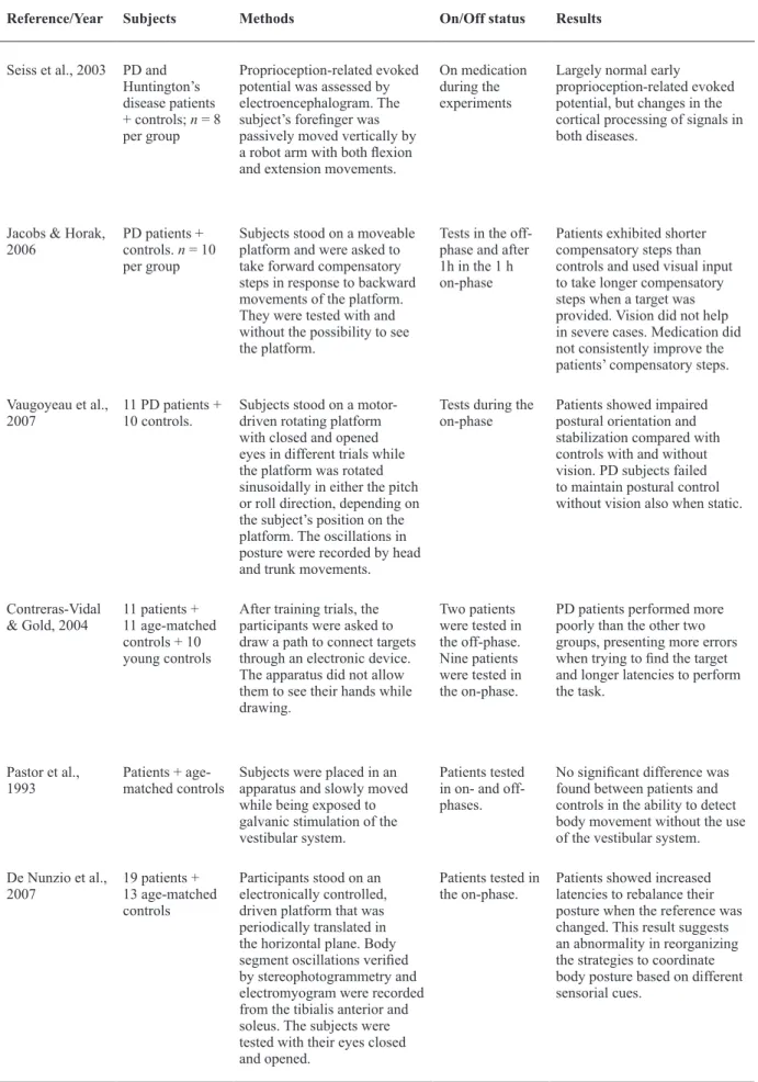
The loss of postural control is the symptom that most affects the quality of life in PD. A characteristic posture in which the forward position of the head and torso affects the ability to control other movements appears in more advanced stages of the disease, leading to impaired balance and falls (Keranen et al., 2003). Although some evidence indicates that cortical proprioceptive information processing is more critical for equilibrium control, the vestibular system exerts a fundamental role in this function, especially with regard to static equilibrium (Vaugoyeau et al., 2007). Through galvanic stimulation of the vestibular system in healthy elderly and PD patients during a body movement perception task, Pastor and colleagues found no difference between patients and controls regarding body movement change perception (Table 2). These results provide evidence that possible vestibular dysfunction is not the cause of postural control impairments in PD, reinforcing the role of proprioception (Pastor, Day, & Marsden, 1993).
Studies that have focused on tracking proprioceptive deicits through postural control have demonstrated the extent of this damage in PD patients. By asking PD patients and controls to reach a target point while seated, Schenkman and collaborators (2001) found that PD patients had reduced control over the spine rotation that is needed for lengthwise movement, showing deicits in postural balance. When asked to stand on a unidirectional spinning platform with lateral disturbances, PD patients also showed signiicant dificulty stabilizing their posture (Vaugoyeau et al., 2007). Another study used a similar device and found a similar increase in the latency to reorganize posture in PD patients (De Nunzio, Nardone, & Schieppati, 2007). Postural dificulties also affect gait quality. Parkinson’s disease may cause a decreased ability to use the lower limbs to recover equilibrium through compensatory steps (Jacobs & Horak, 2006; Rocchi et al., 2004). Proprioceptive deicits may lead to an underestimation of body position, reducing movement amplitude (Berardelli et al., 2001). In order to verify balance capacity through adequate compensatory steps, PD patients were asked to stand on a mobile platform and move towards a target without leg visibility while the base of the device underwent lateral disturbances. The subjects made steps with a smaller amplitude than controls because of a failure of postural reorganization (Jacobs & Horak, 2006). However, parkinsonian gait is also affected by proprioceptive lower limb deicits. Parkinson’s disease patients may also present impairments in applied leg strength during movement. This might be attributable to changes in the threshold or bias of extensor load receptor function and occurs because of a loss of sensitivity of load receptors, which normally occurs with age but reaches pathological levels in PD. No evidence has revealed how nigrostriatal denervation may affect load receptor sensitivity (Dietz & Duysens, 2000).
Reference/Year Subjects Methods On/Off status Results
Seiss et al., 2003 PD and Huntington’s disease patients + controls; n = 8
per group
Proprioception-related evoked potential was assessed by electroencephalogram. The subject’s foreinger was passively moved vertically by a robot arm with both lexion and extension movements.
On medication during the experiments
Largely normal early proprioception-related evoked potential, but changes in the cortical processing of signals in both diseases.
Jacobs & Horak,
2006 PD patients + controls. n = 10
per group
Subjects stood on a moveable platform and were asked to take forward compensatory steps in response to backward movements of the platform. They were tested with and without the possibility to see the platform.
Tests in the off-phase and after 1h in the 1 h on-phase
Patients exhibited shorter compensatory steps than controls and used visual input to take longer compensatory steps when a target was provided. Vision did not help in severe cases. Medication did not consistently improve the patients’ compensatory steps.
Vaugoyeau et al.,
2007 11 PD patients + 10 controls. Subjects stood on a motor-driven rotating platform with closed and opened eyes in different trials while the platform was rotated sinusoidally in either the pitch or roll direction, depending on the subject’s position on the platform. The oscillations in posture were recorded by head and trunk movements.
Tests during the
on-phase Patients showed impaired postural orientation and stabilization compared with controls with and without vision. PD subjects failed to maintain postural control without vision also when static.
Contreras-Vidal
& Gold, 2004 11 patients + 11 age-matched controls + 10 young controls
After training trials, the participants were asked to draw a path to connect targets through an electronic device. The apparatus did not allow them to see their hands while drawing.
Two patients were tested in the off-phase. Nine patients were tested in the on-phase.
PD patients performed more poorly than the other two groups, presenting more errors when trying to ind the target and longer latencies to perform the task.
Pastor et al.,
1993 Patients + age-matched controls Subjects were placed in an apparatus and slowly moved while being exposed to galvanic stimulation of the vestibular system.
Patients tested in on- and off-phases.
No signiicant difference was found between patients and controls in the ability to detect body movement without the use of the vestibular system.
De Nunzio et al.,
2007 19 patients + 13 age-matched controls
Participants stood on an electronically controlled, driven platform that was periodically translated in the horizontal plane. Body segment oscillations veriied by stereophotogrammetry and electromyogram were recorded from the tibialis anterior and soleus. The subjects were tested with their eyes closed and opened.
Patients tested in
the on-phase. Patients showed increased latencies to rebalance their posture when the reference was changed. This result suggests an abnormality in reorganizing the strategies to coordinate body posture based on different sensorial cues.
The inability to reach a target or to correctly estimate the amplitude of a movement applied to their own limbs without visual clues indicates that this illness can impair the mental representation of limb position, a resource that enables movement planning (Contreras-Vidal & Gold, 2004; Flowers, 1976; Jacobs & Horak, 2006; Klockgether et al., 1995; Seiss et al., 2003). The mental representation of body and space is linked to cortical structures, especially the parietal, motor, and premotor cortices, which are responsible for the integration of peripheral proprioceptive and visual inputs involved in movement planning (Montoya, Campbell-Hope, Pemberton, & Dunnett, 1991). The impairments observed in limb estimation tasks suggest a possible degenerative process in these cortical areas in PD (Jacobs & Horak, 2006; Seiss et al., 2003; Toma et al., 1999). Altogether, this body of evidence strengthens the possibility that the origin of proprioceptive deicits in PD may be attributable to peripheral receptor damage or cortical degeneration through basal ganglia denervation. Additionally, there is evidence to indicate the involvement of not only the dopaminergic system because the central cholinesterase inhibitor donepezil was able to reduce falls in advanced PD patients (Chung, Lobb, Nutt, & Horak, 2010).
Although some evidence indicates the peripheral deafferentation of sensory receptors (Nolano et al., 2008) and abnormal functioning of extensor load receptors in the legs (Dietz & Duysens, 2000), most researchers believe that proprioceptive deicits in PD depend on cortical integration dysfunction of these impulses (Seiss et al., 2003). This idea is mainly reinforced by experimental data in animal models based on induced dopaminergic death in the basal ganglia (Meredith, Sonsalla, & Chesselet, 2008; Simola et al., 2007), which is discussed in the following section.
Proprioceptive deicits in animal models of
Parkinson’s disease
Preclinical PD protocols were developed mainly by using rodents to reproduce dopaminergic neuronal loss. The primary adopted techniques include genetic manipulation and, more frequently, neurotoxin-induced dopamine depletion (Meredith et al., 2008; Simola et al., 2007).
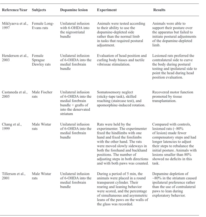
Unilateral 6-hydroxydopamine (6-OHDA) infusion into the medial forebrain bundle (MFB) in rodents is arguably the oldest and most widespread experimental model of PD (Simola et al., 2007). Such lesions impair posture and induce deicits in skilled reaching tasks (Henderson, Watson, Halliday, Heinemann, & Gerlach, 2003; Miklyaeva et al., 1997; Montoya et al., 1991). Sprague Dawley rats lesioned unilaterally in the MFB and tested for postural control showed a signiicant tendency to curve their bodies to the contralateral side
of the lesion while the head was preferably pointed to the ipsilateral side compared with controls (Henderson et al., 2003; Table 3).
Gait impairments have also been shown in animals with similar lesions. In a task in which animals are moved over a horizontal surface and forced to use their forelimbs to regain balance and body posture, animals normally produce compensatory steps. Researchers have reported that MFB-lesioned rats took fewer compensatory steps than controls (Chang, Wachtel, Young, & Kang, 1999). Approximately 80% dopamine depletion in the STR, a level that mimics early PD, is suficient to affect an animal’s ability to adjust posture through compensatory steps (Chang et al., 1999). This task is similar to tasks adopted in human experiments. Parkinson’s disease patients fail in tasks that require postural and gait reorganization, and rats with 6-OHDA-induced MFB lesions present similar dificulties.
Unilateral 6-OHDA lesions of the nigrostriatal system also affect the sensory-motor skills of the paw that is contralateral to the encephalic lesion, leading the animal to use the ipsilateral paw more than the contralateral paw ( Montoya et al., 1991; Simola et al., 2007). Dopamine loss of 50% in the SNpc induces these deicits. Rats tend to choose the ipsilateral paw to achieve their goals in tasks such as the staircase test, which demands excellent forelimb motor skills (Simola et al., 2007). The same preference for the ipsilateral paw has been reported in tasks in which rats are exposed to forced postural adjustments that require the use of their forelimbs. In a skilled reaching task with a movable platform, unilaterally lesioned Long-Evans rats failed to initiate postural adjustments of the limb controlled by the dopamine-depleted side. However, these unilateral behavioral effects can only be seen in partial lesions of the SNpc (Whishaw, Pellis, & Gorny, 1992). Substantial (~95%) reductions of dopamine in the striatum may lead to sensory-motor skill damage in both forelimbs. This result may be attributable to compensatory effects of the dopamine system (Whishaw et al., 1992).
Preference for the ipsilateral paw was also observed in both the cylinder test and sticky tape test. In the cylinder test, the frequency of forelimb leaning on the walls is measured during exploratory behavior in a glass cylinder (Simola et al., 2007). The sticky tape test consists of sticking tape to the animal’s forelimbs and recording the latency to touch and remove the tape. In both tasks, lesioned animals tend to show a preference for the paw on the same side of the lesion by leaning it on the walls or quickly removing the tape, suggesting proprioceptive deicits in the paw affected by the nigrostriatal lesion (Castaneda et al., 2005; Tillerson et al., 2001).
based on genetic manipulation aim to reproduce the dysfunction found in some cases of PD. These studies utilize mutations in parkin, DJ-1, PINK1, a-synuclein, LRRK2, and ATP13A2) (Lee & Trojanowski, 2006). Mice with DJ-1 mutations usually present classic motor disturbances at approximately 11 months of age. Proprioception, however, was reported to affect animals within 5 months. When submitted to the sticky tape test, these mice exhibited an increase in the latency to remove the tape from the paws compared with controls (Meredith et al., 2008). This earlier manifestation of proprioceptive deicits compared with other motor impairments indicates that proprioception may be damaged prior to
the manifestation of other symptoms (Snider et al., 1976). Animal models have provided evidence of the neurocircuitry involved in the pathophysiological mechanisms of proprioceptive damage in PD. Dopaminergic depletion in the basal ganglia helped researchers glimpse the complex network of these structures and the manifestation of proprioceptive loss. The observation of similar gait and postural impairments in nigrostriatal dopamine-depleted subjects reinforces the hypothesis of encephalic proprioceptive loss rather than peripheral damage. What has not yet been addressed is whether the nigral DA loss is involved in peripheral deafferentation.
Reference/Year Subjects Dopamine lesion Experiment Results
Miklyaeva et al.,
1997 Female Long-Evans rats Unilateral infusion with 6-OHDA into the nigrostriatal bundle
Animals were tested according to their ability to use the dopamine-depleted side rather than the normal limb in tasks that required postural adjustment.
Animals were able to support their posture over the apparatus but failed to initiate postural adjustments of the dopamine-depleted limb.
Henderson et al.,
2003 Female Sprague Dawley rats
Unilateral infusion of 6-OHDA into the medial forebrain bundle
Evaluation of head position and curling body biases and tactile vibrissae stimulation.
Lesioned rats preferred the contralateral side to curve the body during postural testing and ipsilateral side to point the head during head position evaluation.
Castaneda et al.,
2005 Male Fischer rats Unilateral infusion of 6-OHDA into the medial forebrain bundle + grafts of into the denervated striatum
Somatosensory neglect (sticky-tape task), skilled reaching (staircase test), and apomorphine-induced rotation.
Recovered motor function promoted by tissue transplantation.
Chang et al.,
1999 Male Wistar rats Unilateral infusion of 6-OHDA into the medial forebrain bundle
Rats were held by the
experimenter. The experimenter ixed the hindlimbs with one hand and ixed the forelimbs with the other hand. The rats were moved slowly sideways in both the forehand and backhand positions. The number of adjusting steps in both directions and with both paws was counted.
Compared with controls, lesioned rats (~80% of lesion) made fewer compensatory steps and had longer latencies to adjust their steps to rebalance the initial posture. Animals with lesions smaller than 80% showed no deicits in this task.
Tillerson et al.,
2001 Male Wistar rats Unilateral infusion of 6-OHDA into the medial forebrain bundle
During a period of 5 min, the animals were placed in a round transparent cylinder. Their rearing and leaning behavior were scored, and the percentage of simultaneous and asymmetric leans of the paws on the walls of the glass was recorded.
Dopamine depletion of ~80% in the striatum caused ipsilateral preference rather than the use of contralateral paws to lean during exploratory behavior.
Cortical degeneration and proprioception
deicits in Parkinson’s disease
Proprioception is a basic condition for planning adequate motor responses that relies on the involvement of premotor and motor cortices. Therefore, dopamine rising from nigrostriatal pathway modulates striatal neurons that regulate the function of the cortical motor area. Dense projections from the primary somatosensory cortex (S1) to the primary motor cortex (M1) reinforce the assumption that a motor cortical mechanism plays a role in proprioceptive modulation (Jacobs & Horak, 2006). Neuroanatomical and electrophysiological indings reported efferent pathways from these two cortices to the putamen, the region most affected by dopaminergic depletion in PD. The putamen promotes subsequent changes in the information lux to the globus pallidus, another structure that plays a crucial role in the motor disturbances seen in PD (Flaherty & Graybiel, 1991). Chemical alterations in this pathway followed by dopamine depletion affect the way peripheral proprioceptive afferents are interpreted, possibly causing disturbances in the ability to correctly estimate member position (Jacobs & Horak, 2006).
Peripheral (tendon) stimulation in PD patients reduces the activation of contralateral somatosensory and premotor lateral cortices, in addition to altering the contralateral posterior cingulate cortex, bilateral prefrontal cortex, and basal ganglia contralateral to the peripheral stimulation (Boecker et al., 1999). Therefore, altered sensory processing might contribute to the motor deicits observed in PD.
Recent studies have shown that a complex interaction exists between the pathways that link cortical regions, the basal ganglia, and the thalamus. The supplementary motor area (SMA) might contribute to proprioceptive loss in PD (Jacobs & Horak, 2006; Seiss et al., 2003). The SMA is a part of the premotor cortex (area 6) that receives proprioceptive and visual afferents from the posterior parietal cortex (areas 5 and 7) and sends descending corticospinal tract projections. This region is considered an indispensable junction for body position perception to understand the requested movement and how it will be performed (Toma et al., 1999). The SMA also likely supports highly abstract visuospatial computations (Leek & Johnston, 2009).
The SMA receives a rich dopaminergic innervation, and a degeneration of this region is observed in the advanced stages of PD (Braak et al., 2002). This area is overactive in dyskinetic patients with PD and might play a central role in the development of dyskinesia induced by dopaminergic stimulation (Bezard, Brotchie, & Gross, 2001). Indeed, in a positron emission tomography study of patients with dyskinetic PD, the degree of overactivation of the SMA, motor cortical areas, and the basal ganglia correlated with the severity of dyskinesia
(Brooks, Piccini, Turjanski, & Samuel, 2000). Moreover, dyskinetic patients showed overactivity of motor and premotor areas, including the SMA, compared with nondyskinetic patients in a single-photon emission computed tomography study (Rascol et al., 1998). Further research is needed to elucidate the interaction between the proprioceptive system and sensorimotor system in the planning and execution of movement.
Final considerations
Proprioceptive loss is not strictly considered a symptom but rather a sign of PD because of its conscious complaint in PD patients. The dificulty of perceiving these deiciencies impairs clinical investigation. Therefore, lack of awareness of the problem and compensatory psychological mechanisms may help patients compensate for mild proprioceptive deicits when executing movement. These occurrences may lead to an underestimation of the presence, prevalence, and magnitude of these alterations.
The involvement of proprioception in PD is important and requires additional exploration to help patients overcome their limitations. Exercise and other physio- and psychotherapeutic strategies may improve proprioception while helping patients deal with their symptoms. The testing of new PD treatment drugs may also improve symptom perception by focusing on proprioception. According to recent evidence of peripheral deafferentation in PD, more studies are necessary to elucidate the magnitude, prevalence, and course of this damage. The relationship between peripheral deafferentation and nigral dopamine loss also needs to be revealed. Dopaminergic impairment in the basal ganglia may also be involved in other neurotransmitter imbalance, such as that involved in nociception, thermal perception, and postural control (Tinazzi et al., 2008). The complex mechanism that leads to this encephalic chemical imbalance of peripheral receptors has yet to be discovered.
Proprioception may be a key to understanding the progression of motor disease pathology. Therefore, studies of proprioceptive changes may help understand the course of proprioceptive alterations and develop strategies that minimize their impact on other systems and diminish the severity of PD symptoms.
References
Abbruzzese, G., & Berardelli, A. (2003). Sensorimotor integration in movement disorders. Movement Disorder, 18, 231-240.
Adamovich, S. V., Berkinblit, M. B., Hening, W., Sage, J., & Poizner, H. (2001). The interaction of visual and proprioceptive inputs in pointing to actual and remembered targets in Parkinson’s disease.
Neuroscience, 104, 1027-1041.
Bendrups, A. P., & McKenzie, J. S. (1974). Suppression of extralemniscal thalamic unit responses by substantia nigra stimulation. Brain Research, 80, 131-134.
Berardelli, A., Rothwell, J. C., Thompson, P. D., & Hallett, M. (2001). Pathophysiology of bradykinesia in Parkinson’s disease. Brain, 124, 2131-2146.
Bezard, E., Brotchie, J. M., & Gross, C. E. (2001). Pathophysiology of levodopa-induced dyskinesia: potential for new therapies. Nature Review Neuroscience, 2, 577-588.
Boecker, H., Ceballos-Baumann, A., Bartenstein, P., Weindl, A., Siebner, H. R., Fassbender, Conrad, B. (1999). Sensory processing in Parkinson‘s and Huntington‘s disease: investigations with 3D H(2)(15)O-PET. Brain, 122, 1651-1665.
Braak, H., Del Tredici, K., Bratzke, H., Hamm-Clement, J., Sandmann-Keil, D., & Rub, U. (2002). Staging of the intracerebral inclusion body pathology associated with idiopathic Parkinson’s disease (preclinical and clinical stages). Journal of Neurology, 249
(Suppl 3), III/1-5.
Brooks, D. J., Piccini, P., Turjanski, N., & Samuel, M. (2000). Neuroimaging of dyskinesia. Annals of Neurology, 47(4 Suppl 1),
S154-158; discussion S158-159.
Castaneda, E., Fleming, S., Paquette, M. A., Boat, K., Moffett, J., Stachowiak, E. K., Stachowiak, M.K. (2005). Assessment of recovery in the hemiparkinson rat: drug-induced rotation is inadequate. Physiology and Behavior, 84, 525-535.
Chang, J. W., Wachtel, S. R., Young, D., & Kang, U. J. (1999). Biochemical and anatomical characterization of forepaw adjusting steps in rat models of Parkinson‘s disease: studies on medial forebrain bundle and striatal lesions. Neuroscience, 88, 617-628.
Chung, K. A., Lobb, B. M., Nutt, J. G., & Horak, F. B. (2010). Effects of a central cholinesterase inhibitor on reducing falls in Parkinson disease. Neurology, 75, 1263-1269.
Contreras-Vidal, J. L., & Gold, D. R. (2004). Dynamic estimation of hand position is abnormal in Parkinson’s disease. Parkinsonism & Related Disorders, 10, 501-506.
De Nunzio, A. M., Nardone, A., & Schieppati, M. (2007). The control of equilibrium in Parkinson’s disease patients: delayed adaptation of balancing strategy to shifts in sensory set during a dynamic task.
Brain Research Bulletin, 74, 258-270.
Dietz, V., & Duysens, J. (2000). Signiicance of load receptor input during locomotion: a review. Gait Posture, 11, 102-110.
Duffau, H., & Capelle, L. (2001). [Functional recuperation following lesions of the primary somatosensory ields. Study of compensatory mechanisms]. Neurochirurgie, 47, 557-563.
Flaherty, A. W., & Graybiel, A. M. (1991). Corticostriatal transformations in the primate somatosensory system. Projections from physiologically mapped body-part representations. Journal of Neurophysiology, 66, 1249-1263.
Flowers, K. A. (1976). Visual “closed-loop” and “open-loop” characteristics of voluntary movement in patients with Parkinsonism and intention tremor. Brain, 99, 269-310.
Goodwin, G. M., McCloskey, D. I., & Matthews, P. B. (1972). The contribution of muscle afferents to kinaesthesia shown by vibration induced illusions of movement and by the effects of paralysing joint afferents. Brain, 95, 705-748.
Gunal, D. I., Nurichalichi, K., Tuncer, N., Bekiroglu, N., & Aktan, S. (2002). The clinical proile of nonmotor luctuations in Parkinson’s disease patients. Canadian Journal of Neurological Science, 29, 61-64.
Hattori, N., Kobayashi, H., Sasaki-Hatano, Y., Sato, K., & Mizuno, Y. (2003). Familial Parkinson’s disease: a hint to elucidate the mechanisms of nigral degeneration. Journal of Neurology, 250 (Suppl 3), III2-10.
Henderson, J. M., Watson, S., Halliday, G. M., Heinemann, T., & Gerlach, M. (2003). Relationships between various behavioural abnormalities and nigrostriatal dopamine depletion in the unilateral 6-OHDA-lesioned rat. Behavior Brain Research, 139, 105-113.
Jacobs, J. V., & Horak, F. B. (2006). Abnormal proprioceptive-motor integration contributes to hypometric postural responses of subjects with Parkinson’s disease. Neuroscience, 141, 999-1009.
Keijsers, N. L., Admiraal, M. A., Cools, A. R., Bloem, B. R., & Gielen, C. C. (2005). Differential progression of proprioceptive and visual information processing deicits in Parkinson’s disease.
TheEuropean Journal of Neuroscience, 21, 239-248.
Keranen, T., Kaakkola, S., Sotaniemi, K., Laulumaa, V., Haapaniemi, T., Jolma, T., Takala, A.(2003). Economic burden and quality of life impairment increase with severity of PD. Parkinsonism & Related
Disorders, 9, 163-168.
Klockgether, T., Borutta, M., Rapp, H., Spieker, S., & Dichgans, J. (1995). A defect of kinesthesia in Parkinson’s disease. Movement Disorder, 10, 460-465.
Koller, W. C. (1984). Sensory symptoms in Parkinson’s disease.
Neurology, 34, 957-959.
Lee, V. M., & Trojanowski, J. Q. (2006). Mechanisms of Parkinson’s disease linked to pathological alpha-synuclein: new targets for drug discovery. Neuron, 52, 33-38.
Leek, E. C., & Johnston, S. J. (2009). Functional specialization in the supplementary motor complex. Nature Review of Neuroscience, 10, 78; author reply 78.
Marsden, C. D. (1982). The mysterious motor function of the basal ganglia: the Robert Wartenberg Lecture. Neurology, 32, 514-539.
Meredith, G. E., Sonsalla, P. K., & Chesselet, M. F. (2008). Animal models of Parkinson’s disease progression. Acta Neuropathologica, 115, 385-398.
Miklyaeva, E. I., Woodward, N. C., Nikiforov, E. G., Tompkins, G. J., Klassen, F., Ioffe, M. E., Whishaw, I.Q. (1997). The ground reaction forces of postural adjustments during skilled reaching in unilateral dopamine-depleted hemiparkinson rats. Behavioural Brain Research, 88, 143-152.
Montoya, C. P., Campbell-Hope, L. J., Pemberton, K. D., & Dunnett, S. B. (1991). The “staircase test”: a measure of independent forelimb reaching and grasping abilities in rats. Journal of Neuroscience Methods, 36, 219-228.
Nakashima, K., Nitta, T., & Takahashi, K. (1992). Recovery functions of somatosensory evoked potentials in parkinsonian patients.
Journal of Neurology Science, 108, 24-31.
Nolano, M., Provitera, V., Estraneo, A., Selim, M. M., Caporaso, G., Stancanelli, A., Santoro, L. (2008). Sensory deicit in Parkinson’s disease: evidence of a cutaneous denervation. Brain, 131, 1903-1911.
Nutt, J. G., & Carter, J. H. (1984). Sensory symptoms in parkinsonism related to central dopaminergic function. Lancet, 2, 456-457.
O’Suilleabhain, P. (2004). Sensory symptoms and sensorimotor distortion Parkinson´s disease. Washington D.C.: CRC Press.
Pastor, M. A., Day, B. L., & Marsden, C. D. (1993). Vestibular induced postural responses in Parkinson’s disease. Brain, 116, 1177-1190.
Rascol, O., Sabatini, U., Brefel, C., Fabre, N., Rai, S., Senard, J. M.,... Cholet, F. (1998). Cortical motor overactivation in parkinsonian patients with L-dopa-induced peak-dose dyskinesia. Brain, 121,
527-533.
Rieu, I., Boirie, Y., Morio, B., Derost, P., Ulla, M., Marques, A., Durif, F. (2010). [The Idiopathic Parkinson’s disease: A metabolic disease?]. Revue Neurologique (Paris), 166, 822-828.
Rocchi, L., Chiari, L., Cappello, A., Gross, A., & Horak, F. B. (2004). Comparison between subthalamic nucleus and globus pallidus internus stimulation for postural performance in Parkinson’s disease. Gait Posture, 19, 172-183.
Schenkman, M. L., Clark, K., Xie, T., Kuchibhatla, M., Shinberg, M., & Ray, L. (2001). Spinal movement and performance of a standing reach task in participants with and without Parkinson disease.
Physical Therapy, 81, 1400-1411.
Seiss, E., Praamstra, P., Hesse, C. W., & Rickards, H. (2003). Proprioceptive sensory function in Parkinson’s disease and Huntington’s disease: evidence from proprioception-related EEG potentials. Experimental Brain Research, 148, 308-319.
Shulman, L. M., Singer, C., Bean, J. A., & Weiner, W. J. (1996). Internal tremor in patients with Parkinson’s disease. Movement Disorder, 11, 3-7.
Simola, N., Morelli, M., & Carta, A. R. (2007). The 6-hydroxydopamine model of Parkinson’s disease. Neurotox Research, 11, 151-167.
Snider, S. R., Fahn, S., Isgreen, W. P., & Cote, L. J. (1976). Primary sensory symptoms in parkinsonism. Neurology, 26, 423-429.
St George, R. J., Nutt, J. G., Burchiel, K. J., & Horak, F. B. (2010). A meta-regression of the long-term effects of deep brain stimulation on balance and gait in PD. Neurology, 75, 1292-1299.
Tatton, W. G., & Lee, R. G. (1975). Evidence for abnormal long-loop relexes in rigid Parkinsonian patients. Brain Research, 100, 671-676.
Tillerson, J. L., Cohen, A. D., Philhower, J., Miller, G. W., Zigmond, M. J., & Schallert, T. (2001). Forced limb-use effects on the behavioral and neurochemical effects of 6-hydroxydopamine.
Journal of Neuroscience, 21, 4427-4435.
evoked potentials. Pain, 136, 117-124.
Toma, K., Honda, M., Hanakawa, T., Okada, T., Fukuyama, H., Ikeda, A., Shibasaki, H. (1999). Activities of the primary and supplementary motor areas increase in preparation and execution of voluntary muscle relaxation: an event-related fMRI study.
Journal of Neuroscience, 19, 3527-3534.
Varraine, E., Bonnard, M., & Pailhous, J. (2002). Interaction between different sensory cues in the control of human gait. Experimental Brain Research, 142, 374-384.
Vaugoyeau, M., Viel, S., Assaiante, C., Amblard, B., & Azulay, J. P. (2007). Impaired vertical postural control and proprioceptive integration deicits
in Parkinson’s disease. Neuroscience, 146, 852-863.
Voight, M. I., & Cook, G. (2001). Impaired neuromuscular control: reactive neuromuscular training. In W. E. Prentice & M. I. Voight (Eds.), Techniques in musculoskeletal rehabilitation (pp. 213-240).
New York: McGraw-Hill.
Whishaw, I. Q., Pellis, S. M., & Gorny, B. P. (1992). Skilled reaching in rats and humans: evidence for parallel development or homology.
Behavioural Brain Research, 47, 59-70.