House dust fungal communities’ characterization: a double take on the six by sixty
by six project (6x60x6)
Raquel Amaro - [email protected]
Departamento de Biologia, Universidade de Aveiro, Aveiro, Portugal Sónia D. Coelho - [email protected]
Departamento de Biologia & CESAM, Universidade de Aveiro, Portugal M. Ramiro Pastorinho - [email protected]
Departamento de Ciências Médicas, Faculdade de Ciências da Saúde, Universidade da Beira Interior Luís Taborda-Barata - [email protected]
Departamento de Ciências Médicas, Faculdade de Ciências da Saúde, Universidade da Beira Interior Maria Assunção Vaz-Pato - [email protected]
Departamento de Ciências Médicas, Faculdade de Ciências da Saúde, Universidade da Beira Interior Marisa Monteiro - [email protected]
LABSED, Faculdade de Engenharia, Universidade da Beira Interior Miguel Nepomuceno - [email protected]
LABSED, C-made, Faculdade de Engenharia, Universidade da Beira Interior João C. G. Lanzinha - [email protected]
LABSED, C-made, Faculdade de Engenharia, Universidade da Beira Interior João Paulo Teixeira - [email protected]
Instituto Nacional de Saúde Dr. Ricardo Jorge, Departamento de Saúde Ambiental Cristiana C. Pereira - [email protected] Instituto Nacional de Saúde Dr. Ricardo Jorge, Departamento de Saúde Ambiental Ana C.A. Sousa - [email protected]
CICS-UBI Centro de Investigação em Ciências da Saúde, Universidade da Beira
Abstract
Fungi are a group microbes, that are found with particular incidence in the indoor environment. Their direct toxicity or capability of generating toxic compounds has been associated with a large number of adverse health effects, such as infectious diseases, allergies and other toxic effects. Given that in modern society people spend a large part of their time indoors; fungal communities’ characterization of this environmental compartment assumes paramount importance in the comprehension of health effects. House dust is an easy to obtain, time-integrative matrix, being its use in epidemiological studies on human exposure to environmental contaminants highly recommended. Furthermore, dust can carry a great variety of fungal content that undergoes a large number of processes that modulate and further complexify human exposure. Our study aims to quantify and identify the fungal community on house dust samples collected using two different methodologies (an approach not often seen in the literature): active (vacuum cleaner bags) and passive sampling (dust settled in petri dishes). Sampling was performed as part of the ongoing 6X60X6 Project in which six houses from Covilhã (Portugal), with building dates representative of six decades, were studied for a period of sixty days.
Keywords
House dust fungal communities’ characterization:
a double take on the six by sixty by six project
(6x60x6)
1. Introduction
In modern society, most people spend a large part of their time indoors, being exposed to a broad number of contaminants, which may come from the outdoors or be locally generated as the result of household activities and building materials as well as from the decay of consumer products [1]. The built environment air pollution is considered a major cause of morbidity and mortality all over the world [2] and as such the study of indoor environmental quality is of great importance.
Fungi are a group of well-known microbes, that are easily found in all types of environments [3] with particular incidence in the indoor environment. Their direct toxicity or capability of generating toxic compounds (e.g., mycotoxins and harmful antigens) has been associated with a large number of adverse health effects in humans, such as infectious diseases, allergies and other toxic effects [4]. Fungi produce tiny spores with those smaller than 10 µm being particularly hazardous to human health, as they can enter the respiratory tract and reach the alveoli (the gaseous exchange areas of the lung), which may lead to respiratory infections and allergic reactions [5,6].
Spores can be suspended in the air, deposited on various surfaces and included within different matrices such as house dust [7]. This matrix results essentially from materials tracked indoors and the settling of airborne particles, a process that can take weeks or even months (especially the latter), being therefore regarded as a time-integrated sample [8,9]. Furthermore, house dust is an easy sample to obtain and its use in epidemiological studies on human exposure to environmental contaminants, has been highly recommended [10,11]. Its relevance as an important exposure pathway is exacerbated by the fact that in general, adults may ingest 50 mg of dust per day and inhale 0.8 mg, and children (a risk group) may ingest 100 mg per day and inhale 2 mg (see for example Coelho et al., 2014 [12]).
Dust can carry a great variety of fungal content - intact fungal conidia, spores, hyphae and other. This microbial content undergoes processes of deposition, removal, proliferation, death and degradation, contributing towards the content and diversity of fungi in this type of sample [8].
To date several papers have been published on the fungal community in house dust samples (see e.g. the review by Rintala et al.[8]) but there is still limited information on this topic, particularly for Portuguese households. Furthermore, comparisons between sampling strategies are scarce in the literature. Hence, our study aims to quantify and identify the fungal community on house dust samples collected using two different methodologies: active and passive sampling. For this purpose we analysed dust collected from vacuum cleaner bags and dust settled in petri dishes. The surveyed houses are part of the ongoing 6X60X6 Project in which six houses from Covilhã (Portugal), with building dates representative of six decades, were studied for a period of sixty days.
2. Materials and Methods
2.1. Sampling
Under the framework of the 6x60x6 project, six houses built from 1960 to 2010 in the urban area of Covilhã were studied for a period of sixty days. Covilhã is located in the interior center of Portugal in the Cova da Beira Region at an average altitude of 7000m. For decades the Municipality had a very strong textile industry, and to this day Covilhã is synonym of
fabrics. However, the crisis experienced by the sector in the 1980’s, led to a profound reconversion of the local economy, being led nowadays by the tertiary sector [13]. The houses were selected by convenience and each participant signed an informed consent and completed a questionnaire about the household characteristics. At each house the master bedroom temperature and humidity values were recorded continuously using a temperature and relative humidity data logger (EasyLog - EL-GFX-2).
During the period of the study the wind regime varied. May was characterized by a dominant wind direction from NW with an average speed of 6.3 Km/h. In June the predominant wind direction was WNW with average speed of 3.9 Km/h, shifting in July to a W dominance an d an average speed of 3.3 Km/h [14].
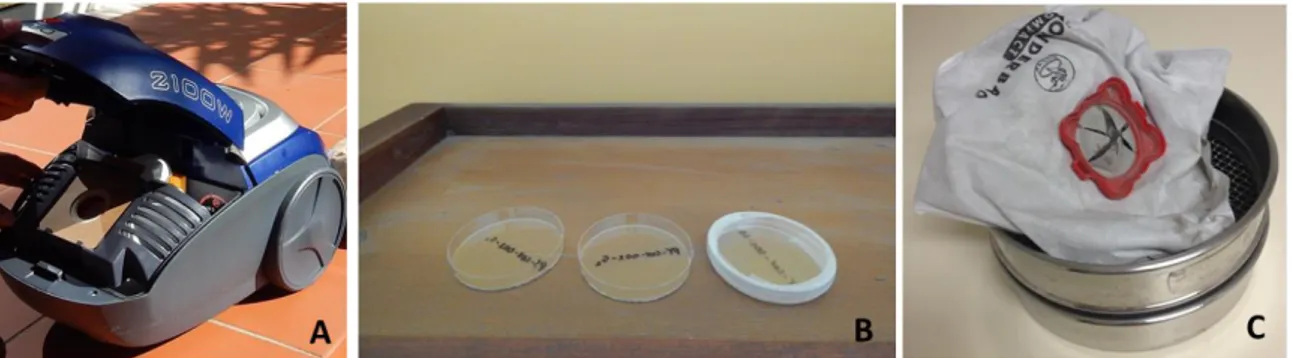
House dust samples were collected by means of active and passive sampling. Active sampling included the use of the household vacuum cleaner. At the beginning of the study a new vacuum cleaner bag (Wonderbag Compact WB 305120) was fitted and the participants were asked to vacuum only inside the house (excluding e.g. garage and cars). At the end of the 60 days the bag was removed, sealed and transported to the CICS-UBI laboratory (Figure 1).
Figure 1. House dust sampling. (A) Household vacuum cleaner used for active sampling; (B) Petri dishes placed on the top of a shelf; (C) Vacuum cleaner bag retrieved after 60 days of sampling.
Passive sampling was performed in the master bedroom using sterile glass petri dishes that remained unlidded at the selected sampling sites during 60 days. The petri dishes were placed at sites that minimized possible disturbances by the normal routine of the inhabitants (e.g. on top of shelves, figure 1B). At the end of the sampling period the dishes were retrieved by the researchers, sealed and transported to the reference laboratory for fungal analysis at the National Institute of Health (INSA), in thermal bags and processed immediately upon arrival. Table 1 describes the characteristics of the houses and the sampling details.
2.2. Treatment of samples
In the laboratory the vacuum cleaner bags were opened and the samples sieved twice through stainless steel sieves of decreasing mesh (5 mm and 500 μm) to remove fibrous material and large pieces of debris in order to obtain a suitable degree of homogeneity. Samples were then stored in polyethylene tubes and transported to the INSA laboratory where they were analysed.
2.3. Culture Methods: Fungal Culture and Identification
For vacuum cleaner samples, we followed the procedure proposed by Verhoeff and collaborators [15]. Three different culture methods were used, in order to achieve an optimal growth for analysis purposes: i) Direct plating: 30 mg representative aliquot of sieved dust was plated directly onto Malt Extract Agar with 1% cloramphenicol (MEA) plates using a sterile plastic spreader; ii) Suspension: 100 mg representative aliquot of sieved dust was mixed with 5 ml of liquid Sabouraud medium. The solution was shaken for 10 minutes. Subsequently, 100 µl of the prepared suspension was plated onto MEA plates with a sterile plastic spreader; iii) Dilution: 1 ml of the previous suspension was diluted in 9 ml of liquid Sabouraud and shaken
for 10 minutes. Afterwards, 100 µl of the diluted suspension was plated onto MEA plates through a sterile plastic spreader.
As a measure of quality assurance, duplicates were made for each method/sample. All samples were incubated at 25±3ºC for 72 ± 3 hours.
For passive samples, each petri dish was washed with 1 mL of liquid culture medium – sabouraud with 1% cloramphenicol, and the obtained suspension was used for seeding over malt extract agar (MEA) and dichloran glycerol agar (DG-18) plates. Five plates of MEA and 5 plates of DG-18 were seeded with 100 uL of the suspension each, and incubated for 72h ± 3h at 25ºC±3ºC.
Quantification was performed by naked eye count. Fungal identification was performed either on the original sampling media (MEA) plates or after subculturing procedures, whenever colony isolation and growth observation were needed. Subculture was made on MEA plates and incubated, at 25 ± 3 ºC, for periods ranging from 3 days to 3 weeks (Figure 2).
Fungal samples were mounted on lactophenol blue and visualized under optical microscope and identification of fungal colonies was based upon phenotypic characteristics and followed standard mycological procedures according to their micro and macro-morphological characteristics [16].
Figure 2. Cladosporium sp., 20 days growth on MEA plate
3. Results and Discussion
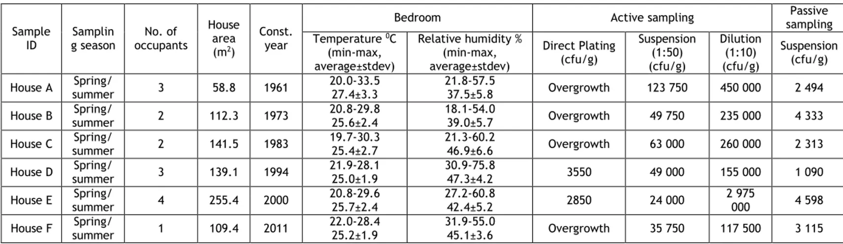
The total number of cultivable fungi found in the analysed dust samples along with some of the house characteristics are depicted in Table 1. The average temperature was 25ºC in the majority of the houses whereas the relative humidity varied from 37.5% in house A to 47.3% in house D. Despite such differences in relative humidity and in the number of CFUs (Table 1) there was no significant correlation between the average humidity found in bedrooms and the number of CFUs at the same location (Spearman correlation, p=0.242).
Table 1. Characteristics of the surveyed houses with the indication of: sampling season, number of occupants, area (m2),construction year (Const. year), temperature (0C) and relative humidity (%) registered in the master bedroom
(min-max, average ± stdev) and the number of total Colony Forming Units (CFU) using active and passive sampling methods. For the active sampling method the results are shown for the three different culture techniques used
Sample
ID g season Samplin occupants No. of
House area (m2)
Const. year
Bedroom Active sampling sampling Passive
Temperature 0C (min-max, average±stdev) Relative humidity % (min-max, average±stdev) Direct Plating (cfu/g) Suspension (1:50) (cfu/g) Dilution (1:10) (cfu/g) Suspension (cfu/g) House A summer Spring/ 3 58.8 1961 20.0-33.5 27.4±3.3 21.8-57.5 37.5±5.8 Overgrowth 123 750 450 000 2 494 House B summer Spring/ 2 112.3 1973 20.8-29.8 25.6±2.4 18.1-54.0 39.0±5.7 Overgrowth 49 750 235 000 4 333 House C summer Spring/ 2 141.5 1983 19.7-30.3 25.4±2.7 21.3-60.2 46.9±6.6 Overgrowth 63 000 260 000 2 313 House D summer Spring/ 3 139.1 1994 21.9-28.1 25.0±1.9 30.9-75.8 47.3±4.2 3550 49 000 155 000 1 090 House E summer Spring/ 4 255.4 2000 20.8-29.6 25.7±2.4 27.2-60.8 42.4±5.2 2850 24 000 2 975 000 4 598 House F summer Spring/ 1 109.4 2011 22.0-28.4 25.2±1.9 31.9-55.0 45.1±3.6 Overgrowth 35 750 117 500 3 115
A
B
C
(direct plating, suspension and dilution).
When comparing the two sampling methods, there are clear differences between them, with a higher amount of CFU per gram of dust when dust is collected by means of active sampling. Such differences are easily explained when one considers the differences between the two methods: passive sampling reflects only the airborne fungi from the main bedroom settled in the petri dish during 60 days, whereas the vacuum cleaner samples concern the entire house and even though the sampling period was the same (60 days), the collected dust might corresponded to a longer period as for example carpets and rugs tend to trap dust for several months.
Overall our results are consistent with other studies on fungal communities’ in house dust (Table 2).
Table 2. Comparison of the total amount of fungi detected in different surveys worldwide. Total CFU/g: Total number of Colony Forming Units (CFU) per gram of dust. *average values. na: information not available
Location Sampling and culture method N0. samples Total
CFU/g Reference
Boston, USA Portable canister vacuum cleaner with
a cellulose thimble; suspension na 355 756*
Chao et al., 2002 [17]
Boston, USA
Portable canister vacuum cleaner with a cellulose extraction thimble;
suspension 397 200 473* Chew et al., 2003 [18] Baden-Württemberg, Germany
Vacuum cleaner with special filter
holder and gelatin filter; suspension 397
1 500 – 1 235
000
Jovanovic et al., 2004 [19]
Brittany, France Dustream Collector sampler-fitted
vacuum cleaner; Suspension 133
1 000 – 3 800
000
Dallongeville et al., 2015 [9]
Covilhã, Portugal Vacuum cleaner bags; suspension 6 24 000 –
123 750 This study
Covilhã, Portugal Passive sampling; suspension 6 1 090 -
4 598 This study
Generally, the most frequent fungi genera found in all samples were Alternaria sp.,
Aspergillus sp., Cladosporium sp., Penicillium sp., and yeasts (Figure 3). Aspergillus sp. and Penicillum sp. are found both in outdoor and indoor environments, where they are considered
common fungi species [20]. Nevertheless, these genera also comprise species that are important allergic agents with implications in human respiratory health [5,8].
In a previous study conducted by our team in two Portuguese cities (Aveiro and Coimbra, n= 28), Aspergillus and Penicillium were also the most abundant genera found [11]. However,
Alternaria sp., present in all the houses in the present study, was not detected in our
previous study. Furthermore, when comparing samples obtained by active sampling in both studies, a higher diversity in the present study is evident. Such outcome is probably a consequence of an optimization of the protocol used in the current study, especially the aspect concerning dust samples being processed immediately after collection (instead of being preserved at -20ºC).
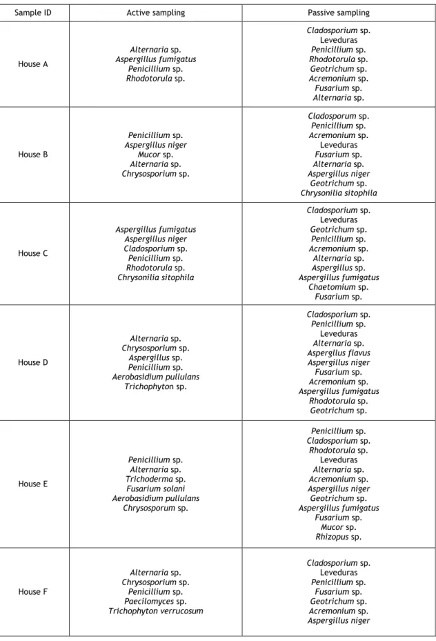
Table 3. Identification of fungi found at each house using dust samples from the vacuum cleaner bag (active sampling) and from the deposited dust on petri dishes (passive sampling).
Sample ID Active sampling Passive sampling
House A Alternaria sp. Aspergillus fumigatus Penicillium sp. Rhodotorula sp. Cladosporium sp. Leveduras Penicillium sp. Rhodotorula sp. Geotrichum sp. Acremonium sp. Fusarium sp. Alternaria sp. House B Penicillium sp. Aspergillus niger Mucor sp. Alternaria sp. Chrysosporium sp. Cladosporum sp. Penicillium sp. Acremonium sp. Leveduras Fusarium sp. Alternaria sp. Aspergillus niger Geotrichum sp. Chrysonilia sitophila House C Aspergillus fumigatus Aspergillus niger Cladosporium sp. Penicillium sp. Rhodotorula sp. Chrysonilia sitophila Cladosporium sp. Leveduras Geotrichum sp. Penicillium sp. Acremonium sp. Alternaria sp. Aspergillus sp. Aspergillus fumigatus Chaetomium sp. Fusarium sp. House D Alternaria sp. Chrysosporium sp. Aspergillus sp. Penicillium sp. Aerobasidium pullulans Trichophyton sp. Cladosporium sp. Penicillium sp. Leveduras Alternaria sp. Aspergllus flavus Aspergillus niger Fusarium sp. Acremonium sp. Aspergillus fumigatus Rhodotorula sp. Geotrichum sp. House E Penicillium sp. Alternaria sp. Trichoderma sp. Fusarium solani Aerobasidium pullulans Chrysosporum sp. Penicillium sp. Cladosporium sp. Rhodotorula sp. Leveduras Alternaria sp. Acremonium sp. Aspergillus niger Geotrichum sp. Aspergillus fumigatus Fusarium sp. Mucor sp. Rhizopus sp. House F Alternaria sp. Chrysosporium sp. Penicillium sp. Paecilomyces sp. Trichophyton verrucosum Cladosporium sp. Leveduras Penicillium sp. Fusarium sp. Geotrichum sp. Acremonium sp. Aspergillus niger
Regarding the taxon characterization, the passive sampling method proved to be more effective for the identification of fungi found in each sample (Table 3). Such results are foremost a consequence of the lower counts of fungi obtained with this method, thus enabling a greater rate of success in obtaining isolated and identifiable colonies. Also, suspension procedures may lead to breakage of suspended fungal spores, preventing their growth. Furthermore the low diversity of fungi found using the active method might be a consequence of the complex matrix that we are dealing with. Besides fungi this dust also includes a large variety of other biological and chemical contaminants that may work as inhibitors and affect the viability of some fungal species.
The passive sampling technique using petri dishes provides a useful, simple and cost effective alternative for the fungal characterization of a particular set of the indoor environment and it should be considered in future monitoring studies.
Conclusions and future perspectives
House dust is a repository and concentrator of many contaminants including biological ones such as fungi. The obtained results showed that house dust samples obtained through active sampling are very complex and should not be assessed by direct plating. Based on the results from the suspension and dilution methods we recommend the use of the dilution method. When aiming to analyse specific locations inside a house, passive sampling using Petri dishes is a cost-effective and useful technique and should be used as a complement to vacuum cleaner bags (that are able to integrate the dust borne fungi of the entire household).
A future sampling campaign will be performed in the studied houses during winter in order to evaluate the seasonal trends in dustborn fungi. Furthermore, the obtained results (in terms of species distribution and richness) will be correlated with the respiratory health of the participants and a set of recommendations in order to reduce exposure will be prepared.
Acknowledgments
The authors also wish to deeply acknowledge all the volunteers’ that participated in this study.
This work was supported by European Funds through COMPETE and by National Funds through the Portuguese Science Foundation (FCT) within project PEst-OE/SAU/UI0709/2014. Ana C. A. Sousa and Sónia D. Coelho acknowledge FCT for the grants SFRH/BPD/65884/2009 and SFRH/ BD/78168/2011 (supported by funding from the Human Potential Operational Programme POPH, inscribed in the National Strategic Reference Framework and partially subsidized by the European Social Fund).
References
[1] WHO (2010). WHO guidelines for indoor air quality : Selected pollutants. p. 454,World Health Organization Regional Office for Europe.
[2] WHO (2009). WHO guidelines for indoor air quality: dampness and mould. p. 228, World Health Organization Regional Office for Europe.
[3] M.T. Madigan; J.M. Martinko; P.V. Dunlap; D.P. Clark. Brock Biology of Microorganisms,
12th edition. p. 1061, Pearson Education, 2009.
[4] Méheust D.; Le Cann P.; Reboux G.; Millon L.; Gangneux J-P.: "Indoor fungal
contamination: health risks and measurement methods in hospitals, homes and workplaces" Crit Rev Microbiol, Vol. 40 n°3 (2014), ISSN 1549-7828, pp. 248–260.
[5] Araujo R.; Cabral J.P.Chapter 7 – Fungal air quality in medical protected environments. In A. Kumar, Air Quality. p. 390, Sciyo, 2010.
[6] Stetzenbach L.D.; Buttner M.P.; Cruz P.: "Detection and enumeration of airborne biocontaminants" Curr Opin Biotechnol, Vol 15 n°3 (2004), ISSN 0958-1669, pp. 170–174. [7] Nevalainen A.; Täubel M.; Hyvärinen A.: "Indoor fungi: companions and contaminants"
Indoor Air, Vol 25 nº 2 (2015), ISSN 1600-0668, pp.125–156.
[8] Rintala H.; Pitkäranta M.; Täubel M.: “Microbial communities associated with house dust”
Adv Appl Microbiol, Vol 78 (2012), ISSN 0065-2164, pp. 75-120.
[9] Dallongeville A.; Le Cann P.; Zmirou-Navier D.; Chevrier C.; Costet N.; Annesi-Maesano I.; et al.: "Concentration and determinants of molds and allergens in indoor air and house dust of French dwellings" Sci Total Environ, (2015), ISSN 1879-1026.
[10] Sousa A.C.; Takahashi S.; Tanabe S.: "Organic Contaminants in House Dust" Curr Org
Chem, Vol 18 n°17 (2014), ISSN 1875-5348, pp. 2181.
[11] Sousa A.C.; Almeida J.R.S.L.; Pereira C.C.; Ramiro Pastorinho M.; Pereira Â.M.C.; Nogueira A.J.a.; et al.: "Characterization of fungal communities in house dust samples collected from central Portugal-a preliminary survey" J Toxicol Environ Health, A Vol 77 n°14-16 (2014), ISSN 1087-2620, pp. 972–982.
[12] Coelho S.D.; Sousa A.C.A.; Isobe T.; Tanabe S.; Nogueira A.J.A.: "Flame Retardants in Indoor Dust - A Review on the Levels of Polybrominated Diphenyl Ethers and Hexabromocyclododecanes" Curr Org Chem, Vol 18 (2014), ISSN 1087-2620, pp. 2218–2230. [13] PORDATA, Base de Dados Portugal Contemporâneo.
http://www.pordata.pt/DB/Municipios/Ambiente+de+Consulta/Tabela (24/10/2015).
[14] GOA-UVa, GOA-UVa in situ measurement station. http://webx.ubi.pt/~goa (26/10/2015).
[15] Verhoeff A.P.; Hoekstra E.S.; Samson R.A.; Flannigan B.; Flannigan M.E.; Adan O.C.G.: "Fungal propagules in house dust.1. Comparison of analytic methods and their value as estimators of potential exposure" Heal Implic fungi indoor Environ, Vol 29 (1994), ISBN 0-444-81997-5, pp. 49–63.
[16] F. Fisher; N.B. Cook. Fundamentals of Diagnostic Mycology. p. 1-11, Saunders, 1998. [17] Chao H.J.; Milton D.K.; Schwartz J.; Burge H.A.: "Dustborne fungi in large office buildings" Mycopathologia, vol 154 nº 2 (2002), ISSN 1573-0832, pp. 93–106.
[18] Chew G.L.; Rogers C.; Burge H. A.; Muilenberg M.L.; Gold D.R.: "Dustborne and airborne fungal propagules represent a different spectrum of fungi with differing relations to home characteristics" Allergy, Vol 58 nº 1 (2003), ISSN 1398-9995, pp. 13–20.
[19] Jovanovic S.; Felder-Kennel A.; Gabrio T.; Kouros B.; Link B.; Maisner V.; et al.: "Indoor fungi levels in homes of children with and without allergy history" Int J Hyg Environ Health, Vol 207 nº 4 (2004), ISSN 1438-4639, pp.369–378.
[20] Fairs A.; Wardlaw A.J.; Thompson Jr.; Pashley C.H.: "Guidelines on Ambient Intramural Airborne Fungal Spores" Investig Allergol Clin Immunol, Vol 20 n°6 (2010), pp.490–498.