Luís Carlos Sá Loureiro
janeiro de 2015
Functional characterization of mitochondrial
suspensions by cytofluorimetric techniques
towards the screening of mitochondrial
target drugs
UMinho|20
15
Luís Carlos Sá Lour
eir
o
Functional characterization of mitochondrial suspensions by cytofluorimetric techniques tow
ards t
he screening of mitochondrial t
arget drugs
Universidade do Minho
Luís Carlos Sá Loureiro
janeiro de 2015
Dissertação de Mestrado
Mestrado em Bioquímica Aplicada
Functional characterization of mitochondrial
suspensions by cytofluorimetric techniques
towards the screening of mitochondrial
target drugs
Universidade do Minho
Escola de Ciências
Trabalho efetuado sob a orientação da
Professora Doutora Maria Manuela Sansonetty
Gonçalves Côrte-Real
e do
ii
DECLARAÇÃO
Nome: Luís Carlos Sá Loureiro
Endereço eletrónico: luis_saloureiro@hotmail.com Telefone: + 351 916252928
Número do Bilhete de Identidade: 14015811
Título da dissertação de mestrado:
Functional characterization of mitochondrial suspensions by cytofluorimetric techniques towards the screening of mitochondrial target drugs
Orientadores: Professora Doutora Maria Manuela Sansonetty Gonçalves Côrte-Real e Professor Doutor Hernâni Varanda Gerós
Ano de conclusão: 2015
Designação do Mestrado: Mestrado em Bioquímica Aplicada
1. É AUTORIZADA A REPRODUÇÃO INTEGRAL DESTA DISSERTAÇÃO APENAS PARA EFEITOS DE INVESTIGAÇÃO, MEDIANTE DECLARAÇÃO ESCRITA DO INTERESSADO, QUE A TAL SE COMPROMETE;
Universidade do Minho, ___/___/______
iii
Acknowledgements
Durante o mestrado em Bioquímica Aplicada foram muitas as pessoas que me ajudaram e fizeram com que o caminho se torna-se mais fácil, mais simples. Foram dois anos de crescimento não só académico como pessoal.
Agradeço aos pilares da minha vida, os meus pais, não só construíram a melhor família do mundo, como sempre lutaram pelo melhor para mim. Todos os valores que me transmitiram, a integridade na educação que me deram, todos os esforços para que hoje possa ter o que tenho. Um muito obrigado por tudo.
Estou eternamente grato à minha conselheira, ao meu auxílio, o meu exemplo. Uma irmã sempre disposta a ajudar-me em tudo que podia.
Aos meus avós, que não passam um dia sem saber se tudo está a correr bem, se vão bem os estudos, se me alimento, se tenho dinheiro. O carinho que a palavra “avós” por si só transmite, não é suficiente para demonstrar o que vocês são.
Agradeço à minha Madrinha, aos meus tios/tias e primos/primas, somos realmente uma família especial.
Um obrigado aos meus amigos, a todos eles. Estiveram sempre do meu lado neste meu percurso com tantos altos e baixos a nível pessoal. Tenho que destacar a minha grande família escutista (os meus exploradores) e os meus grandes companheiros destes longos anos, André, Cátia, Ilda, Marília e Mário.
Agradeço aos meus fantásticos orientadores, Professora Manuela Côrte-Real e Professor Hernâni Gerós, por todo o apoio, prestabilidade, motivação, compreensão e amizade ao longo deste trabalho. Agradeço-lhes as oportunidades que me deram de crescer tanto a nível académico em tão pouco tempo. O meu muito obrigado.
A todo os colegas de laboratório, quer da Micro I como do Laboratório de Biologia Vegetal, um muito obrigado por toda a ajuda prestada.
Se é possível dedicar este meu trabalho a alguém, faço-o à minha prima. Edina, foste e és o exemplo da força que temos que ter para enfrentar todos os desafios que a vida nos propõe. Perder-te foi muito difícil, mas é por ti que fiz este meu ultimo esforço. Muito muito obrigado por tudo.
v
Abstract
Besides its role in respiration, mitochondria control other key metabolic pathways, cell proliferation and regulated cell death, with a crucial role in the regulation of the intrinsic apoptotic pathway. Recent studies have focused on the screen for anti-cancer agents that induce apoptosis through permeabilization of the mitochondrial outer membrane, leading to the cytosolic release of pro-apoptotic proteins and to the activation of the caspase cascade and consequently to cell dismantling.
In the present study, staining protocols were developed and optimized aiming at the structural and functional characterization of isolated mitochondrial populations from yeast cells and rat liver by flow cytometry. Mitotracker Green and Nonyl Acridine Orange were used to measure changes in mitochondrial mass, 3,3'-Dihexyloxacarbocyanine Iodide and Mitotracker Red CMXROS to monitor the electrical
potential of the inner membrane (Δψm), Mitotracker Red CM-H2XROS and
Dihydroethidium to evaluate the accumulation of ROS, and 2',7'-Bis-(2-Carboxyethyl)-5-(and-6)-Carboxyfluorescein and Acetoxymethyl Ester (BCECF-AM) to measure the pH of the mitochondrial matrix. Subsequent flow cytometry studies were designed to evaluate the effect in mitochondria function of two well-known apoptosis inducers in both mammalian cell lines and yeast cells, namely bovine lactoferrin (bLf) and acetate, and to understand whether or not they act directly on mitochondria independently of
upstream cellular pathways.The results showed that both bLf and acetate promoted
changes in mitochondrial mass, matrix mitochondrial pH, Δψm and ROS levels of rat liver mitochondria. Moreover, the assessment of the activity of the complexes of electron transport chain (ETC) showed that bLf and acetate affect their activity and trigger mitochondrial swelling. These results are particularly interesting since the two compounds appear to induce in isolated mitochondria mainly the same alterations as those occurring in whole cells, and which have been proposed to be involved in the induction of cell death. Altogether, this study show that flow cytometry is a valuable tool to study key mitochondrial functional parameters and to screen for mitochondria-targeted drugs, including the anticancer agents, such as acetate and bLf.
vii
Resumo
Para além do seu papel na respiração celular, as mitocôndrias controlam outros aspetos chaves do metabolismo, bem como a proliferação celular e a morte celular regulada, tendo um papel crucial na regulação da via apoptótica intrínseca. Estudos recentes têm-se focado na procura de agentes anticancerígenos capazes de induzir apoptose mediada pela permeabilização da membrana mitocondrial externa, conduzindo à libertação de proteínas pró-apoptóticas e à ativação da cascata de caspases e consequente desmantelamento celular. No presente estudo, foram desenvolvidos e otimizados protocolos de marcação com vista à caracterização estrutural e funcional por citometria de fluxo de populações de mitocôndrias isoladas de células de levedura e de hepatócitos de rato. As sondas Mitotracker Green e laranja de nonil-acridina foram usadas para medir variações da massa mitocondrial, o iodeto de 3,3'-Dihexiloxacarbocianina e o Mitotracker Red CMXROS para medir o potencial
eléctrico da membrana mitocondrial interna (Δψm), o Mitotracker Red CM-H2XROS e
dihidroetídeo para avaliar a acumulação de ROS e o acetoximetil éster de (2',7'-Bis-(2-Carboxietil)-5-(and-6)-carboxifluoresceína e(BCECF-AM) para medir o pH da matriz mitocondrial. Os estudos subsequentes de citometria de fluxo foram desenhados para avaliar o efeito de diferentes drogas na função mitocondrial, em particular da lactoferrina bovina (bLf) e do acetato, dois reconhecidos indutores de apoptose, no sentido de se compreender se atuam diretamente na mitocôndria independentemente de outras vias celulares a montante. Os resultados mostraram que tanto a bLf como o acetato promovem alterações na massa mitocondrial, pH da matriz mitocondrial, Δψm e nos níveis de ROS em mitocôndrias isoladas de fígado de rato. Além disso, a avaliação da atividade dos complexos da cadeia de transportadora de eletrões (ETC) mostrou que a bLf e o acetato afetam a sua atividade e desencadeiam a tumefação mitocondrial. Estes resultados são particularmente interessantes uma vez que os dois compostos parecem induzir, em mitocôndrias isoladas as mesmas alterações que ocorrem em células inteiras, e que têm sido propostas como estando envolvidas na indução da morte celular. No seu conjunto, este estudo mostra que a citometria de fluxo é uma ferramenta valiosa para o estudo da funcionalidade mitocondrial e para o rastreio de drogas que têm como alvo a mitocôndria, incluindo agentes anticancerígenos, tais como a bLf e o acetato.
ix
Index
Acknowledgements ... iii Abstract ... v Resumo ... vii Index ... ix Abbreviations ... xi Introduction ... 131. Mitochondria in life and death... 13
2. Role, features, and organization of the mitochondria ... 14
2.1. The mitochondrial transmembrane electrochemical potential (Δp) ... 15
2.2. Accumulation of reactive oxygen species ... 16
2.3. Mitochondrial matrix pH ... 17
2.4. Mitochondrial calcium storage ... 19
3. The role of mitochondria in regulated cell death ... 19
4. Mitochondria as cellular target for cancer therapy ... 23
5. Drug mitochondrial targets in cancer therapy ... 24
Aims ... 26
Materials and methods ... 27
Reagents ... 27
Yeast strains and growth conditions ... 27
Isolation and purification of mitochondria from yeast cells ... 27
Isolation and purification of mitochondria from rat liver ... 28
Western Blot ... 29
Fluorochrome solutions ... 29
Flow cytometry analysis ... 30
Enzymatic assays ... 30
Succinate dehydrogenase assay ... 30
NADH-cytochrome c reductase ... 31
Cytochrome c oxidase ... 31
Mitochondrial Swelling Assay ... 31
Results... 32
Functional studies of mitochondrial suspensions by flow cytometry ... 32
Characterization of the purity of mitochondrial fractions ... 32
x
Response of resting mitochondria from rat liver to ADP, oligomycin and CCCP ... 35
Response of mitochondria from rat liver to energization with succinate ... 36
Effect of bovine lactoferrin (bLf) on mitochondrial function ... 38
Effect of acetate on mitochondrial function ... 42
Discussion ... 46
Isolated mitochondria from yeast and rat liver were structurally intact and functionally active ... 46
Flow cytometry revealed a valuable tool to study key mitochondrial functional parameters ... 47
BLf and acetate induce loss of mitochondrial mass ... 50
BLf and acetate promote mitochondrial dysfunction ... 51
Concluding Remarks ... 53
xi
Abbreviations
ACD - Accidental cell death ADP - Adenosine diphosphate
ANT - Adenine nucleotide translocase ATP - Adenosine triphosphate
BCECF-AM - 2', 7'-Bis-(2-Carboxyethyl)-5-(and-6)-Carboxyfluorescein, Acetoxymethyl Ester
bLf – Bovine lactoferrin COX – Cytochrome c oxidase cyt c - Cytochrome c
DHE - Carbonyl cyanide m-chlorophenyl hydrazine
DiOC6(3) - Dihexyloxacarbocyanine Iodide
DNA – Deoxyribonucleic acid DTT – Dithiothreitol
EGTA – Ethylene glycol tetraacetic acid ER - Endoplasmic reticulum
ETC - Electron transport chain
IMM - Inner mitochondrial membrane IMS – Intermembrane space
Lf - Lactoferrin
MOMP - Mitochondrial outer membrane permeabilization MPT - Mitochondrial permeability transition
mPTP - Mitochondrial permeability transition pore mtDNA - Mitochondrial DNA
MTG - MitoTracker Green
NADH – Nicotinamide adenine dinucleotide NAO - Acridine Orange 10-Nonyl Bromide OMM - Outer mitochondrial membrane PCD - Programmed cell death
xii
pHIMS - Intermembrane space pH
pHmito - Mitochondrial pH
Pi – Inorganic phosphate
PTP - Permeability transition pore RCD – Regulated cell death ROS - Reactive oxygen species SCFA - Short chain fatty acid TCA - Tricarboxylic acid
VDAC - Voltage-dependent anion channel YNB - Yeast Nitrogen Base
Δp – Electrochemical gradient ΔpHm - Mitochondrial pH gradient
13
Introduction
1. Mitochondria in life and death
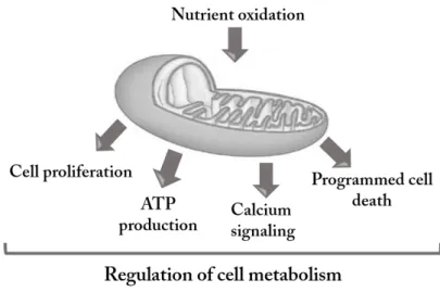
Mitochondria are organelles currently viewed as a cell dynamic compartment resulting from the equilibrium between fusion and fission processes. They play crucial roles in eukaryotic aerobic cells as the primary producers of ATP, regulators of ion homeostasis or redox state, and producers of free radicals [1]. Mitochondria are also key organelles for regulation of cell metabolism [2]. They produce the adenosine triphosphate (ATP) mainly from glucose and fatty acid oxidation, and to a lesser extend from amino acid oxidation. Nutrient oxidation within the mitochondrial matrix or the cytosol results in acetyl-CoA production, the main substrate of the Tricarboxylic Acid (TCA) cycle, which takes place in the mitochondrial matrix. These oxidative pathways are coupled to the mitochondrial electron transport chain (ETC), whose enzyme content and activity together define the mitochondrial oxidative capacity [3].
Mitochondria are involved in the maintenance of cell viability and vitality, but also play a central role in the modulation of regulated cell death (RCD) [4, 5]. There is continuous cross talk exists between cell and mitochondria, which allows the cell adaptation to physiological changes or to commit suicide [2]. Thus, mitochondria may be regarded as the guardians of the gate between life and death [4, 6].
Figure 1: Mitochondria are responsible for ATP production through metabolism and respiration. They also play key roles in several signalling processes such as Ca2+ trafficking,
14
2. Role, features, and organization of the mitochondria
Mitochondria are double-membrane organelles. While the outer mitochondrial membrane (OMM) is rather permeable, allowing the free diffusion of small proteins and contains specialized channels for import of larger proteins, the inner mitochondrial membrane (IMM) is highly impermeable. Additionally, the IMM has a higher superficial area than the OMM and thus possesses characteristic folding morphology forming typical cristae [7]. Mitochondria contain their own mitochondrial DNA (mtDNA), which encodes a subset of proteins essential for the synthesis of adenosine triphosphate (ATP) through oxidative phosphorylation coupled with the transport of electrons by the electron transport chain (ETC) [8, 9]. The ETC consists of four large enzyme complexes (Complex I, NADH-CoQ oxidoreductase; Complex II, succinate dehydrogenase; Complex III, CoQ cytochrome c oxidoreductase; and Complex IV, cytochrome c oxidase, COX). Mobile electron carriers (e.g. coenzyme Q or ubiquinone, and cytochrome c) transport the electrons from one complex to the next with oxygen acting as the final electron acceptor allowing COX reoxidation [10]. The
transfer of the high-energy electrons through the ETC results in the pumping of H+ ions
across the inner membrane, creating an electrochemical proton gradient that provides
the energy required to drive the synthesis of ATP, thanks to a fifth complex called F0F1
ATP synthase [3]. This complex consists of an H+ ion channel (F
0) connected to a
catalytic subunit (F1). The mechanism of oxidative phosphorylation consists in the
utilization of the energy delivered by the flux of H+ ions through F
0 to drive ATP
synthesis from ADP and Pi by F1 [10]. ATP production in mitochondria has to strictly
match cellular needs and therefore is strongly controlled by different factors, namely
the ATP/ADP-Pi ratio, the NAD+/NADH-H+ ratio and local oxygen pressure. The transfer
of ATP, ADP and Pi between the cytosol and the mitochondrial matrix is mediated by the Voltage Dependent Anion Channel (VDAC) and by the ATP/ADP carrier (adenine nucleotide translocase, ANT) [10].
15 Figure 2: Schematic overview about the mechanism of the mitochondrial respiratory chain. The mitochondrial electron transport chain is composed of five complexes. Electron transport between complexes I to IV is coupled to extrusion of protons from complexes I, III and IV into the intermembrane space, creating an electrochemical gradient (Δp) across the inner mitochondrial membrane. Protons then flow through complex V (ATP synthase), which utilizes the energy to synthesize ATP from ADP (Adapted from [11]).
The mitochondrial transmembrane electrochemical potential (Δp)
As referred above the transfer of electrons through protein complexes I - IV in the inner mitochondria membrane, associated with the mitochondrial respiration establishes the mitochondrial transmembrane electrochemical potential or protonmotive force (Δp) [12, 13]. This results in a net accumulation of H+ outside the
membrane, constituted by a charge or electrical gradient (Δψm) and by a chemical or concentration gradient (ΔpHm) [12]. Using a simplified Nernst factor for the second term, Δp can be represented at 37°C by the equation: Δp (mV) = Δψm − 60ΔpHm [13, 14].
Together, the Δψm and ΔpHm contribute to mitochondrial control over energy metabolism, intracellular ion homeostasis, and cell death in eukaryotic cells [12]. While Δp provides the bioenergetic driving force for and regulates ATP production, the Δψm
16
regulates reactive oxygen species (ROS) production, and thus is also a central regulator of cell health [15].
Accumulation of reactive oxygen species
The accumulation of ROS by mitochondria is important because it underlies oxidative damage in many pathologies and contributes to retrograde redox signalling from the organelle to the cytosol and nucleus [16]. During the electron transfer through the respiratory chain, a small percentage of electrons may prematurely reduce oxygen, forming ROS [17]. It was demonstrated that the main sites of ROS production are located at complexes I and III, the iron-sulphur centres of complex I being potentially the most important ROS generators [3].
The reduction of oxygen by one electron at a time produces relatively stable
intermediates. Superoxide anion (O2•-) is the precursor of most ROS and a mediator in
oxidative chain reactions. Dismutation of O2•-, either spontaneously or through a
reaction catalysed by superoxide dismutases, produces hydrogen peroxide (H2O2),
which in turn may be fully reduced to water or partially reduced to hydroxyl radical
(OH•), one of the strongest oxidants in nature [17].
The rate of mitochondrial ROS accumulation is mainly determined by the
membrane potential, the ratio between NADH and NAD+, the ratio between CoQH
2
and CoQ and the local O2 concentration. It is well established that there is a strong
positive correlation between Δψm and ROS production [16]. A slight mitochondrial hyperpolarization has been associated to a large stimulation of ROS accumulation [16]. Similarly, a slight mitochondrial depolarization deeply reduces ROS accumulation [18]. Thus, ROS accumulation by mitochondria can lead to oxidative damage of mitochondrial proteins, lipids and DNA, impairing the ability of mitochondria to synthesize ATP and to carry out their wide range of metabolic functions, including the function of TCA cycle, fatty acid oxidation, urea cycle, amino acid metabolism and FeS centre assembly that are central to the normal operation of most cells [16]. Consequently, it is predictable that mitochondrial oxidative damage contributes to a wide range of pathologies [16-18].
17
Mitochondrial oxidative damage can also increase the tendency of mitochondria to release IMS proteins such as cytochrome c (cyt c) to the cytosol by mitochondrial outer membrane permeabilization (MOMP) and thereby activate the cell’s intrinsic apoptotic machinery [16], as discussed below.
Figure 3: Overview of mitochondrial ROS production. ROS production by mitochondria can lead to oxidative damage to mitochondrial proteins, lipids and DNA, impairing the ability of mitochondria to synthesize ATP and to carry out their wide range of metabolic functions (Adapted from [16]).
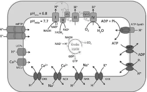
Mitochondrial matrix pH
The regulation of protons and other ions is crucial for mitochondrial functionality [4]. The generation of an electrochemical proton gradient across the inner mitochondrial membrane (IMM) is an essential energy conservation event that couples the oxidation of carbohydrates and fat to the synthesis of ATP. Studies in isolated mitochondria have established that the proton chemical gradient (ΔpHm) and the proton electrical gradient (Δψm) contribute independently to the Δp that drives the synthesis of ATP [19].
18
ΔpHm drives the fluxes of metabolic substrates required for mitochondrial respiration, and the activity of electroneutral ion exchangers that maintain mitochondria osmolarity and volume. ΔpHm reflects the pH difference between the
mitochondrial matrix pH (pHmito) and the pH within the IMS (pHIMS). A difference of 0.9
pH units make the pHIMS more acidic than pHmito [19]. The pH on the IMS side of
mitochondria cristae might be even more acidic than the bulk IMS pH because respiratory complexes are concentrated on these invaginations, which are connected to the IMS by small tubular junctions that constrain the diffusion of solutes [20].
Recent studies indicate that the pHmito plays an important and underappreciated role
in physiological and pathological situations such as apoptosis, neurotransmission, and insulin secretion [19]. From a chemiosmotic point of view, Δψm and ΔpHm are
independent components that equally contribute to the Δp as H+ ions return to the
matrix at the level of complex V [21].
Figure 4: Determinants of the mitochondrial matrix pH. Protons are pumped from the matrix to the IMS by the respiratory chain complexes I, III, and IV as electrons flow from reduced substrates in the matrix to O2. The pumping of electrically charged protons generates a Δψm
19
Mitochondrial calcium storage
Calcium (Ca2+) is an essential ion for cell life, acting as a key second messenger
in almost all cellular functions. Cytosolic Ca2+ concentration is important in signalling
and regulation of many essential responses in practically all cell types [22]. To make this signalling possible, cells maintain cytosolic calcium at low levels and have different transporters that regulate the concentration of this cation in the intracellular compartments, causing the oscillations required for cell signalling [23]. Increase of
intracellular [Ca2+] can be elicited through two fundamental mechanisms: i) Ca2+
mobilization from intracellular stores, mainly the (ER), or ii) entry from the
extracellular milieu through the opening of plasma membrane Ca2+ channels [24].
Mitochondria are able to store significant amount of Ca2+ bound to phosphate
within their matrix. Ca2+ taken up by the mitochondria regulates mitochondrial
endoplasmic reticulum functioning in response to a variety of extracellular stimuli [25]. It activates three dehydrogenases of the TCA cycle, namely pyruvate dehydrogenase, isocitrate dehydrogenase, and α-keto glutarate dehydrogenase. It may also regulate
the ETC, the F0F1 ATP synthase and ANT [26]. This induces an increased substrate
uptake by mitochondria, enhanced mitochondrial NADH/NAD+ ratio and increased ATP
production. The high capacity to accumulate Ca2+ confers to mitochondria a key role in
the regulation of intracellular Ca2+ signalling, which includes regulation of gene
expression (including those involved in mitochondrial biogenesis and glucose uptake), cell functioning and control of protein trafficking between organelles [27].
3. The role of mitochondria in regulated cell death
Cell death instances can be classified into two categories: ‘accidental’ and ‘regulated’. Accidental cell death (ACD) is caused by severe stimulus, including physical (e.g., elevated temperatures or high pressures), chemical (e.g., potent detergents or extreme variations in pH) and mechanical (e.g., shearing). ACD is practically immediate and is insensitive to pharmacologic or genetic interventions of any kind [28]. In contrast to its accidental counterpart, regulated cell death (RCD) involves a genetically encoded molecular machinery. RCD often occurs in a relatively tardy manner and is
20
initiated in the context of adaptive responses that attempt to restore cellular homeostasis. Thus, while ACD is completely unpreventable, RCD can be modulated not only by inhibiting the transduction of lethal signals but also by improving the capacity of cells to mount adaptive responses to stress [28]. Importantly, RCD occurs not only as a consequence of microenvironmental perturbations but also in the context of (post-) embryonic development, tissue homeostasis and immune responses. Such completely physiologic instances of RCD are generally referred to as ‘programmed cell death’ (PCD) [28]. Of note, apoptosis, autophagy and programmed necrosis are the three main forms of RCD, easily distinguished by their morphological differences [29]. Apoptosis, designated before as type I cell death, is characterized by specific morphological and biochemical changes of dying cells, including cell shrinkage, nuclear condensation and fragmentation, dynamic membrane blebbing and loss of adhesion to neighbours or to extracellular matrix [30]. Biochemical changes include chromosomal DNA cleavage into inter-nucleosomal fragments, phosphatidylserine externalization and a number of intracellular substrate cleavages by specific proteolysis [31].
Autophagic cell death, designated before as type II cell death, is an evolutionarily conserved catabolic process beginning with formation of autophagosomes, double membrane-bound structures surrounding cytoplasmic macromolecules and organelles, destined for recycling [32]. Besides apoptosis and autophagic cell death, there exists a type III cell death termed programmed necrosis, which involves cell swelling, organelle dysfunction and cell lysis [33]. Since RCD processes have an important role during preservation of tissue homoeostasis and elimination of damaged cells, systems biology of cell death has a wide range of clinical applications for drug treatment of various human diseases [29]. Decrypting the labyrinth of cell death pathways has important implications in choosing the right drug targets that will prevent the death of cells in pathological conditions such as, ischemia, neurodegenerative diseases and stroke [28]. Conversely, drug treatments designed for cancer and autoimmune diseases aim at achieving maximum cell death. Because tumors carry a collection of genetic perturbations that block different functional modules within the RCD network, efficient tumor cell killing depends on activating
21
alternative pathways and bypassing blocks in the pathways mapped individually for each tumor [29].
The most frequent and well-defined form of RCD is apoptosis. Apoptotic processes are of a widespread biological importance, being involved in the development, differentiation of multicellular organisms, proliferation, homeostasis, regulation and function of immune system and in the removal of defective cells [34]. Numerous extra- or intracellular factors are associated with apoptotic signaling pathways have been identified, such as attachment of death ligands to membrane death receptors, DNA damage caused by defaults in DNA repair mechanisms, treatment with cytotoxic compounds or irradiation, lack of survival signals, or development of death signals [35].
Two main signalling pathways lead to apoptosis: the extrinsic or death receptor
pathway, and the intrinsic or mitochondrial pathway.The extrinsic pathway is defined
as mitochondria independent, although mitochondria can be involved in the amplification of the death signal. It involves the activation of receptors in the plasma membrane through binding of ligands that trigger a proteolytic chain responsible for the characteristic morphological features of apoptosis. The mitochondrial pathway involves the permeabilization of the mitochondrial outer membrane allowing the release into the cytosol of a variety of pro-apoptotic proteins [35]. Mitochondria have a central role in this apoptotic pathway and dysfunction of this organelle induced by DNA damage and other genotoxic factors leads to apoptotic cell death [36]. Mitochondrial fragmentation during apoptosis was connected with the collapse of the mitochondrial membrane potential (ΔΨm) that was considered a point of no return in the death cascade. Members of the Bcl-2 family of proteins play a key role regulate outer mitochondrial membrane (OMM) integrity and function [37].
One of the crucial events in the process linking lethal signals to controlled death of mammalian cells is the mitochondrial outer membrane permeabilization (MOMP) [38]. During early stages of apoptosis, the pro-apoptotic protein Bax translocates to the OMM and, almost instantly after translocation, concentrates into sub-mitochondrial punctate foci. In addition, Bak co-localizes with Bax in these foci.
22
Bax and Bak induce cell death via MOMP that leads to the release of small pro-apoptotic molecules such as cytochrome c [39].
Figure 5: Schematic presentation of activation of mitochondrial pathway of apoptosis (Adapted from [41]).
Mitochondrial permeability transition (MPT) is a mitochondrial state in which the proton-motive force is disrupted. This disruption involves the mitochondrial membrane permeability transition pore (mPTP) [40]. The PTP is a transmembrane channel formed at the contact sites between the mitochondrial inner membrane (MIM) and the mitochondrial outer membrane (MOM). The components of the PTP are the voltage-dependent anion channel (VDAC) in the outer mitochondrial membrane and the adenine nucleotide translocator (ANT) in the inner mitochondrial membrane [41]. During normal mitochondrial function, the intermembrane space separates the inner and outer mitochondrial membranes and the VDAC and the ANT do not interact. When mitochondrial permeability is activated by the formation of the PTP, increase in
MIM permeability to solutes up to ~1500 Da, leading to a reduction of the Δψm,
swelling of the mitochondrial matrix, and OM permeabilization, presumably due to physical rupture of OM [41].
23
The release of mitochondrial factors is widely accepted as the initiating event of the intrinsic pathway of apoptosis, but it can also serve as an amplification mechanism of the extrinsic pathway of cell death. The proteins released can either activate a group of enzymes of the cysteine protease family known as caspases or act in a caspase-independent fashion to bring about cell death [35].
4. Mitochondria as cellular target for cancer therapy
The evasion/resistance of human cancers cells to cell death is a major cause of treatment failure. The lack of efficacy of established therapeutic regimens is due, at least in part, to the oncogenic blockade of cell death pathways [42]. Thus, drugs designed to activate the cell death machinery may represent a more effective therapeutic option. This machinery is composed of catabolic hydrolases, mostly proteases and nucleases, which are held in check by specific inhibitors or by the sequestration of their activators. The permeabilization of the mitochondrial outer membrane is a potent way of unleashing such activators. Multiple apoptosis- and necrosis-inducing biochemical cascades converge on mitochondria to cause their deregulation and permeabilization [43].
Mitochondria exert both vital and lethal functions in physiological and pathological scenarios. On the one hand, mitochondria are indispensable for energy production and hence for the survival of eukaryotic cells. On the other hand, mitochondria are crucial regulators of the intrinsic pathway of apoptosis [44]. Mitochondria control the activation of apoptotic effector mechanisms by regulating the translocation of pro-apoptotic proteins from the mitochondrial inter-membrane space to the cytosol. As mitochondria are key regulators of cell death and as mitochondrial functions are often altered in neoplasia, mitochondrial-targeted compounds represent a promising approach to eradicate chemotherapy-refractory cancer cells [45].
There is ample evidence of metabolic alterations affecting the capacity of malignant cells to engage in apoptosis, necrosis and autophagy [46]. Moreover, modifications in the levels of ROS have recently been linked to the intrinsic
24
chemotherapy resistance of cancer stem cells. These changes are intricately linked to the bio-energetic functions of mitochondria, making these organelles attractive drug targets [46]. Accordingly, multiple hallmarks of cancer cells, including limitless proliferative potential, insensitivity to anti-growth signals, impaired apoptosis, enhanced anabolism and decreased autophagy, have been linked to mitochondrial dysfunctions [47].
The correction of cancer-associated mitochondrial dysfunctions and the (re)activation of cell death programmes by pharmacological agents that induce or facilitate mitochondrial membrane permeabilization represent attractive strategies for cancer therapy [48].
5. Drug mitochondrial targets in cancer therapy
A large number of agents with anti-cancer activity that act on mitochondria, mitocans (an acronym for “mitochondria and cancer”), hold a substantial promise to be developed into efficient anti-cancer drugs, based on their selectivity for cancer cells [48]. Thus, it is propose the study of anti-cancer agents that act via mitochondrial destabilization (mitocans) based on the site of action of the individual agents from the surface of the mitochondrial outer membrane (MOM) to the mitochondrial matrix. The selection of the sites also stems from their importance as targets for the development of drugs that hold substantial promise to be utilized in the clinical practice [65].
Acetate and lactoferrin (Lf), are two known anti-cancer agents that induce an apoptotic process mediated by the mitochondria in colorectal carcinoma cells [49, 50] and in the yeast Candida Albicans [51].
Lf is an 80-kDa, non-heme iron-binding glycoprotein that belongs to the transferrin family. It is found in most mucosal sites and secondary granules of neutrophils in mammals [52]. Several functions have been attributed to Lf as a key component in the host’s first line of defense, contributing to a variety of physiological changes at both the cellular and organ levels. Lf is a major pleiotropic mediator that plays an important role in the development of inflammatory responses [53]. Lf also contributes to tumour growth inhibition might be related to apoptosis of these cells
25
induced by the activation of the Fas signaling pathway (extrinsic apoptotic pathway). Nevertheless, the exact mechanism of these functions has not been discovered so far [54].
Acetate is a short chain fatty acid (SCFA) which has been described as an endogenous metabolite of beta-oxidation of fatty acids in liver [55]. SCFAs are final product of carbohydrate´s fiber fermentation by Propionibacteria that live in the human intestine [49, 50]. These natural compounds have been the subject of increased research over the past few decade. SCFAs besides mediating the ions transport for the colonic epithelium, may act as modulators of intracellular pH, cell volume, and other functions associated with ion transport, and as regulators of proliferation, differentiation and gene expression [56, 57]. For SCFAs, palmitate, propionate and acetate induced apoptosis occurred via the stereotyped biochemical events, including mitochondrial alterations, caspase activation and nuclear degradation [50]. Studies indicated that SCFAs can act as anticancer agent, through inhibition of colon cancer cell growth and differentiation, and enhancement of apoptosis in coloretal cancer cells [49]. Therefore, a diet rich in fibers has been associated with a decreased incidence and growth of colon cancers [58].
26
Aims
The aim of the present thesis was to explore flow cytometric analysis to perform a functional characterization of isolated mitochondria from the yeast model system S. cerevisiae and rat liver, towards the screening of mitochondrial target drugs. For such purpose, a protocol for the isolation and purification of yeast and rat liver mitochondria was implemented and optimized. Then, flow cytometry analysis and western blot were used to characterize the purity and functional integrity of mitochondria. A set of staining protocols were optimised for the analysis by flow cytometry of different functional features of the isolated organelles namely, their mass, mitochondrial matrix pH, Δψm and ROS accumulation.
The effect of Lf and acetate on mitochondria function was tested in isolated mitochondria from rat liver using specific fluorescent probes combined with flow cytometry. Complementary studies were also performed to understand if the biochemical activity of the complexes of ETC and osmotic swelling were affected in response to Lf and acetate. Further investigation of the role of these drugs in mitochondria-dependent death pathways will offer new perspectives for an enhanced understanding of the effect of Lf and acetate in mammalian apoptosis.
27
Materials and methods
Reagents
The mitochondrial markers Mitotracker Green, Mitotracker Red CMXROS;
Mitotracker Red CM-H2XROS, 2', 7'-Bis-(2-Carboxyethyl)-5-(and-6)-Carboxyfluorescein,
Acetoxymethyl Ester (BCECF-AM), Acridine Orange 10-Nonyl Bromide (NAO) and
3,3'-Dihexyloxacarbocyanine Iodide (DiOC6(3)) were purchased from Invitrogen-Molecular
Probes (Eugene, OR, USA). Yeast nitrogen base, peptone and yeast extract were purchased from Difco. Dihydroethidium (DHE), cytochrome c, carbonyl cyanide m-chlorophenyl hydrazone (CCCP), oligomycin, succinate, adenosine 5′-diphosphate sodium salt (ADP) were purchased from Sigma-Aldrich.
Yeast strains and growth conditions
Saccharomyces cerevisiae wild type strain W303-1A was grown in YNB medium
with 2% (w/v) lactate, pH 5.5. Frozen aliquots of yeast strain were streaked onto plates containing the adequate culture medium and incubated for 48 h at 25°C prior to each experiment. All strains were inoculated at an OD of about 0.005 and grown overnight
to log phase (OD640nm = 0.5-0.8) in 500 mL flasks, in an orbital shaker, at 30 °C, 200
rpm.
Isolation and purification of mitochondria from yeast cells
Yeast cells were pelleted at 4oC by centrifugation at 5000 rpm for 5 min and
washed with distilled water. The yeast pellet was resuspended in DTT buffer (100 mM Tris, 30 mM dithiothreitol (DTT), pH 7.0) and incubated at 30°C for 15 min. After
centrifugation at 5000 rpm for 5 min at 4oC, the yeast pellet was incubated for 50-60
min at 30°C with Zymolyase buffer containing 1.35 M sorbitol, 30 mM disodium phosphate, 10 mM citric Acid, 1 mM ethylene glycol tetraacetic acid (EGTA), and 10 mg Zymolyase per g of cells wet weight, pH 5.8. After the digestion of the cell walls, cells
were harvested by centrifugation at 7700g for 5 min at 4oC and the pellet was washed
with Zymolyase buffer without the enzyme. Spheroplasts were centrifuged at 7700g
for 5 min at 4 oC and the pellet was resuspended in ice-cold homogenization buffer
28
bovine serum albumin (BSA). The lysed material was carefully maintained at low temperature to avoid proteolysis. Spheroplasts were homogenized in a final volume of 15 mL with 10 strokes using a glass-Potter homogenizer. After homogenization, the sample was diluted to 40 mL with homogenization buffer, centrifuged at 500g for 10
min at 4oC and the pellet was discarded. To isolate mitochondria, the supernatants
were collected and centrifuged at 17000g for 10 min. To achieve a greater degree of purification the last two centrifugations steps were repeated. The crude mitochondrial pellet was resuspended in 2 mL of homogenization buffer. Protein quantification was determined by Lowry method [59].
Isolation and purification of mitochondria from rat liver
All manipulations were performed on ice with ice-cold buffers (IB; 250 mM sucrose, 1 mM EDTA, 50 mM Tris-HCl, pH 7.2). Animal experiments were performed according to the Portuguese law for animal welfare (Diário da República, Portaria 1005/92). Rattus norvegicus, strain Sprague-Dawley OFA (Charles-River, Spain) were housed in an accredited mouse house and treated as specified by the recommendations of the Helsinki convention (World Medical Association - ethical principles for medical research involving humans) and the Guide for the Care and Use of Laboratory Animals, published by the National Academy Press. Animals were sacrificed with 150mg/kg of pentobarbital (Eutasil®, Ceva, Algés, Portugal) and the liver was rapidly dissected out and stored in IB buffer.The tissue was manually homogenized in IB buffer (10%, wt/vol) with 8-10 strokes using a glass-Potter homogenizer. The homogenate was diluted two-fold with
IB and centrifuged at 1000g for 10 min at 4oC to pellet erythrocytes, cell debris and
nuclei. Supernatants were collect and centrifuged at 10000g for 10 min at 4oC. The last
two steps were repeated to increase the degree of purification. The crude mitochondrial pellet was gently resuspended in 2 mL of IB. Protein quantification was determined by Lowry method [59].
29
Western Blot
For western blot assay, protein quantification was determined by Bradford method [60]. Fifty μg of protein was resolved in SDS-polyacrylamide gel and then electroblotted to a polyvinylidene difluoride membrane (Millipore, Billerica, MA, USA). Membranes were blocked in TPBS (PBS with 0.05% Tween-20) containing 5% (w/v) nonfat dry milk, washed in TPBS and incubated with primary antibody (TOM 20, GAPDH and V-ATPase) overnight. Then, after washing, membranes were incubated with secondary antibody and immunoreactive bands were detected using the Immobilon solutions (Millipore, Billerica, MA, USA) under a chemiluminescence
detection system, the ChemiDoc XRS (Bio-Rad Laboratories).
Fluorochrome solutions
Mitochondria were selectively stained using 10-nonyl acridine orange (NAO), which binds to cardiolipin in the inner mitochondrial membrane. NAO is one of the most commonly used dyes for the monitoring of mitochondrial mass [61]. A concentrated stock solution of NAO (2.5 mM) was prepared in DMSO, and stored at -20 °C.
MitoTracker Green (MTG) is a mitochondrial-selective fluorescent label commonly used in flow cytometry. It is expected that this dye selectively accumulates in the mitochondrial matrix where it covalently binds to mitochondrial proteins by reacting with free thiol groups of cysteine residues [62]. A stock solution (50 μM) was prepared in DMSO and stored at -20°C.
Mitotracker Red CMXROS was used for monitoring the membrane potential (Δψm). Mitotracker Red CMXROS binds irreversibly to the polarized mitochondrial membrane and does not require reduction or oxidation for emission of fluorescence [63]. Mitotracker Red CMXROS was dissolved in DMSO at a concentration of 500 µg/mL and stored at -20 °C until use.
DiOC6(3) has been also widely used as a cytofluorometric Δψm indicator. When
DiOC6(3) is used at low concentrations (10–20 nM) it rapidly reaches equilibrium in the
mitochondria with low quenching effects [4]. The stock solution of DiOC6(3) (0.2 µM)
30
Mitochondrial ROS accumulation was evaluated with the probe Dihydroethidium (DHE). This is a fluorescent probe that up on oxidation by superoxide anion gives converted into ethydium which intercalates in the double bound nucleic acids and emits red fluorescence [64]. A stock solution of DHE (2 µg/mL) was prepared in DMSO and stored at -20°C.
Mitotracker Red CM-H2XROS is a reduced, nonfluorescent version of
MitoTracker Red (M-7512) that fluoresces upon oxidation [65]. The probe was dissolved in DMSO at a concentration of 200 µM and stored at -20 °C until use.
BCECF (2',7'-Bis-(2-Carboxyethyl)-5-(and-6)-Carboxyfluorescein, Acetoxymethyl
Ester) was used to evaluate the mitochondrial matrix pH as it is sensitive to H+. After
the hydrolytic cleavage of its ester bonds by esterases within the matrix the probe becomes entrapped. A concentrated stock solution of BCECF-AM (1.6 mM) was prepared in DMSO, and stored at -20ºC.
Flow cytometry analysis
Sample analysis by flow cytometry was performed in an Epics® XL™ (Beckman Coulter) flow cytometer, equipped with an argon-ion laser emitting a 488 nm beam at
15 mW. Monoparametric detection of red fluorescence from Mitotracker CMXROS, H2
-XROS, and DHE was performed using FL-4 (525/620 nm); for the Nonyl Acridine Orange war performed using FL-3 (488/620 nm); and detection of green fluorescence from
DIOC6(3), Mitotracker Green FM and BCECF-AM was performed using FL-1 (488/525
nm). For each sample, at least 20000 events were analysed at low flow rate. An acquisition protocol was defined to measure forward scatter (FS), side scatter (SS) and green fluorescence (FL1) on four decade logarithmic scales.
Enzymatic assays
Succinate dehydrogenase assay
Enzyme activity was measured following the reduction of the hexacyanoferrate (III). Intact mitochondria (150 µg protein), 40 mM succinate, 0.10% BSA were added to the 100 mM potassium phosphate, pH 7.8 (final volume 3 mL). The reaction was
31
started by the addition of potassium hexacyanoferrate (II) (0.5 mM), and the decrease in absorbance at 420 nm was recorded.
NADH-cytochrome c reductase
Cytochrome c reductase activity was recorded in a spectrophotometer set at 550 nm. Samples of homogenate were diluted with cold 10 mM phosphate buffer (pH 7.5) to yield a final protein concentration of 0.25 mg/mL. Cytochrome c solution was obtained by dilution of 1% cytochrome c in 10 mM phosphate buffer (pH 7.5). To obtain a baseline, 50 mg protein were added in the absence of substrate and the reaction was measured for 0.5 min at 550 nm. The reaction was started with 0.1 mM NADH solution and the rate of cytochrome c reduction was measured for additional 1-2 min.
Cytochrome c oxidase
Samples of homogenate were diluted with cold 20 mM phosphate buffer (pH 7.5), to yield a final protein concentration of 0.25 mg/ml. Cytochrome c solution was obtained by dilution of 1% cytochrome c in 20 mM phosphate buffer (pH 7.5). The rate of cytochrome c oxidation was recorded at 550 nm and a reference of total oxidation of cytochrome c was obtained with addiction of a small amount of potassium ferricyanide in aliquot of cytochrome c solution. To start reaction of cytochrome c oxidase was added 50 µg of mitochondrial protein in 1mL of cytochrome c solution. The absorbance was registered at 550 nm every 15 seconds or using a recorder.
Mitochondrial Swelling Assay
Mitochondrial swelling was followed spectrophotometrically at 540 nm. Isolated liver rat mitochondria were resuspended in the swelling buffer containing 120
mM KCl, 10 mM Tris-HCl (pH 7.4) and 5 mM KH2PO4, to a final protein concentration of
Results
Functional studies of mitochondrial suspensions by flow
cytometry
Mitochondrial suspensions isolated from rat liver and yeast cells were prepared in order to optimise their structural and functional characterization by flow cytometry. This characterization was corroborated by biochemistry approaches including western blot as well as enzymatic assays.
Characterization of the purity of mitochondrial fractions
Biparametric histograms (FS log vs SS log) of a mixture of yeast cells and isolated yeast mitochondria revealed differences in complexity and size between both biological samples. Yeast cells are far more complex and larger than isolated mitochondria (Fig. 1A). In addition, mitochondrial suspensions isolated from either yeast or rat liver formed homogeneous populations suggesting an homogeneous population which reflects a good degree of purity (Fig. 1B and C).
Figure 1: Flow cytometric analysis of intact yeast cells (A) and mitochondrial suspensions isolated from yeasts (A and B) and rat liver (C). Scattergrams with distinguishable populations of cells (blue) and purified mitochondrial suspensions (red). In A yeast cells and purified mitochondria were mixed prior to analysis
Western blot analysis with antibodies against proteins representative of important subcellular compartments was used to further characterize the purity of the mitochondrial population. TOM20 was used as mitochondrial marker, GAPDH as marker of the cytosol, and the subunit C of the V-ATPase as marker of the lysosome. As
33
can be seen in Fig. 2 both mitochondrial batches from rat liver were enriched in TOM20 and depleted in GAPDH and in the subunit C of the V-ATPase, further confirming that they were highly pure.
Optimization of the staining protocols
To perform structural and functional measurements by flow cytometry of intact mitochondria isolated from either yeast or rat liver, in a first stage of the work, several probes were tested and the corresponding staining protocols were optimized in order to monitor the mitochondrial memebrane potential (Δψm), the pH of the mitochondrial matrix, ROS accumulation, and mitochondrial mass. Both the incubation
time (15, 30, 45 and 60 min), the temperature (room temperature, 30oC and 37oC), the
concentration of each probe and the amount of mitochondrial protein per each assay were optimized.
Mitotracker Green and Nonyl Acridine Orange
Both probes were used to analyse changes in mitochondrial mass. The best staining conditions were as follows: for Mitotracker Green (MTG), 50 µg of
mitochondrial protein (in 500 µL) incubated in the dark for 30 min at 37 oC with 0.4 µM
of the probe; for Nonyl Acridine Orange (NAO), 50 µg of mitochondrial protein (in 500 µL) incubated in the dark for 15 min at room temperature with 0.5 µM of the probe. In these conditions the mitochondrial population exhibited a green positive fluorescence (FL1; 488/525 nm) with MTG staining, and an orange positive fluorescence (FL3; 488/620 nm) with NAO staining (Fig. 3).
Figure 2: Western blots of protein extracts from isolated mitochondria from rat liver and from the corresponding homogenate, using specific antibodies raised against the mitochondrial protein Tom 20, the subunit C of the V-ATPase from the lysosome, and the protein GAPDH resident in the cytosol.
34
3,3'-Dihexyloxacarbocyanine Iodide and Mitotracker Red CMXROS
Both probes were used to assess changes in the Δψm and the best staining conditions were: for Mitotracker Red CMXROS (MTR), 50 µg of mitochondrial protein
(in 500 µL) incubated in the dark for 30 min at 37 oC with 0.2 µM of the probe; for
3,3'-Dihexyloxacarbocyanine Iodide (DiOC6(3)), 50 µg of mitochondrial protein (in 500 µL)
incubated in the dark for 30 min at room temperature with 10 nM of the probe. As shown in Fig. 3 clear green positive fluorescence (FL1; 488/525 nm) was observed with
DiOC6(3) staining and a red positive fluorescence (FL4; 525/620 nm) with Mitotracker
Red CMXROS staining.
Mitotracker Red CM-H2XROS and dihydroethidium
Mitotracker Red CM-H2XROS and dihydroethidium (DHE) were selected to
evaluate the accumulation of ROS. The best staining conditions were found when 50 µg protein of mitochondrial suspension (in 500 µL) was incubated during 30 min at 37
oC in the dark with 0.5 µM Mitotracker Red CM-H
2XROS or 2ng/mL of DHE. A clear red
positive fluorescence of mitochondria (FL4; 525/620 nm) can be observed in Fig. 3
after staining whith Mitotracker Red CM-H2XROS and DHE.
2',7'-Bis-(2-Carboxyethyl)-5-(and-6)-Carboxyfluorescein, Acetoxymethyl Ester
The staining protocol with
(2',7'-Bis-(2-Carboxyethyl)-5-(and-6)-Carboxyfluorescein, Acetoxymethyl Ester (BCECF-AM) was optimized to study how the pH of the mitochondrial matrix is changed after challenging isolated mitochondria with a specific substrate or drug. Results showed that the best staining condition was found when 50 µg of mitochondrial protein (in 500 µL) was incubated with 5 µM BCECF-AM during 30 min at 37°C in the dark. As shown in Fig. 3, a green positive fluorescence was clearly observed (FL1; 488/525 nm) after staining with BCECF-AM.
35 Figure 3: Flow cytometry analysis of isolated mitochondrial suspensions isolated from yeast cells (A) and rat liver (B) stained with DIOC6(3) (0.4 µM), Mitotracker Red CMXROS (0.2 µM),
NAO (0.5 µM), Mitotracker Green BCECF-AM (1.6 mM), DHE (2ng/mL) and Mitotracker Red CM-H2XROS (0.5 µM). Histograms in white correspond to the autofluorescence
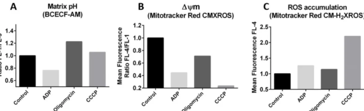
Response of resting mitochondria from rat liver to ADP, oligomycin
and CCCP
In these assays staining with BCECF-AM, Mitotracker Red CMXROS, or Mitotracker
Red CM-H2XROS was used to evaluate changes in the pH of the mitochondrial matrix,
mitochondrial mass, Δψm and ROS accumulation in the mitochondrial compartment, respectively. The addition of 500 nmol of ADP to rat liver isolated resting mitochondria stained with BCECF-AM resulted in a decrease of fluorescence within 5 min, suggesting
36
an acidification of the mitochondrial matrix due to coupling between endogenous respiration and proton driven ATP synthesis. On the contrary, the addition of 0.9 µg/mL oligomycin promoted alkalinisation of the matrix. No significant changes in mitochondrial pH were observed upon addition of 50 µM CCCP (Fig. 4A).
The addition of either 500 nmol ADP or 0.9 µg/mL oligomycin, or 50 µM CCCP to rat liver mitochondria stained with Mitotracker Red CMXROS promoted a reduction of the red fluorescence suggesting that each compound was able to induce a transient depolarization of the mitochondrial membrane. The strongest effect was observed upon addition of the protonophore CCCP (Fig. 4B).
When the effect of ADP, oligomycin, or CCCP were tested in mitochondria labelled with
Mitotracker Red CM-H2XROS, only CCCP was able to promote a significant increase in
the red fluorescence suggesting that the accumulation of ROS was induced following enhanced endogenous respiration due to the collapse of the transmembrane proton gradient by the protonophore (Fig. 4C).
Figure 4: Flow cytometry analysis of resting mitochondria from rat liver after incubation with ADP (500 nmol), oligomycin (0.9 µg/ml) and CCCP (50 µM) for 5 min. Mitochondrial suspensions were pre-stained with BCECF-AM (A) Mitotracker Red CMXROS (B), and Mitotracker Red CM-H2XROS (C).
Response of mitochondria from rat liver to energization with
succinate
Incubation of resting mitochondria with succinate led to an increase in the pH of the matrix, which was prevented by ADP that promoted a slight acidification of the matrix. The addition of oligomycin to this mixture induced a slight alkalinisation (Fig. 5A).
37
Figure 5B shows that succinate induced a very small depolarization of the mitochondrial membrane within 60 min that was slightly more pronounced in the presence of 500 nmol ADP. The addition of oligomycin prevented membrane depolarization and cause a slight hyperpolarization when compared to resting mitochondria.
The activation of the electron transport chain with 6 mM succinate caused an increase in ROS production as shown in mitochondria suspensions labelled with
Mitotracker CM-H2XROS. The presence of either 500 nmol ADP or 500 nmol ADP + 0.9
µg/mL mM oligomycin did not affect the rate of ROS production induced by succinate (Fig. 5C).
Figure 5: Flow cytometry analysis of resting mitochondria from rat liver after incubation with succinate (6mM), succinate (6mM) + ADP (500 nmol) and succinate (6mM) + ADP (500 nmol) + oligomycin (0.9 µg/ml) for 5 min. Mitochondrial suspensions were pre-incubated for 30 min with 1.6 mM of BCECF-AM (A), 0.2 µM of Mitotracker Red CMXROS (B), and 0.5 µM of Mitotracker Red CM-H2XROS (C).
38
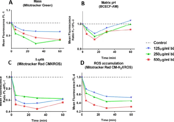
Effect of bovine lactoferrin (bLf) on mitochondrial function
Results presented in figure 6 clearly show that 125 - 500 µg/mL bLf strongly affected rat liver mitochondrial structure/function within 60 min of incubation. In resting mitochondria labelled with Mitotracker Green a clear loss in mitochondrial mass was evident 30 min after the addition of bLf (Fig. 6A). Similar results were obtained for mitochondria stained with NAO after 60 min. incubation with bLf (Fig. 7).
The three concentrations of bLf tested also promoted a clear acidification of the mitochondrial matrix, which was dose-dependent. However, this phenomenon was transient being more evident 15 min after incubation (Fig. 6B). As shown in Fig. 6C, the addition of 125 - 500 µg/mL bLf caused a strong reduction of the Δψm within 5 min of incubation. ROS levels were also strongly reduced after addition of bLf being the effect more evident till 30 min of incubation and clearly dose-dependent (Fig. 6D).
Figure 6: Time course analysis of effect of bLf on mitochondrial function. Mitochondrial suspensions from rat liver were pre-stained with fluorescent probes to assess changes in mitochondrial mass, matrix pH, Δψm and ROS accumulation.
39
Subsequent experiments were performed to study how bLf affects the matrix pH, Δψm and ROS homeostasis of mitochondria energized with 6 mM succinate/500 nmol ADP. Results are depicted in Fig. 8. As can be seen, the pre-incubation of mitochondria suspensions with 125 µg/mL bLf for 60 min induced an acidification of the mitrochondrial matrix measured 5 min after the energization with succinate in all experimental conditions tested (succinate alone, succinate + ADP, succinate + ADP + oligomycin). The same result was obtained when the incubation with bLf was followed by the addition of oligomycin alone. Thus, the pre-incubation with bLf counteracted the alkalinization effect promoted by the addition of succinate alone, succinate + ADP + oligomycin or oligomycin alone (Fig. 8A).
Figure 8B shows that the pre-treatment of isolated mitochondria with bLf promoted a strong depolarization of the inner membrane in all experimental conditions tested (succinate alone, succinate + ADP, succinate + ADP + oligomycin or oligomycin alone), potentiating the dissipation effect of the Δψm promoted by the addition of either succinate alone, succinate + ADP, or oligomycin alone. In what concerns ROS homeostasis, the pre-treatment of isolated mitochondria with bLf promoted a strong decrease in ROS accumulation in all energization conditions tested and in the presence of oligomycin alone, counteracting the observed increase in ROS production promoted by each condition in control samples (Fig. 8C).
Figure 7: Flow cytometry mass analysis of rat liver isolated mitochondria after incubation with different concentration of bLf, 125 µg/mL, 250 µg/mL and 500 µg/mL for 60 min.
40
As referred in material and methods, to study the effect of bLf on the activity of electron transport complexes of the inner mitochondrial membrane isolated mitochondria from rat liver were pre-incubated with 125, 250 and 500 µg/mL of bLf for 60 min. As can be seen in figures 9A and B bLf inhibited by 65-70% the activity of the complex II, the effect being similar for all concentrations tested. The complex III was inhibited by 38% to 44% (Fig. 9C and D) and the cytochrome c oxidase (complex III) by 35% to 58% (Fig. 9E).
Mitochondrial swelling was followed by the decrease in absorbance of
mitochondrial suspensions at 540 nm. The addition 250 µM CaCl2 (positive control)
promoted a prompt mitochondrial swelling (Fig. 10). Results showed that 50 – 500 µg/mL bLf also induced an immediate mitochondrial swelling, which was much stronger at the lowest concentration of bLf used (50 µg/mL).
Figure 8: Flow cytometry analysis of mitochondrial suspensions from rat liver stained with BCECF-AM, Mitotracker Red CMXROS and Mitotracker CM-H2XROS and pre-incubated with 125
µg/mL bLf for 60 min before energization with either 6 mM of succinate or 6 mM of succinate + 500 nmol of ADP. Flow cytometry analysis was performed 5 min after energization. Concentration of oligomycin: 0.9 µg/mL of oligomycin.
41 Figure 9: Effect bLf on the activity of Complex II (succinate dehydrogenase) (A and B), Complex III (cytochrome c – oxireductase) (C and D) and Complex IV (cytochrome c oxidase) (E) from rat liver mitochondria. Mitochondrial suspensions were pre-incubated with 125 – 500 µg/ml bLf for 60 min before the enzymatic analysis.
42
Effect of acetate on mitochondrial function
Flow cytometry experiments were also performed to evaluate mitochondrial mass, the pH of the matrix, Δψm and ROS accumulation in mitochondria in response to 15 – 90 mM acetate. As can be seen in Fig. 11, these mitochondrial parameters were very sensitive to acetate. Similarly to that observed after incubation with bLf, acetate promoted a decrease in the mitochondrial mass, the acidification of matrix, a depolarization of the inner membrane and a decrease in ROS production within 60 min of incubation in rat liver mitochondria. A clear dose-response effect was not observed in all experiments but the highest concentration of acetate used (90 mM) was always the most effective.
To study the effect of acetate in energized mitochondria suspensions from rat liver were pre-incubated with 30 mM of acetate for 60 min before the addition of 6 mM of succinate or succinate + 500 nmol of ADP, and flow cytometry analysis was performed 5 min after energization. Oligomycin alone or in combination with succinate and ATP was used to inhibit F-ATPase. As can be seen in Fig. 12, acetate induced acidification of the mitrochondrial matrix, promoted the depolarization of the inner membrane and prevented ROS accumulation in all experimental conditions tested much like the effects of Lf shown above (Fig. 8).
Figure 10: Effect of bLf on the swelling (SW) of rat liver mitochondria as assessed by the decrease in absorbance at 540 nm of the mitochondrial suspension. Positive control, 250 µM CaCl2.
43 Figure 12: Flow cytometry analysis of mitochondrial suspensions from rat liver stained with BCECF-AM, Mitotracker Red CMXROS and Mitotracker CM-H2XROS and pre-incubated with
30mM of acetate (pH7) for 60 min before energization with either 6 mM of succinate or 6 mM of succinate + 500 nmol of ADP. Flow cytometry analysis was performed 5 min after energization. Concentration of oligomycin: 0.9 µg/mL of oligomycin.
Figure 11: Time course analysis of effect of acetate (pH7) on mitochondrial function. Mitochondrial suspensions from rat liver were pre-stained with fluorescent probes to assess changes in mitochondrial mass, matrix pH, Δψm and ROS accumulation.
44
To study the effect of acetate on the activity of electron transport complexes of the inner mitochondrial membrane, isolated mitochondria from rat liver were pre-incubated with 15 - 90 mM acetate, pH 7.0, for 60 min. As can be seen in Fig. 13 over the concentration range tested acetate strongly inhibited the activity of the complex II (up to 77 %) and complex III (up to 99%). Comparatively, the inhibitory effect of acetate over the activity of cytochrome c oxidase was much weaker, from 7% to 26%, with 30 and 90 mM acetate, respectively.
Figure 13: Effect acetate (pH7) on the activity of Complex II (succinate dehydrogenase) (A and B), Complex III (cytochrome c – oxireductase) (C and D) and Complex IV (cytochrome c oxidase) (E) from rat liver mitochondria. Mitochondrial suspensions were pre-incubated with 125 – 500 µg/ml bLf for 60 min before the enzymatic analysis.
45
As can be seen in Fig. 14, acetate also induced a strong mitochondrial swelling over all concentrations tested. The rates and amplitude of mitochondrial swelling in response to 15- 90 mM acetate were of the same magnitude as compared with the
well known mitochondrial swelling inducer CaCl2.
Figure 14: Effect of acetate (pH7) on the swelling of rat liver mitochondria. Positive control: 250 µM CaCl2.
46
Discussion
Isolated mitochondria from yeast and rat liver were
structurally intact and functionally active
Due to their pivotal role in the metabolism of eukaryotic cells, in particular in the bioenergetics metabolism, mitochondria have been the focus of intensive studies
through the 2nd half of last century till now. The Nobel Prize in Chemistry was awarded
in 1978 to Peter Mitchell "for his contribution to the understanding of biological energy transfer through the formulation of the chemiosmotic theory" and more recently, in 1997, it was awarded (2/3) to Paul D. Boyer and John E. Walker "for their
elucidation of the enzymatic mechanism underlying the synthesis of adenosine triphosphate (ATP)" (www.nobelprize.org/). Among hundreds of other studies,
important ones have focused on monitoring the mitochondrial Ca2+ homeostasis [66],
mitochondria involvement in Programmed Cell Death induced [67, 68], and, more recently, on the mechanisms involved in the fusion and fission dynamics of mitochondria [69].
Studies of mitochondrial function in intact cells have meet some problems as the organelle is not directly accessible to substrates and potential inhibitors added to the extracellular space, and the complexity of cytosolic metabolism, together with the presence of separate pools of adenine nucleotides, nucleotides and calcium in the cytosol and mitochondrial matrix, impair accurate studies. In addition, many reagents and substrates are cell-impermeant, restricting experimental options [70]. In this context, isolated mitochondria preparations offer a number of advantages over intact cells [71].
In the present study two different batches of mitochondria suspensions were routinely isolated from yeasts and rat liver to study the effect of specific compounds on mitochondrial function. The yeast has a high degree of conservation of basic molecular and cellular mechanisms of human cells, and represents an inexpensive and simple alternative cell system. Importantly, yeast is a easily genetically tractable system for several functional studies [72]. Important work in our laboratory on mitochondria involvement in cell death [67, 73, 74] and mitochondrial dysfunction [75]

![Figure 5: Schematic presentation of activation of mitochondrial pathway of apoptosis (Adapted from [41])](https://thumb-eu.123doks.com/thumbv2/123dok_br/17678142.826209/23.892.185.704.185.523/figure-schematic-presentation-activation-mitochondrial-pathway-apoptosis-adapted.webp)