UNIVERSIDADE DE LISBOA
FACULDADE DE MEDICINA VETERINÁRIA
PHENOTYPIC AND GENETIC CHARACTERIZATION OF CULICOIDES (DIPTERA:
CERATOPOGONIDAE) IN PORTUGAL AND COMPARISON OF THE EFFECT OF
PYRETHROID INSECTICIDES IN THEIR CONTROL
DAVID WILSON RUSSO RAMILO
Orientadores: Doutora Isabel Maria Soares Pereira da Fonseca de Sampaio Doutor Javier Lucientes Curdi
Tese especialmente elaborada para obtenção do grau de Doutor em Ciências Veterinárias na Especialidade de Sanidade Animal
Júri:
Presidente: Doutor Rui Manuel de Vasconcelos e Horta Caldeira Vogais:
- Doutora Maria Rosa Santos de Paiva - Doutor António Paulo Gouveia de Almeida
- Doutora Isabel Maria Soares Pereira da Fonseca de Sampaio - Doutor Fernando Jorge Silvano Boinas
- Doutora Maria Teresa Ferreira Ramos Nabais de Oliveira Rebelo
To my immortal grandparents, Helena Ramilo and Joaquim Ramilo, To my beloved parents, Lúcia Russo and Luis Ramilo, To my adorable sister, Raquel Reis, To my godchildren, Sara Rodrigues, Tiago Reis and Sofia Reis, To my closest family, To my soulmate, Joana Simão, To my most dear friends, To Alpha and Omega (as source of knowledge…)
i
Acknowledgements
I would like to thank my supervisor, Professor Isabel Pereira da Fonseca, who tirelessly helped me at any time, giving the necessary advice and wisdom for all matters concerning both doctoral work and life. It was for me a pleasure to develop the present work under your supervision, together with all the friendship and trust shown, hoping that our collaboration can give more results in the near future. Thank you also for listening me when the world seems to vanish beneath my feet.
My special thanks to my co-supervisor, Professor Javier Lucientes Curdi, who kindly shared his experience and knowledge during the development of this doctoral work, and also for his quick help when time was running out.
Thanks to the coordinator of the National Entomologic Surveillance Program (NESP) for Culicoides, Professor Fernando Boinas, and to Direção-Geral de Alimentação e Veterinária (DGAV) for both 2010 and 2011 financial support and the opportunity to study Culicoides biting midges during and after the abovementioned program. Thanks to all the people who together collaborated with the NESP since 2005, in both laboratorial and data analysis. I would also like to thank Professor Graça Alexandra-Pires and Dr. Telmo Nunes (FCUL) for their friendship, patience, availability, advice and help on scanning electron microscopy images for Chapters 2 and 5, hoping that our collaboration still be productive in the near future.
Thanks to: Doctor Elisabete Silva for her PCR support (in Chapter 2), sharing of knowledge and friendship, M.Sc. Telmo Nunes (FMV) for his availability in statistical evaluation (in Chapters 3 and 4); to Professor Manuela Oliveira for the opportunity to show part of my work to her students, for precious help during critical nerves and friendship; finally, to Professor Berta São Braz for her total trust in my person and friendship. It is and it will always be a pleasure to work with you all.
Thanks to Professor Jean-Claude Delécolle and Ph.D. Bruno Mathieu from University of Strasbourg and Ph.D. Claire Garros and her team from CIRAD, Montpellier, who received me with open arms and were extremely kind and available for me during three weeks. Thank you for sharing your facilities and laboratory material to perform my work. Thanks to Ph.D. Roger Venail from EID Méditerranée, Montpellier, France, who kindly provided Culicoides biting midges exposed to insecticides for the work performed in Chapter 5. Thank you all for your eternal friendship and sharing of knowledge.
ii
Also from Montpellier, I would like to thank Doctor Christine Le Roux and Doctor Alain for their hospitability, friendship and trust during my French journey. You were very important during those days and my memory will always be with you.
My thanks go also for: Dr. Lidia Gomes, who readily showed her help and knowledge in all matters concerning laboratorial work, and also for her friendship; M.Sc. Marcos Santos, for his all-the-time availability, precious drawings and photo editing, PCR help and advice and for precious and rare friendship both as roommate and laboratory colleague; M.Sc. Ana Valente for image scaling, precious opinions and special friendship; M.Sc. Solange Pacheco, M.Sc. Rita Ribeiro and M.Sc. Sara Madeira for their availability and help during these four years of intense work from both sides, as well as for their special friendship; M.Sc. Cátia Marques for her unconditional help, mutual work, eternal friendship and huge laughs/deep thoughts during our car travels – your existence in my life is essential.
I cannot forget all the precious help given from my colleagues and students during these four years concerning insect mounting and preparation of laboratorial solutions – my deep and sincere thanks to all of you.
Thanks to all the people, who, together, constitute the Faculty of Veterinary Medicine, for their friendship, smiles, trust, knowledge, help, inspiration and source of strength in all the moments, for they are my second family. Thanks to CIISA and Faculty of Veterinary Medicine for all the financial support during this doctoral work.
A special thanks to a big friend of mine, Dr. Ana Lúcia Ferreira, who picked up all the little pieces that were sparse in my soul and bring it all together for a better comprehension of the world and of myself. Thank you for saving my life once.
Thanks to my soulmate, Joana Simão, for her precious help in this work, patience, comprehension, love and trust.
Finally, thanks to my family for all the trust and support during my entire life and also as a source of comfort and love when things go wrong.
Financial Support
This study was supported by:
National Entomologic Surveillance Program of Culicoides biting midges, vectors of Bluetongue virus, in national territory, funded by Direção Geral de Veterinária/Faculdade de Medicina Veterinária, Universidade Técnica de Lisboa.
iii
Centro de Investigação Interdisciplinar em Sanidade Animal (CIISA), Faculdade de Medicina Veterinária, Universidade de Lisboa (FMV-ULisboa), through the Projects UID/CVT/00276/2013 and Pest-OE/AGR/UI0276/2014, both funded by FCT.
FCT – Fundação para a Ciência e Tecnologia (FCT), through the fellowship SFRH/BD/77268/2011.
VectorNet: A European network for sharing data on the geographic distribution of arthropod vectors, transmitting human and animal disease agents, through Project OC/EFSA/AHAW/2013/02.
This work was partially funded by the Centre de Coopération Internationale en Recherche Agronomique pour le Développement (CIRAD), Ministère en charge de l’Agriculture (France) and by the convention Cherchuer d’Avenir 2011 from Languedoc-Roussillon region (France).
v
Phenotypic and genetic characterization of Culicoides (DIPTERA:
CERATOPOGONIDAE) in Portugal and comparison of the effect of
pyrethroid insecticides in their control
Abstract
Culicoides genus is of major importance in animal and human health since hematophagous females are vectors of several pathogens, like viruses (Bluetongue, African Horse Sickness and Schmallenberg) and filarial nematodes, among others. As a consequence, female biting midges are responsible for huge economical losses worldwide. A deeper knowledge of Culicoides fauna present in each country and their ecological preferences is required, so different control strategies can be applied efficiently.
The present work was based on Culicoides species captures during the National Entomologic Surveillance Program for Bluetongue disease in mainland Portugal and Azores and Madeira archipelagos (2005-2013), using miniature CDC light traps, and in 2015, with OVI traps, in the framework of VectorNet European network.
Biting midges were evaluated phenotypically and genetically, showing the variation of Culicoides species in Portuguese territory. Twenty-two Culicoides species were mentioned for the first time in Portugal, including C. dewulfi and C. montanus in mainland Portugal and species of Obsoletus group in the islands of Azores archipelago where they were never reported, as well as a description of Culicoides paradoxalis, a new species for science. Moreover, a detailed study focused the morphology, genetics and ecology of Obsoletus group, showing that the distribution of those species was unequal in mainland Portugal. Plus, a redefinition of the 3rd palpus segment length/width ratio and spermathecae size intervals,
aiming Obsoletus group species identification, together with the reference of anatomical aberrations in these species, was performed, allowing the reduction of errors during midges studies. An identification key for all known Portuguese midges was also created.
In this thesis, elaboration of risk assessment maps based on the association of some abiotic variables with the occurrence of C. imicola, C. pulicaris, C. punctatus, C. newsteadi and species from Obsoletus group in mainland Portugal were also performed.
Finally, evaluation of C. imicola morphological modifications in sensorial organs localized in the 3rd palpus segment, used for host detection, was performed after an assay with
pyrethroid insecticides (permethrin and deltamethrin) at different concentrations, showing complete destruction of sensorial organs with probable feeding implications.
The results presented in this scientific work contributed to a better knowledge of Culicoides genus in Portugal, being some of them of worldwide relevance.
vii
Caracterização fenotípica e genética de Culicoides (DIPTERA:
CERATOPOGONIDAE) em Portugal e comparação do efeito de
inseticidas piretróides no seu controlo
Resumo
Os insetos do género Culicoides (Diptera: Ceratopogonidae) possuem uma grande importância em Saúde Animal e Humana, uma vez que as fêmeas hematófagas são vetores de vários agentes patogénicos, como vírus (da Língua Azul, da Peste Equina Africana, da Doença Hemorrágica Epizoótica e de Schmallenberg), filarídeos, entre outros. Consequentemente, as fêmeas do género Culicoides são responsáveis por elevadas perdas económicas a nível mundial. Um conhecimento profundo da fauna de Culicoides presente em cada país é necessário, assim como as suas preferências ecológicas, de modo a que possam ser aplicadas de modo mais eficaz diferentes estratégias de controlo.
Uma das doenças que nos últimos anos afetou gravemente os ovinos em Portugal foi a Doença da Língua Azul. O primeiro foco desta doença em Portugal foi reportado em julho de 1956, afetando os animais da região sul do território abaixo do rio Tejo, excluindo a região algarvia, tendo sido causado pelo serótipo 10 (BTV-10). O país foi considerado livre da doença em 1960 depois da aplicação de uma vacina em ovinos. Nesse período de 4 anos, a doença matou mais de 179 mil ovinos.
A Peste Equina Africana foi detetada em Portugal em Agosto de 1989 em Castro Marim, tendo-se alastrado pelo Alentejo, ao longo da zona fronteiriça. As medidas de controlo incluiram o estudo entomológico de insetos do género Culicoides, vacinação massiva dos cavalos, burros e mulas, aspersão de inseticidas nas cavalariças e estábulos e divulgação de informação aos proprietários dos animais sobre prevenção. Até ao final de 1989 a doença vitimou 202 cavalos na zona do rio Guadiana, tendo sido Portugal considerado livre da doença em 1992.
Após um silêncio epizoótico de 44 anos, a doença da Língua Azul voltou a surgir em território nacional, nas regiões centro-oeste e sul, em novembro de 2004, sendo desta vez causada pelo serótipo BTV-4. EM 2005, foi criado o Programa Entomológico de Vigilância Nacional para a doença da Língua Azul em Portugal Continental e nos arquipélagos dos Açores e da Madeira (DGV/FMV, 2005-2013) com o objetivo de conhecer a distribuição de Culicoides em território português, reportando às autoridades competentes a presença de espécies com capacidade vetorial, por forma a que se pudesse atuar em tempo real e prevenir a dispersão da Língua Azul em caso de um surto da mesma.
Em setembro de 2007, o serótipo BTV-1 foi detetado num concelho do Alentejo perto da fronteira com Espanha. Entre setembro e dezembro de 2007, o serótipo 1 (BTV-1) foi assinalado nas regiões do centro e sul de Portugal continental, tendo-se expandido para o
viii
norte do território desde 2008 e mostrando uma diferente distribuição quando comparada com a dos serótipos BTV-4 e BTV-10.
As campanhas de vacinação contra os serótipos 4 (2005-2008) e 1 (2007-2010) do vírus da doença da Língua Azul, em ovinos e bovinos, contribuíram para o controlo da emergência e dispersão da doença. Em 2013, o serótipo BTV-4 reemergiu no nosso país, tendo sido adotadas medidas de controlo da doença, que passaram pela vacinação obrigatória dos ovinos e facultativa dos bovinos na região do Algarve contra o referido serótipo. A vacinação contra o mesmo serótipo foi permitida na região do Alentejo como medida profilática.
Finalmente, em setembro de 2015, e após um silêncio epizoótico de 3 anos, o serótipo 1 foi novamente detetado na região do Alentejo, nos concelhos de Serpa, Moura e Barrancos. Presentemente, o serótipo 1 encontra-se presente em todo o território português enquanto que o BTV-4 encontra-se em circulação apenas na região do Algarve.
Durante o Programa Entomológico de Vigilância Nacional para a doença da Língua Azul em Portugal Continental e nos arquipélagos dos Açores e da Madeira (2005-2013), com recurso a armadilhas do tipo CDC, e em 2015 com armadilhas OVI, no âmbito do grupo de trabalho europeu VectorNet, foram realizadas capturas de insetos do género Culicoides que permitiram a realização do presente trabalho.
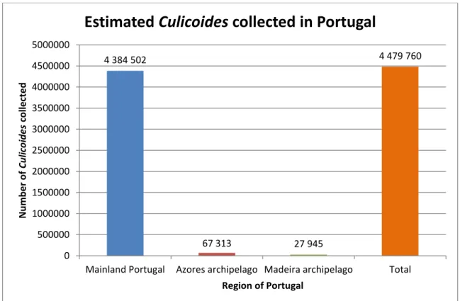
Os insetos capturados foram analisados fenotípica e geneticamente, mostrando a variação das espécies de Culicoides em território português. C. imicola foi a espécie mais capturada, representando 70,92% do total estimado de insetos capturados. Seguidamente, surgiu C. achrayi (10,34%), C. punctatus (9,34%), espécies do grupo Obsoletus (4,93%) e C. newsteadi (1,93%) como as espécies mais capturadas. C. pulicaris, espécie vetor do vírus da Língua Azul, perfez 0,08% do total estimado de insetos capturados (N=4.384.502).
A distribuição espacial das espécies de Culicoides em Portugal Continental apresentou padrões diferentes. Assim, enquanto que algumas espécies deste género estavam dispersas por todo o território (e.g., C. achrayi, C. punctatus), outras localizaram-se preferencialmente nas regiões norte e centro (e.g., C. deltus, C. heliophilus), diversas nas regiões centro e sul (e.g., C. nubeculosus, C. sahariensis) e algumas foram capturadas consistentemente em regiões específicas (e.g., C. impunctatus e C. lupicaris).
Vinte e duas espécies de Culicoides foram mencionadas pela primeira vez em Portugal: C. alazanicus, C. deltus, C. dewulfi, C. heliophilus, C. jumineri near C. bahrainensis, C. jurensis, C. kingi, C. lupicaris, C. malevillei, C. montanus, C. paolae, C. picturatus, C. remmi, C. riebi, C. santonicus, C. semimaculatus, C. simulator e C. subfagineus em Portugal continental, C. obsoletus, C. scoticus nas ilhas do arquipélago dos Açores onde ainda não tinham sido referidas e C. circumscriptus e C. newsteadi em todas as ilhas do mesmo arquipélago, com exceção das Flores e do Corvo. Para além destas, foi realizada a descrição de uma nova espécie para a ciência, Culicoides paradoxalis.
ix
Outro objetivo deste trabalho focou a morfologia, a genética e a ecologia do grupo Obsoletus, mostrando que a distribuição destas espécies é diferente em Portugal continental. Entre as quatro espécies presentes, C. dewulfi foi apenas identificada em três explorações no Norte do território português. As restantes três espécies do grupo Obsoletus encontravam-se presentes em todo o país, sendo C. obsoletus a espécie mais prevalente no Norte, Centro Sul e Sul de Portugal continental, enquanto que C. scoticus foi a espécie mais prevalente na região Centro Norte. Esta espécie foi menos comum que C. obsoletus e C. montanus nas regiões Centro Sul e Sul. Estas três espécies estão bem adaptadas ao território de Portugal Continental, com a exceção da região Centro Sul.
A correta identificação de espécies do complexo Obsoletus nem sempre é possível devido às suas elevadas semelhanças morfológicas. Nesse sentido, foi realizado um estudo com base em várias estruturas anatómicas destes insetos, que levaram à redefinição dos intervalos relativos ao rácio comprimento/largura do 3.º segmento do palpo e ao comprimento das espermatecas. Para além disso, a menção de várias aberrações anatómicas nestas espécies (fossetas sensoriais aberrantes, fossetas sensoriais duplas, segmentos do palpo fundidos, segmentos do palpo com alterações morfológicas, número irregular de espermatecas, espécimens com genitália masculina e simultaneamente com espermatecas, comprimento do 3.º artículo do palpo desigual dentro do mesmo exemplar, flagelómeros antenares fundidos e aberrantes) permitirá uma redução nos erros associados ao estudo destes insetos.
Uma chave de identificação para todas as espécies de Culicoides presentes em Portugal foi também elaborada durante este estudo que incidiu sobre cerca de 93.100 exemplares deste género.
Nesta tese, a produção de mapas de análise de risco baseados na associação de algumas variáveis abióticas climáticas (temperatura média no período mais seco e no período mais húmido) e edáficas (territórios artificializados, áreas agrícolas e agroflorestais, florestas e meios naturais e seminaturais, zonas húmidas e corpos de água) com a ocorrência de C. imicola, C. pulicaris, C. newsteadi, C. punctatus e espécies do grupo Obsoletus em Portugal Continental foi também efetuada. A partir destes mapas observou-se que a probabilidade de capturar C. imicola é maior nas regiões a sul do rio Tejo e na Beira Baixa, enquanto que C. pulicaris ocorre com maior frequência nas regiões do norte de Portugal continental, na linha costeira e em zonas com elevada altitude. A probabilidade de ocorrência de C. newsteadi é semelhante à de C. imicola, embora aquela espécie possa ser capturada em regiões ligeiramente mais a norte que esta última. C. punctatus pode ocorrer com uma probabilidade maior que 50% em qualquer parte de Portugal Continental e em quase todas as estações do ano. As espécies do grupo Obsoletus surgem nas regiões do norte de Portugal continental, estando estas espécies praticamente ausentes na região Centro Sul durante o outono e inverno. A distribuição do grupo Obsoletus é aproximadamente a oposta de C. imicola.
x
Finalmente, a avaliação de modificações morfológicas dos órgãos sensoriais localizados no 3.º segmento do palpo de C. imicola, usados na deteção do hospedeiro, foi realizada através de microscopia eletrónica de varrimento após um ensaio com inseticidas piretróides (permetrina e deltametrina) em diferentes concentrações. Este estudo revelou a completa destruição dos órgãos sensoriais, com uma provável influência na alimentação destes insetos.
Tendo em conta o impacto económico associado aos agentes patogénicos que transmitem, aliado à descoberta recente de novos serótipos do vírus da Língua Azul na Europa (BTV-25 e BTV-27), os resultados apresentados neste trabalho científico evidenciam e sustentam a importância do estudo entomológico dos insetos do género Culicoides.
xi
Table of Contents
Acknowledgements ... i Financial Support ... ii Abstract ... v Resumo ... vii Table of Contents ... xiIndex of Publications ... xvii
Index of Figures ... xxi
Index of Tables ... xxiii
Index of Annexes ... xxv
List of Abbreviations ... xxvi
Chapter 1: General Introduction ... 1
1.1. The importance of vector-borne diseases ... 3
1.2. Culicoides taxonomy – a classification far from finished ... 6
1.2.1. Culicoides genus and species identification ... 6
1.2.2. Culicoides subgenera ... 8
1.2.3. Culicoides groups and complexes ... 8
1.2.4. Culicoides species in Portugal – a historical review (1952-2005) ... 10
1.3. Culicoides and Bluetongue disease – a double-connected problem ... 12
1.3.1. Brief historical review of Bluetongue disease ... 12
1.3.2. Bluetongue disease ... 13
1.3.2.1. Etiology... 13
1.3.2.2. Affected species and pathogeny ... 13
1.3.2.3. Clinical signs ... 14
1.3.2.4. Control ... 15
1.3.3. Culicoides as vectors of Bluetongue disease ... 16
1.3.4. Bluetongue disease in mainland Portugal ... 17
1.4. African Horse Sickness disease in mainland Portugal ... 18
1.5. Adult Culicoides morphology ... 18
xii
1.5.1.1. Eyes ... 19
1.5.1.2. Antennas ... 20
1.5.1.3. Palpi ... 22
1.5.1.4. Mouth parts ... 23
1.5.1.5. Pharynx (cibarium) and posterior pharynx... 24
1.5.2. Thorax ... 24 1.5.2.1. Wings ... 25 1.5.2.2. Legs... 26 1.5.3. Abdomen ... 27 1.5.3.1. Females ... 27 1.5.3.2. Males ... 29
1.5.4. Morphological alterations in Culicoides genus ... 30
1.6. Culicoides biology ... 31
1.6.1. Life cycle... 31
1.6.2. Circadian rhythm and seasonality ... 33
1.6.3. Larval feeding behaviour ... 35
1.6.4. Adult feeding behaviour ... 36
1.6.4.1. Host preferences ... 37
1.6.5. Vector competence and vector capacity ... 38
1.6.6. Culicoides saliva ... 40
1.6.7. Female parity status ... 40
1.7. Factors influencing Culicoides occurrence ... 41
1.7.1. Climatic variables ... 41
1.7.2. Edaphic and topographical factors ... 43
1.7.2.1. Larvae ... 43
1.7.2.2. Pupae ... 44
1.7.2.3. Adults ... 44
1.7.2.4. Species habitat preferences ... 44
1.7.3. Host detection and availability ... 46
xiii
1.8.1. Microscopic techniques ... 47
1.8.2. Molecular biology techniques ... 48
1.9. Culicoides biting midges control ... 50
1.9.1. Culicoides control in larval and adult phases ... 50
1.9.2. Chemical control ... 51
1.9.2.1. Insecticides ... 51
1.9.2.2. Repellents and attractants ... 53
1.9.3. Biological control ... 54
1.9.3.1. Biocontrol agents ... 54
1.9.4. Biotechnological control ... 57
1.9.4.1. Hormones ... 57
Chapter 2: Morphological and molecular study of different Culicoides species in Portugal ... 59
2.1. Introduction... 61
2.2. Objectives ... 61
2.3. Materials and methods ... 62
2.3.1. The NESP and VectorNet European network ... 62
2.3.2. Insect sampling ... 63
2.3.3. Morphological and molecular identification of Culicoides species ... 65
2.3.3.1. Stereoscope and composed optical microscopy ... 65
2.3.3.2. “Culicoides sublupicaris” specimens ... 65
2.3.4. Data assessment of Culicoides species found in Portugal (1952-2005) ... 67
2.3.5. Design of an identification key for Portuguese Culicoides fauna ... 67
2.4. Results ... 67
2.4.1. Distribution of Culicoides species in Portuguese territory ... 67
2.4.1.1. Mainland Portugal... 68
2.4.1.2. Azores archipelago ... 73
2.4.1.3. Madeira archipelago ... 74
2.4.2. Culicoides paradoxalis – a new species for science ... 74
2.4.3. Information obtained after data assessment of Culicoides species found in Portugal (1952-2005) ... 78
xiv
2.4.4. Culicoides species captured in cattle and horse farms ... 81
2.4.5. Identification key for Culicoides female specimens... 82
2.5. Discussion ... 102
Chapter 3: Distribution of different species within Obsoletus group in mainland Portugal (2006-2009) ... 109
3.1. Introduction ... 111
3.2. Objectives ... 111
3.3. Materials and methods ... 112
3.3.1. NESP for BTD and insect sampling ... 112
3.3.2. Farm selection and abiotic factors analysis ... 112
3.3.3. Sample selection and morphological identification ... 113
3.3.4. Statistical analysis ... 115 3.4. Results ... 115 3.4.1. Species identification ... 115 3.4.1.1. Females... 116 3.4.1.2. Males ... 117 3.4.1.3. Culicoides spp. specimens ... 118
3.4.2. Distribution of Obsoletus group species in mainland Portugal ... 118
3.4.3. Abiotic factors ... 120
3.4.4. Anatomical aberrations ... 124
3.4.5. Anatomical measures and other observations ... 126
3.5. Discussion ... 127
Chapter 4: Ecologic characterization of Culicoides species in mainland Portugal ... 133
4.1. Introduction ... 135
4.2. Objectives ... 136
4.3. Material and methods ... 136
4.3.1. NESP for BTD and insect sampling ... 136
4.3.2. Edaphoclimatic information and statistical analysis ... 137
4.4. Results ... 138
xv
4.4.2. Cut-off points and presence/absence probability maps ... 141
4.5. Discussion ... 152
Chapter 5: Morphological modifications in Culicoides sensorial organs exposed to pyrethroid insecticides ... 159
5.1. Introduction... 161
5.2. Objectives ... 162
5.3. Materials and methods ... 162
5.3.1. Culicoides collection and identification ... 163
5.3.2. Selection of insecticides and production of impregnated papers ... 163
5.3.3. Insecticide susceptibility tests ... 164
5.3.4. Specimens preparation for SEM ... 164
5.4. Results ... 165
5.5. Discussion ... 168
Chapter 6: Conclusions and Future Perspectives ... 171
Bibliography ... 175
xvii
Index of Publications
Book Chapters
Alexandre-Pires, G., Ramilo, D., Diaz, S., Meireles, J., Boinas, F. & Pereira da Fonseca, I. (2010). Investigating morphological structures of Culicoides from Obsoletus complex by using Scanning Electron Microscopy and Composed Optical Microscopy. In A. Méndez-Vilas & J.D. Álvarez, Microscopy: Science, Technology, Applications and Education. (pp. 792-802). Badajoz, Spain: Formatex Research Center.
Scientific Articles
Jacquet, S., Garros, C., Lombaert, E., Walton, C., Restrepo, J., Allene, X., Baldet, T., Cetre-Sossah, C., Chaskopoulou, A., Delecolle, J-C., Desvars, A., Djerbal, M., Fall, M., Gardes, L., de Garine-Wichatitsky, M., Goffredo, M., Gottlieb, Y., Gueye Fall, A., Kasina, M., Labuschagne, K., Lhor, Y., Lucientes, J., Martin, T., Mathieu, B., Miranda, M., Pages, N., Pereira da Fonseca, I., Ramilo, D.W., Segard, A., Setier-Rio, M-L., Stachurski, F., Talla Seck, M., Venter, G., Balenghien, T., Guis, H., Chevillon, C., Bouyer, J. & Huber, K. (2015). Colonization of the Mediterranean Basin by the vector biting midge species Culicoides imicola: an old story. (Accepted by Molecular Ecology).
Jacquet, S., Huber, K., Pagès, N., Talavera, S., Burgin, L.E., Carpenter, S., Sanders, C., Djerbal, M., Lhor, Y., Lucientes, J., Miranda, M., Pereira da Fonseca, I., Ramilo, D.W., Setier-Rio, M-L., Bouyer, J., Chevillon, C., Balenghien, T., Guis, H. & Garros, C. (2015). Range expansion of the Bluetongue vector Culicoides imicola in continental France thanks to meteorological events. (Submitted to Scientific Reports). Ribeiro, R., Wilson, A.J., Nunes, T., Ramilo, D.W., Amador, R., Madeira, S., Baptista, F.M.,
Harrup, L.E., Lucientes, J. & Boinas, F. (2015). Spatial and Temporal Distribution of Culicoides Species in Mainland Portugal (2005-2010). Results of the Portuguese Entomological Surveillance Programme. PLoS ONE, 10(4), e0124019.
Ramilo, D., Garros, C., Mathieu, B., Benedet, C., Allène, X., Silva, E., Alexandre-Pires, G., Pereira da Fonseca, I., Carpenter, S., Rádrová, J. & Delécolle, J-C. (2013). Description of Culicoides paradoxalis sp. nov. from France and Portugal (Diptera: Ceratopogonidae). Zootaxa, 3745(2), 243-256.
Ramilo, D.W., Diaz, S., Pereira da Fonseca, I., Delécolle, J-C., Wilson, A., Meireles, J., Lucientes, J., Ribeiro, R. & Boinas, F. (2012). First report of 13 species of Culicoides (Diptera:Ceratopogonidae) in mainland Portugal and Azores by morphological and molecular characterization. PLoS ONE, 7(4), e34896.
Oral presentations
Ramilo, D. “Distribuição de espécies do grupo Obsoletus em diferentes regiões de Portugal Continental entre 2006 e 2009”. Seminars of Investigation by the Interdisciplinary Animal Health Research Center (CIISA). 09 de junho de 2015, Faculdade de Medicina Veterinária, Universidade de Lisboa, Portugal.
Ramilo, D. “Importância da identificação de espécies de Culicoides”. Seminars of Investigation by the Interdisciplinary Animal Health Research Center (CIISA). 07 de
xviii
Fevereiro de 2014, Faculdade de Medicina Veterinária, Universidade de Lisboa, Portugal.
Ramilo D, Alexandre-Pires G, Nunes T, Silva E, Pereira da Fonseca I. “Application of different laboratorial techniques in identification of organisms from Class Arachnida and Class Insecta (Phylum Arthropoda)”. Microscopy in research – An SPMicros Congress, Egas Moniz Cooperativa de Ensino Superior, Monte da Caparica, 9-10 dezembro 2013. Life Sciences: Oral 7, pp: 24-25.
Marques CS, Ramilo D, Meireles J, Duarte A, Gomes GS, Tavares L, Fonseca IP. “Detection of Leishmania DNA in Culicoides (Diptera: Ceratopogonidae) collected in leishmaniosis endemic regions of Portugal”. WorldLeish5, Porto Galinhas, Brasil, 13-17 maio 2013. Oral 220, pp: 168.
Ramilo DW, Pereira da Fonseca I, Meireles J, Lucientes J, Pacheco S, Ribeiro R, Madeira S, Boinas F. “Características ecológicas e climáticas favoráveis à ocorrência de 13 espécies do género Culicoides (DIPTERA: CERATOPOGONIDAE) referidas pela primeira vez em Portugal”. XVI Congresso Português de Parasitologia (SPP). 29 e 30 de Novembro de 2012, Faculdade de Medicina Veterinária, Universidade Técnica de Lisboa, Lisboa, Portugal. pp: 132-133.
Ramilo, D., Alexandre-Pires, G., Nunes, T., Meireles, J., Boinas, F., Pereira da Fonseca, I. “Application of SEM in the study of Culicoides (Diptera: Ceratopogonidae) morphological studies”. Congress of the Portuguese Society For Microscopy: Microscopy – a tool for the development of science (SPMicros 2012). 24-25 September 2012, Hospital D. Estefânia, Lisboa, Portugal. pp: 33.
Meireles, J., Alexandre-Pires, G., Ramilo, D., Diaz, S., Pereira da Fonseca, I. “Studying Morphological Structures of Culicoides Species by using Composed Optical Microscopy and Scanning Electron Microscopy”. 17th European Society for Vector Ecology Conference (ESOVE). 13-17 September 2010, Uniwersytet Wrocławski Wrocław, Poland. pp:63.
Oral presentations by invitation
Ramilo, D. “Existirão outros vetores de Leishmania para além dos Flebotomíneos?”. Comunicação oral a convite da Bastonária da Ordem dos Médicos Veterinários, Professora Doutora Laurentina Pedroso. IV Encontro de Formação e VII Congresso da Ordem dos Médicos Veterinários, 30 de Novembro e 1 de Dezembro de 2013, Centro de Congressos de Lisboa, Portugal.
Ramilo, D. “Estudo morfológico de espécies de insectos Culicoides, por microscopia convencional e de varrimento, no âmbito do Projecto Língua Azul”. Comunicação oral a convite do Centro de Investigação Interdisciplinar em Sanidade Animal (CIISA), no âmbito do ciclo de seminários do CIISA. 18 de Março de 2011, Faculdade de Medicina Veterinária, Universidade Técnica de Lisboa, Portugal.
Ramilo, D., Fonseca, I., Boinas, F. “Projecto Língua Azul – Envio de Amostras ao Laboratório”. Comunicação oral a convite do coordenador do Projeto de Vigilância Entomológica Nacional para a Língua Azul, Professor Doutor Fernando Boinas e da coordenadora do laboratório de Parasitologia e Doenças Parasitárias, Professora Doutora Isabel M. da Pereira Fonseca. 12 de Março de 2010, Direcção Geral de Veterinária – DGV, Lisboa, Portugal.
xix Posters
Ramilo DW, Alexandre-Pires G, Santos M, Pereira da Fonseca I (2014). “Estudo premilinar de padrões de interferência alar em 4 espécies do Género Culicoides (DIPTERA: CERATOPOGONIDAE)”. XVII Congresso Português de Parasitologia (XVII CPP), Faculdade de Farmácia da Universidade de Coimbra (FFUC), Coimbra. 20 e 21 de Novembro de 2014.
Ramilo DW, Venail R, Alexandre-Pires G, Nunes T, Pereira da Fonseca I. (2014). “Estudo preliminar sobre as alterações morfológicas observadas nos órgãos sensoriais dos insetos da espécie Culicoides imicola (Diptera: Ceratopogonidae) expostos a inseticidas piretróides”. VI Congresso da Sociedade Portuguesa de Ciências Veterinárias (SPCV): Ciências Veterinárias – Praxis e Futuro, INIAV/Oeiras, Lisboa. 3 a 5 de Abril de 2014.
Ramilo DW, Meireles J, Boinas F, Lucientes J, Pereira da Fonseca I. (2012). “Caracterização morfológica das espécies Culicoides dendriticus, C. malevillei e C. riebi (DIPTERA: CERATOPOGONIDAE) referidas pela primeira vez em Portugal”. XVI Congresso Português de Parasitologia, Faculdade de Medicina Veterinária, Universidade Técnica de Lisboa. 29 e 30 de Novembro de 2012.
Ramilo DW, Pereira da Fonseca I, Meireles J, Lucientes J, Boinas F. (2012). “Revisão do Género Culicoides (DIPTERA: CERATOPOGONIDAE) em Portugal”. XVI Congresso Português de Parasitologia, Faculdade de Medicina Veterinária, Universidade Técnica de Lisboa. 29 e 30 de Novembro de 2012.
Ramilo, D., Diaz, S., Pereira da Fonseca, I., Delécolle, J-C., Wilson, A., Marques, C., Meireles, J., Lucientes, J., Boinas, F. (2011). “Identificação de espécies de Culicoides (DIPTERA:CERATOPOGONIDAE) referidas pela primeira vez em Portugal Continental e no Arquipélago dos Açores por caracterização morfológica no âmbito do Programa Entomológico Nacional da Língua Azul”. V Congresso da Sociedade Portuguesa das Ciências Veterinárias – As Ciências Veterinárias para uma só Saúde, Instituto Nacional de Recursos Biológicos (INRB), IP, L-INIA/Fonte Boa, Vale de Santarém, Portugal. 13 a 15 de Outubro de 2011.
Ramilo, D., Pereira da Fonseca, I., Delécolle, J-C., Meireles, J., Lucientes, J., Boinas, F. “Preliminary Description of Culicoides (Diptera:Ceratopogonidae) species reported for the first time in mainland Portugal”. Abst. 17th European SOVE Conference. 13th – 17th September 2010, Uniwersytet Wrocławski, Wrocław, Poland. Poster 05; pp. 122.
xxi
Index of Figures
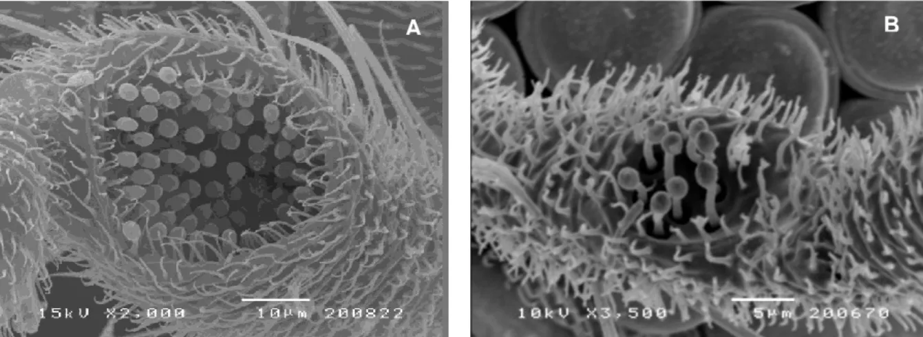
Figure 1.1. – Culicoides Latreille, 1809 (Diptera: Ceratopogonidae) biting midge ... 3 Figure 1.2. – Palearctic region ... 5 Figure 1.3. - Cells and veins of a Culicoides wing ... 7 Figure 1.4. – Culicoides fascipennis wing. ... 7 Figure 1.5. – Culicoides circumscriptus female wings with intraspecific variation ... 8 Figure 1.6. – Wing pattern of Obsoletus group species (original photo) ... 9 Figure 1.7. – Culicoides female specimen from Obsoletus group ... 19 Figure 1.8. – Culicoides derisor female’s head... 19 Figure 1.9. – Holoptic and dichoptic species ... 20 Figure 1.10. – Different kinds of sensilla found in Culicoides genus antenna ... 21 Figure 1.11. – Sensilla basiconica within the sensorial pit of the 3rd palpus segment of a
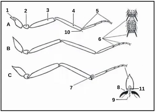
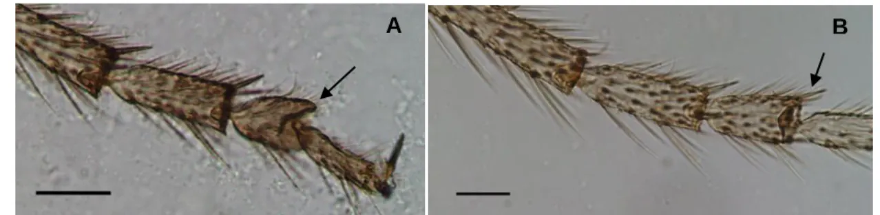
Culicoides circumscriptus female specimen (A) and a Culicoides obsoletus male specimen (B) ... 22 Figure 1.12. – Different conformations of the 3rd palpus segment. ... 23 Figure 1.13. – Sensorial pits of two Culicoides specimens ... 23 Figure 1.14. – Mandibles and maxillae of two Culicoides specimens ... 24 Figure 1.15. – Cibarium and posterior pharynx of three Culicoides specimens ... 24 Figure 1.16. – Adult Culicoides thorax ... 25 Figure 1.17. – Different wing patterns observed in Culicoides biting midges ... 25 Figure 1.18. – Fore, middle and hind legs of Culicoides biting midges ... 26 Figure 1.19. – 3rd and 4th tarsomere of middle legs of two Culicoides specimens ... 27
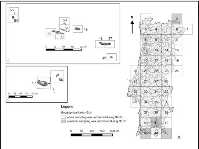
Figure 1.20. – Bifurcated claw of a C. obsoletus male hind leg ... 27 Figure 1.21. – Female Culicoides abdomen ... 28 Figure 1.22. – Spermathecae from two Culicoides specimens ... 28 Figure 1.23. – Sclerotic rings from two Culicoides specimens ... 29 Figure 1.24. – Male Culicoides abdomen ... 29 Figure 1.25. – Abdomen from two males Culicoides specimens ... 30 Figure 1.26. – Life cycle of Culicoides biting midges ... 31 Figure 1.27. – Culicoides female parity status ... 41 Figure 1.28. – A male C. circumscriptus parasitized with H. cataloniensis ... 55 Figure 1.29. – A female C. parroti parasitized with H. cataloniensis ... 56 Figure 1.30. – C. brevitarsis adults uninfected (A) and infected (B) with M. anisopliae ... 56 Figure 2.1. – Geographical Units in mainland Portugal and in Azores and Madeira archipelagos ... 62 Figure 2.2. – Miniature CDC light trap (A) placed within 30 m of animal enclosures, 1.70 m above ground (B). ... 64 Figure 2.3. – Flask containing a performed captured (A) and sample analysis with SM (B) . 64 Figure 2.4. – Onderstepoort light trap ... 65
xxii
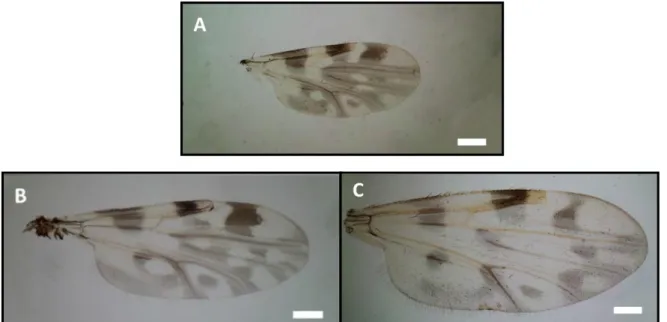
Figure 2.5. – Estimated Culicoides biting midges collected in Portugal during NESP (2005-2013) and VectorNet European network (2015). ... 67 Figure 2.6. – Wing pattern of Culicoides paradoxalis (A) and two close related species: C. newsteadi (B) and C. lupicaris (C) ... 75 Figure 2.7. – Sensilla basiconica of the 3rd palpus segment of C. newsteadi (A), C.
paradoxalis (B) and C. lupicaris (C) ... 76 Figure 2.8. – Sensilla coeloconica on the 1st antennal flagellomere (white arrows) of C.
newsteadi (A), C. paradoxalis (B) and C. lupicaris (C) ... 76 Figure 2.9. – 3rd palpus segment of C. newsteadi (A), C. paradoxalis (B) and C. lupicaris (C).
... 76 Figure 2.10. – 4th tarsomere of the middle legs (white star) of C. newsteadi (A), C.
paradoxalis (B) and C. lupicaris (C) ... 77 Figure 2.11. – Phylogenetic tree for Culicoides inferred from COI sequences ... 78 Figure 2.12. – Identification key for Portuguese Culicoides female specimens ... 83 Figure 3.1. – Division of mainland Portugal into 4 regions and localization of selected farms. ... 112 Figure 3.2. – Annual distribution of analysed specimens from Obsoletus group ... 114 Figure 3.3. – Wing pattern of Obsoletus group species ... 115 Figure 3.4. – Pubescence between ommatids ... 116 Figure 3.5. – Spermathecae of three Obsoletus group species ... 116 Figure 3.6. – 3rd palpus segment conformation and sensorial pit depth of species belonging
to Obsoletus complex ... 117 Figure 3.7. – Chitinous plates conformation: parallel (black arrow) and convergent (white arrow) ... 117 Figure 3.8. – Genital structures of C. obsoletus and C. scoticus males ... 118 Figure 3.9. – Distribution of Obsoletus group species in the four regions of mainland Portugal (2006-2009). ... 122 Figure 3.10. – Anatomical aberrations observed in Obsoletus group specimens ... 125 Figure 3.11. – Cut-off values of the 3rd palpus segment length/width ratio. ... 127
Figure 4.1. – Presence/absence probability maps for C. imicola per season. ... 142 Figure 4.2. – Presence/absence probability maps for Obsoletus group species per season. ... 144 Figure 4.3. – Presence/absence probability maps for C. pulicaris per season. ... 146 Figure 4.4. – Presence/absence probability maps for C. punctatus per season. ... 148 Figure 4.5. – Presence/absence probability maps for C. newsteadi per season. ... 150 Figure 5.1. – Culicoides imicola from control group and those submitted to different concentrations of deltamethrin active ingredients (after 24h of exposure). ... 166 Figure 5.2. – Culicoides imicola from control group and those submitted to different concentrations of permethrin active ingredients (after 24h of exposure) ... 167
xxiii
Index of Tables
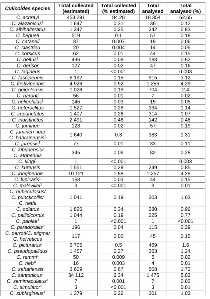
Table 1.1. – Viruses transmitted by Culicoides species present in Western Europe. ... 4 Table 1.2. – Culicoides species referred in mainland Portugal (1952-2005). ... 10 Table 1.3. – Culicoides species referred in Madeira and Azores archipelagos (1990-2005). 12 Table 1.4. – Repellent compounds previously evaluated on impregnated meshes. ... 53 Table 1.5. – Nematodes found parasitizing Culicoides biting midges. ... 55 Table 2.1. – Absolute and relative frequencies of estimated and analysed Culicoides collected during the NESP for BTD (2005-2013) and VectorNet European network (2015) in mainland Portugal. ... 68 Table 2.2. – Absolute and relative frequencies of estimated and analysed Culicoides spp. collected during the NESP for BTD (2005-2013) and VectorNet European network (2015) in mainland Portugal. ... 69 Table 2.3. – Culicoides species distribution in mainland Portugal by GUs. ... 70 Table 2.4. – Culicoides species seasonality in mainland Portugal. ... 72 Table 2.5. – Absolute and relative frequencies of estimated and analysed Culicoides biting midges collected during the NESP for BTD (2005-2012) in each Azores archipelago island. ... 73 Table 2.6. – Absolute and relative frequencies of estimated and analysed Culicoides biting midges collected during the NESP for BTD (2005-2012) in each Madeira archipelago island. ... 74 Table 2.7. – Main morphological characteristics for Culicoides paradoxalis differentiation from close related species (Ramilo et al., 2013). ... 75 Table 2.8. – Mean genetic distance of the COI region between and within species (Ramilo et al., 2013). ... 77 Table 2.9. – Comparison of Culicoides species referred in the 1952-2005 and 2005-2013 periods and 2015 in mainland Portugal ... 79 Table 2.10. - Comparison of Culicoides species referred in the 1952-2005 and 2005-2013 periods in Azores and Madeira archipelagos. ... 80 Table 2.11. – Culicoides species observed by Pena (2003) and Vila-Viçosa et al., (2009) and similar species. ... 80 Table 2.12. – Culicoides species captured near cattle and horse farms during VectorNet field work (2015). ... 81 Table 3.1. – Geographic coordinates of selected farms and their distance in straight line to the closest meteorological station. ... 113 Table 3.2. – 3rd palpus segment L/W ratio, as performed by Nielsen & Kristensen (2011) and
spermathecae size, as performed by Delécolle (1985). ... 115 Table 3.3. – Relative frequency of Obsoletus group midges per region of mainland Portugal (2006-2009). ... 118 Table 3.4. – Relative frequency of each Obsoletus group species per region (2006-2009). 119 Table 3.5. – Distribution of species from Obsoletus complex per region of mainland Portugal in 2006-2009 period. ... 119 Table 3.6. – Regions where no midges from Obsoletus group were captured per month. .. 119 Table 3.7. – Seasonal characterization of mean air temperature and precipitation in mainland Portugal (2006-2009) ... 121
xxiv
Table 3.8. – Relative frequency of Obsoletus group species captured per season and per year in each region of mainland Portugal. ... 124 Table 3.9. – Distribution of anatomical aberrations found in species from Obsoletus group. ... 125 Table 3.10. - Distribution of anatomical aberrations found in Culicoides spp. and proposed species distribution. ... 126 Table 3.11. – 3rd palpus segment L/W ratio and spermathecae length for C. montanus. .... 126
Table 3.12. – Chitinous plates conformation for C. obsoletus and C. scoticus species. ... 126 Table 4.1. – Climate and land cover variables chosen for model analysis. ... 137 Table 4.2. - Variables which influence positively and negatively the occurrence of different Culicoides species per season (2005-2013). ... 139 Table 4.3. – Cut-off points with the correspondent sensitivity and specificity mean values for each species and per season... 141 Table 5.1. – Concentrations of deltamethrin and permethrin at which Culicoides imicola specimens were submitted and number of analysed specimens with SEM (n=101). ... 165 Table 5.2. – Number of C. imicola specimens with morphological modifications in the sensilla basiconica of one or both 3rd palpus segment concerning different concentrations of
xxv
Index of Annexes
Annex 1.1. – First description of Culicoides species from Palearctic and other Earth ecozones by author(s) and year ... 221 Annex 1.2. – Synonymies of Culicoides species referred in Portugal (1952-2005). ... 224 Annex 1.3. – Host preferences of different Culicoides species from Palearctic ecozone. ... 225 Annex 1.4. – Habitat preferences of different Culicoides species from Palearctic ecozone. 230 Annex 2.1. – The nomenclature of territorial units for statistics, subdivision 3 ... 236 Annex 2.2. – Hoyer’s Medium ... 237 Annex 2.3. – Synonyms of Culicoides species referred for the first time in mainland Portugal during the NESP (2005-2013) and VectorNet European network (2015). ... 238 Annex 4.1. – Meteorological data obtained from the closest meteorological stations to the farms. ... 239
xxvi
List of Abbreviations
1-Octen-3-Ol Octenol
16S rRNA 16 Svedberg Ribosomal Ribonucleic Acid, a component of the 30 Svedberg small prokaryotic ribosomal subunit
18S rRNA 18 Svedberg Ribosomal Ribonucleic Acid, a component of the 40 Svedberg small eukaryotic ribosomal subunit
28S rRNA 28 Svedberg Ribosomal Ribonucleic Acid, the eukaryotic nuclear homologue of the prokaryotic 23S ribosomal RNA
AHSD African Horse Sickness Disease AHSV African Horse Sickness Virus AIC Akaike Information Criterion AinoV Aino Virus
BBC British Broadcasting Corporation BTD Bluetongue Disease
BTV Bluetongue Virus
BTV-1 Bluetongue virus serotype 1 BTV-2 Bluetongue virus serotype 2 BTV-3 Bluetongue virus serotype 3 BTV-4 Bluetongue virus serotype 4 BTV-6 Bluetongue virus serotype 6 BTV-8 Bluetongue virus serotype 8 BTV-9 Bluetongue virus serotype 9 BTV-10 Bluetongue virus serotype 10 BTV-11 Bluetongue virus serotype 11 BTV-16 Bluetongue virus serotype 16 BTV-25 Bluetongue virus serotype 25 BTV-26 Bluetongue virus serotype 26 BTV-27 Bluetongue virus serotype 27
C. Culicoides
CAD Carbamoyl-phosphate synthetase 2, aspartate transcarbamylase, and dihydroorotase, a gene which encodes several enzymes
xxvii
involved in pyrimidine biosynthesis CDC Centers of Disease Control
CIISA Centre of Research in Animal Health
cm Centimeter
CO2 Carbon Dioxide
COI Cytochrome Oxidase Subunit I
COII Cytochrome Oxidase Subunit II COM Composed Optical Microscopy
Csa Hot-summer Mediterranean climate
Csb Warm-summer Mediterranean climate
Cytb Cytochrome b
DDT Dichlorodiphenyltrichloroethane DEET N,N-Diethyl-3-methylbenzamide DGV Direção Geral de Veterinária DNA Deoxyribonucleic Acid
dNTPs Deoxynucleoside triphosphates
e.g. Exempli gratia, for example
EFSA European Food Safety Authority EHD Epizootic Hemorrhagic Disease EHDV Epizootic Hemorrhagic Disease Virus
EID Entente Interdépartementale pour la Démoustication ELISA Enzyme-Linked Immunosorbent Assay
et al. et alii, and others
FCUL Faculty of Sciences, University of Lisbon FMV Faculty of Veterinary Medicine
g/m2 Gram per square meter GPS Global Positioning System GUs Geographical Units
h Hour
HDPE High-density Polyethylene
ICZN International Code of Zoological Nomenclature IDIs Insect Development Inhibitors
xxviii
i.e. id est, that is
IGRs Insect Growth Regulators
IPMA Instituto Português do Mar e da Atmosfera ITS1 Internal Transcribed Spacer 1
ITS2 Internal Transcribed Spacer 2 JHAs Juvenile Hormone Analogs
km Kilometers
km/h Kilometers per hour
kV Kilovolt
LC99 Lethal concentration for 99% of a group of test animals
LCS Liquid crystals L/W Length/Width m Meter MgCl2 Magnesium chloride min Minute ML Maximum Likelihood ml Mililiter
MLV Modified Live Virus
mM Millimolar
mm Millimeter
μm Micrometer
mtDNA Mitochondrial Deoxyribonucleic Acid
N North
n Size of statistical sample
n.d. Not defined
NESP National Entomologic Surveillance Program
NUTS III Nomenclature of Territorial Units for Statistics, subdivision 3 OIE Office International des Epizooties
OVI Onderstepoort Veterinary Institute PCR Polymerase Chain Reaction
qPCR Quantitative Real-time Polymerase Chain Reaction |r| Pearson correlation coefficient
xxix rDNA Ribosomal Deoxyribonucleic Acid ROC Receiver Operating Curve
S South
s Second
SBV Schmallenberg Virus
SEM Scanning Electron Microscopy
SM Stereoscope Microscopy
spp. Species
syn. Synonymy
UK United Kingdom
ULisboa University of Lisbon USA United States of America
UV Ultraviolet
V Volt
VBD Vector-borne Diseases VP2 Outer capsid viral protein 2
W Watt
WAHID World Animal Health Information Database WHO World Health Organization
μl Microliter μM Micromolar μm Micrometers ° Degree °C Degree Celsius ' Minute γ Gamma δ Delta
1
3 1.1. The importance of vector-borne diseases
The morbidity and mortality associated to infectious diseases affects, since ever, the society at the politic, economic and cultural levels (Nelson & Williams, 2007). According to the World Health Organization (WHO, 2014), vector-borne diseases (VBD) account for more than 17% of all infectious diseases, causing more than 1 million deaths annually.
VBD cause huge economic harm when livestock is affected and even the threat of infection can prejudice trade (Lemon et al., 2008). The distribution of these diseases depends on complex environmental and social factors such as globalization of travel and trade, unplanned urbanization and environmental challenges. Due to increased human mobility, population growth, trade, climate, ecology and land use changes, some VBD are appearing in countries where they have never been reported before (de Vos, Hoek, Fischer, Koeijer & Bremmer, 2012; WHO, 2014; Faburay, 2015).
An efficient and cost-effective risk management, as well as a deeper insight of the pathways leading to the introduction and spread of VBD, requires an improved knowledge of the mentioned diseases coupled with interdisciplinary collaboration among scientific areas such as epidemiology, virology, entomology, ecology, climatology and economy (de Vos et al., 2012).
Among VBD, those transmitted by arthropods can be referred. There are some kinds of pathogenic agents (e.g., Arboviruses) that need an arthropod vector to complete their life cycle and that are transmitted to the vertebrate host by the arthropod bite or sting (Chippaux, 2003). There are several arthropods that can transmit different kinds of pathogenic agents, and, in this context, biting midges of the genus Culicoides Latreille, 1809 (Diptera: Ceratopogonidae) can be mentioned (Figure 1.1.).
Figure 1.1. – Culicoides Latreille, 1809 (Diptera: Ceratopogonidae) biting midge.
4
Culicoides are of major importance in Veterinary Medicine and Public Health since hematophagous females of this genus are known vectors of viruses, filarial nematodes and protozoans, like Bluetongue virus (BTV), Schmallenberg virus (SBV), African Horse Sickness virus (AHSV), Epizootic Haemorrhagic Disease virus (EHDV), Onchocerca cervicalis, Mansonella spp., Haemoproteus meleagridis, Leucocytozoon caulleryi, among others.
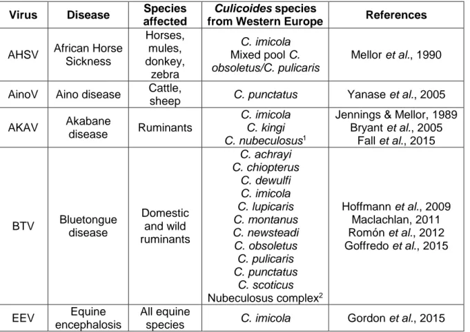
Bluetongue Disease (BTD), African Horse Sickness Disease (AHSD) and Epizootic Haemorrhagic Disease (EHD), all classified as notifiable by the Office International des Epizooties (OIE) (Venter, Labuschagne, Boikanyo, Morey & Snyman, 2011), have huge economical impact to producers and affected countries; as example, BTD, a viral disease which affects sheep and cattle, results in annual losses of approximately $3 billion due to morbidity and mortality of affected animals, trade embargoes and vaccination costs (Osburn, 2008; Maclachlan & Mayo, 2013). Table 1.1. shows some viral diseases transmitted by Culicoides species of Western Europe.
Moreover, they are a major tourist problem and an occupational concern for people working outdoors due to their nuisance and persistent, painful bite (Mullen & Durden, 2009; de Heredia & Lafuente, 2011; Elbers, Meiswinkel, van Weezep, Sloet van Oldruitenborgh-Oosterbaan, Kooi, 2013). Their bite is also responsible for the Equine Allergic Dermatitis, a severe allergic reaction observed in horses (Mullen & Durden, 2009).
Table 1.1. – Viruses transmitted by Culicoides species present in Western Europe. Virus Disease Species
affected
Culicoides species
from Western Europe References
AHSV African Horse Sickness Horses, mules, donkey, zebra C. imicola Mixed pool C. obsoletus/C. pulicaris Mellor et al., 1990
AinoV Aino disease Cattle,
sheep C. punctatus Yanase et al., 2005 AKAV Akabane
disease Ruminants
C. imicola C. kingi C. nubeculosus1
Jennings & Mellor, 1989 Bryant et al., 2005 Fall et al., 2015 BTV Bluetongue disease Domestic and wild ruminants C. achrayi C. chiopterus C. dewulfi C. imicola C. lupicaris C. montanus C. newsteadi C. obsoletus C. pulicaris C. punctatus C. scoticus Nubeculosus complex2 Hoffmann et al., 2009 Maclachlan, 2011 Romón et al., 2012 Goffredo et al., 2015 EEV Equine encephalosis All equine
5
Table 1.1. – Viruses transmitted by Culicoides species present in Western Europe (Continuation).
1Biting midges experimentally infected; 2Includes, at least, C. nubeculosus, C. puncticollis and C.
riethi.
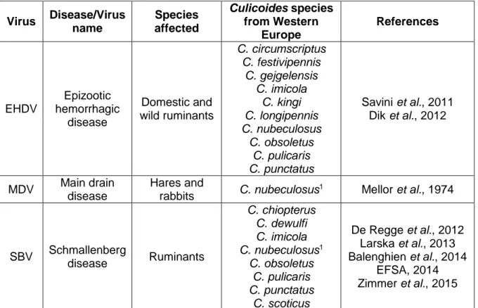
Culicoides are distributed worldwide and can be found in a wide range of habitats, from sea level to 4 000 m in altitude, and in almost all countries in the world, with exception of small regions of New Zealand, Patagonia, Hawaii islands, Iceland and Antarctica (Mellor, Boorman & Baylis, 2000; de Heredia & Lafuente, 2011). Planet Earth is divided into several zoogeographical regions, based on distributional patterns of animal organisms, and Europe is included in the Palearctic region (Figure 1.2.) (Procheş & Ramdhani, 2012). Although Culicoides species are distributed worldwide, some of them are characteristic of certain Earth regions (Wilson & Mellor, 2009; Mathieu et al., 2012) (Annex 1.1.).
Figure 1.2. – Palearctic region (adapted from Procheş & Ramdhani, 2012). Virus Disease/Virus name Species affected Culicoides species from Western Europe References EHDV Epizootic hemorrhagic disease Domestic and wild ruminants C. circumscriptus C. festivipennis C. gejgelensis C. imicola C. kingi C. longipennis C. nubeculosus C. obsoletus C. pulicaris C. punctatus Savini et al., 2011 Dik et al., 2012 MDV Main drain disease Hares and rabbits C. nubeculosus 1 Mellor et al., 1974 SBV Schmallenberg disease Ruminants C. chiopterus C. dewulfi C. imicola C. nubeculosus1 C. obsoletus C. pulicaris C. punctatus C. scoticus De Regge et al., 2012 Larska et al., 2013 Balenghien et al., 2014 EFSA, 2014 Zimmer et al., 2015
6
1.2. Culicoides taxonomy – a classification far from finished
According to Fauna Europaea website (2015), the taxonomic position of Culicoides genus is as follows: Kingdom Animalia Subkingdom Eumetazoa Phylum Arthropoda Subphylum Hexapoda Class Insecta Order Diptera Suborder Nematocera Infraorder Culicomorpha Superfamily Chironomoidea Family Ceratopogonidae Subfamily Ceratopogoninae Tribe Culicoidini Genus Culicoides
Since the first descriptions of these biting midges by William Derham in 1713 (as Culex), Carl von Linné in 1758 (as Culex genus) and finally Latreille in 1809 as Culicoides, many species inside this genus have been described, some of them being synonyms (Delécolle, 1985; Mathieu, 2011). Up to 2015, Culicoides genus was divided into 31 subgenus and 38 species groups (unplaced to subgenus). A total of 1 401 species is identified worldwide (1 355 extant species and 46 fossil species), although a hard systematic work must be done in order to establish some organization inside this genus (Borkent, 2015a).
1.2.1. Culicoides genus and species identification
Ceratopogonidae family includes four subfamilies: Dasyheleinae, Leptoconopinae, Forcipomyiinae and Ceratopogoninae (de Heredia & Lafuente, 2011). Ceratopogonids characteristic wing venation allows us to distinguish these midges from other groups of flies (Mullen & Durden, 2009). Midges from Ceratopogonidae family are sometimes very similar between different genus and an accurate evaluation with stereoscope microscopy (SM) must be performed to separate Culicoides genus from others (de Heredia & Lafuente, 2011). Culicoides biting midges have two open radial cells with similar sizes on the wing (although in some species the second can be bigger than the first) (Figures 1.3. and 1.4.) (Mathieu, 2011). Furthermore, the presence of not aligned spines in the first hind tarsus can also be used to distinguish Culicoides specimens from other ceratopogonids (de Heredia & Lafuente, 2011).
7
Figure 1.3. - Cells and veins of a Culicoides wing (scheme by Santos, M.).
r-m – Radio-medial crossvein; r1 – First radial cell; r2 – Second radial cell; r3 – Third radial cell;
m1 – First medial cell; m2 – Second medial cell; cua1 – Anterior cubital cell; a – Anal cell;
Pst – Poststigmatic pale spot; M1 – First medial vein; M2 – Second medial vein; CuA1 – First
branch of anterior cubital vein.
Figure 1.4. – Culicoides fascipennis wing.
The two radial cells are inside the dark circle. Scale bar: 200 µm. Original photo.
The wing pattern present in most Culicoides species results from the distribution of small and big hairs on wing surface named microtrichia and macrotrichia, respectively. The density of these hairs gives to the wings the appearance of dark and light spots or patches (Figure 1.4.). The shape, disposition or absence of these spots is of most importance, since it is the main characteristic that permits Culicoides species identification (Mullen & Durden, 2009; de Heredia & Lafuente, 2011; Mathieu, 2011; Service, 2012). However, it must be taken into account that wing pattern can show high levels of intraspecific variation (Chaker, Delécolle & Kremer, 1980; Wirth, Dyce & Peterson, 1985; Wirth, Dyce & Spinelli, 1988; Felippe-Bauer, Cáceres, Silva, Valderrama-Bazan & Gonzales-Perez, 2005; Felippe-Bauer & Silva, 2006; Felippe-Bauer et al., 2008; Felippe-Bauer, Damasceno, da Trindade & Py-Daniel, 2010), giving rise to confusing situations and sometimes rendering difficult to allocate similar wing patterns to either the same or to different species (Figure 1.5.).
8
Figure 1.5. – Culicoides circumscriptus female wings with intraspecific variation.
Intraspecific variation can be observed in the spot inside the dark circle. Scale bars: 200 µm. Original photos.
1.2.2. Culicoides subgenera
Some Culicoides species are grouped into the same subgenus, for they share some identic morphological characteristics (Borkent, 2015b,c). In Iberian Peninsula, the following Culicoides subgenera can be found: Avaritia Fox, 1955, Beltranmyia Vargas, 1953, Culicoides Latreille, 1809, Monoculicoides Khalaf, 1954, Oecacta Poey, 1853, Pontoculicoides Remm, 1968, Silvaticulicoides Glukhova, 1977, Synhelea Kieffer, 1925 and Wirthomyia Vargas, 1973 (Iberfauna, 2008; de Heredia & Lafuente, 2011; Borkent, 2015b). Almost all Culicoides species vectors of BTV and SBV in Europe belong to Avaritia subgenus (Wirth & Dyce, 1985; Meiswinkel, Gomulski, Delécolle, Goffredo & Gasperi, 2004a; de Heredia & Lafuente, 2011; Mathieu, 2011). Several species do not have a defined subgenus and are included into the Oecacta subgenus, showing, once more, that Culicoides systematic classification is far from finished (Jones et al., 1985; de Heredia & Lafuente, 2011). Each subgenus has a type species (Harrup, Bellis, Balenghien & Garros, 2014).
1.2.3. Culicoides groups and complexes
Some Culicoides species share the same or have similar wing patterns and, in this way, their differentiation is done under evaluation of other morphological characteristics; sometimes, these species are arranged into the same group, for they exhibit a notable degree of morphological differentiation although they are close related species (Mathieu, 2011; Harrup et al., 2014).
There are many different Culicoides groups mentioned in literature and, from those, Imicola, Obsoletus and Pulicaris groups [defined in the past as complexes (Meiswinkel et al., 2004a; Nolan et al., 2007; Pagès et al., 2009)] can be referred, since they have many Culicoides
9
species proven or incriminated as vectors of several animal and human diseases worldwide (Hoffmann et al., 2009; Mathieu, 2011; Rasmussen et al., 2012; De Regge et al., 2012; Harrup et al., 2014).
‘Species complex’ is composed by isomorphic species, which are extremely difficult to be morphologically differentiated in one or both sexes (Harrup et al., 2014).
As examples:
1) Obsoletus group consists of five species in Western Europe, all of them from Avaritia subgenus, with the same wing pattern: Culicoides obsoletus, C. scoticus, C. chiopterus, C. dewulfi and C. montanus (Figure 1.6.) (Meiswinkel et al., 2004a; Garros, Mathieu, Balenghien, Cêtre-Sossah & Delécolle, 2010; Venail et al., 2012; Meiswinkel et al., 2014a). Inside this group and after composed optical microscopy (COM) evaluation, C. chiopterus and C. dewulfi female specimens can be easily distinguished from those of the other three species. These last ones, due to their similar morphologic conformation and very difficult differentiation, even after COM evaluation, are grouped into the Obsoletus complex (Delécolle, 1985; Meiswinkel et al., 2004a; Garros et al., 2010; Harrup et al., 2014). Concerning males of C. obsoletus and C. scoticus species, they exhibit sufficient and strong differences on their genitalia which permits their differentiation (Delécolle, 1985; Alexandre-Pires et al., 2010; Garros et al., 2010). However, males from C. obsoletus and C. montanus are more difficult to distinguish between them since their genitalia differences are minimal (Mathieu, 2011; Kirkeby & Dominiak, 2014).
Figure 1.6. – Wing pattern of Obsoletus group species.
Scale bar: 200 μm. Original photo.
2) Culicoides subgenus is composed by several groups and complexes: Pulicaris and Newsteadi groups and Pulicaris, Newsteadi, Fagineus and Impunctatus complexes, just to mention some (Meiswinkel et al., 2004a; Gomulski, Meiswinkel, Delécolle, Goffredo & Gasperi, 2006; EFSA, 2008; Pagès et al., 2009; Harrup et al., 2014). Several works have shown a high level of variation within species of these groups and complexes (Meiswinkel et al., 2004a; Pagès et al., 2009), being also a subject far from finished. Both Obsoletus and Pulicaris groups are ubiquitous across the Palearctic region (Wilson & Mellor, 2009).