http://dx.doi.org/10.1590/1806-9061-2016-0406
Author(s)
Chang ZI
Zhang HI
Wu XI
Nabi FI
Rehman MUI
Yuan XI
Mehmood KI,II
Zhou DI
I College of Veterinary Medicine, Huazhong
Agricultural University, Wuhan 430070, People’s Republic of China.
II College of Veterinary and Animal Sciences,
Islamia University of Bahawalpur-63100, Pakistan
Chang Z and Zhang H made equal contribu-tions to this article.
Mail Address
Corresponding author e-mail address Dhongai Zhou
College of Veterinary Medicine, Huazhong Agricultural University, Wuhan 430070, People’s Republic of China.
Email: bigdefoot@163.com
Keywords
AQP2; renal dose dopamine; kidney; broiler chickens.
Submitted: 08/October/2016 Approved: 16/February/2017
ABSTRACT
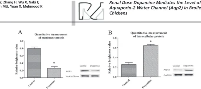
Aquaporin 2 (AQP2) is a small protein located in the collecting tubules of kidneys; it plays an important role in the concentration and production of urine. The aim of this study was to determine the expression level of the AQP2 gene in the kidney of broiler chickens after the administration of renal dose dopamine. Broiler chickens (25 days-old) were randomly divided into two groups (n=20 per group): intravenous administration of saline solution (control group) or renal-dose dopamine (dopamine group). The expression and localization of the AQP2 gene were evaluated by real-time quantitative polymerase chain reaction (RT-qPCR) and immunohistochemistry (IHC), respectively. The protein level of AQP2 was analyzed by western blot analysis. The dopamine group presented no significant difference (p>0.05) in the biochemical criterion or mRNA expression of AQP2 compared with the control group. However, AQP2 protein level was significantly reduced (p<0.05) in the membrane of renal tubular epithelial cells. In contrast, protein level was significantly increased (p<0.05) in the cytoplasm of the dopamine group compared with the control group. Moreover, AQP2 protein was apparently more distributed and localized in the cytoplasmic vacuoles than in the membranes of the kidney in the renal-dose dopamine administered chickens group. In conclusion, present findings suggest that renal dose dopamine mediates the level of AQP2 protein via shuttle from the cell membrane to the cytoplasm rather than changing the expression of AQP2 gene to adjust the secretion and absorption of water in kidney.
INTRODUCTION
Aquaporins (AQP) are important integral membrane proteins for rapid and selective transport of water and some small solutes across cell membranes in mammals and plants. AQPs have strong water permeability, and water molecules across renal tubules through various channels (Preston et al., 1992). In the last decade, 13 water-channel proteins were found in mammals, namely AQP0 to AQP12 (Yang, 2000; Verkman, 2002; Hatakeyama et al., 2001). AQPs present an open pore that can control the channel insertion and retrieval from the cell surface for transmembrane water transport (Marinelli et al.,
1997; Nielsen et al., 1993). AQP2 (water channel of the collecting duct, WCH-CD) is one of the best-characterized water channels located in the kidney collecting tubules and duct, distributed in cell apical plasma membrane and intracellular vesicles (Verkman, 2002). AQP2 water channel proteins are affected by hormones in the body, and also are the main target of the hormone arginine-vasopressin (AVP) for regulation of collecting duct water permeability (Deen et al., 1994, Digiovanni
et al., 1994). Various studies have confirmed that altered expression
disorders in animals (Nielsen et al., 2002). Inadequate cell-surface expression of AQP2 results in nephrogenic diabetes insipidus, whereas increased AQP2 cell surface expression and excessive water reabsorption are observed in congestive heart failure and preeclampsia in animals (Marples et al., 1995).
Dopamine (DA) is an important catecholamine neurotransmitter in mammals, and it is also synthesized in plants and tubule cells. DA is an important sodium homeostasis regulator, and plays a critical role in memory and behavior. Renal dose DA can induce natriuresis and diuresis via acute inhibition of most sodium reabsorption by the renal tubules, and it is widely used to improve renal function in the treatment of a variety of renal diseases (Jeon et al., 2014). Based on pharmacological characteristics, DA is divided into two subtypes of receptors (D1 and D2) and signal transduction pathways. D1 dopamine receptors play an important role in diuresis via combine with dopamine. As a diuretic and natriuretic hormone, dopamine can act on renal tubules to inhibit sodium reabsorption (Nielsen & Pedersen, 1997).
AQP2 has been investigated in several animal models of kidney diseases and kidney tubule defects. In addition of maintaining and regulating water balance, AQP2 is also important for maintaining the structural and functional integrity of the kidney tubules. Identifying changes in AQP2 expression levels may allow understanding the function of DA in water-electrolyte metabolism. In this context, the reviewed literature shows that there is little research conducted on the role of dopamine on the expression of genes encoding AQP2 in the collecting ducts of broiler chickens. The objective of the present study was to explore the influence of dopamine on the AQP2 gene expression and protein level to understand the mechanism of water and salt metabolism regulation by dopamine in broiler chickens.
MATERIALS AND METHODS
Experimental design and intravenous administration methods
Forty 1-day-old AA broiler chicks were purchased from a commercial hatchery (Chia Tai Animal Husbandry co., ltd., Wuhan, China), and were reared under recommended temperature and standard hygienic conditions. Birds were fed a regular diet ad libitum.
After 25 days, the birds were randomly and equally divided into two groups and anesthetized. The control group (n=20) received intravenous saline solution
by micro-injection (Hong zhan SK-500II, Shenzhen) at constant speed (2 mL/h) for 4 hours, whereas the dopamine group (n=20) received intravenous renal-dose dopamine (5 µg/kg/min) by micro-injection at constant speed (2 mL/h) for four hours. At the end of the injection period, all birds from both groups were sacrificed by cervical dislocation. Blood was collected. Blood samples were centrifuged at 3000×g for 20 min for the separation of serum, and stored at -20 °C until subsequent use. Kidney samples were removed, and fixed in 4% paraformaldehyde. Some of the kidney tissues were dissected out, immediately frozen in liquid N2, and then stored at -70 °C until analyses.
Biochemical criterion determination
The concentration of sodium, potassium, chlorine, creatinine, and urea nitrogen in the serum were measured using a semi-automatic biochemical equipment (COULTER®LH 750, Guangdong).
Immunohistochemistry (IHC)
Formalin-fixed kidney tissues were embedded in paraffin, and 4-μm thick histological sections were cut and mounted on polylysine-coated slides. After de-waxing with xylene and antigen recovery, the slides were washed thrice in peroxidase blocking solution (DakoCytomation, Carpinteria, CA, USA). The slides were then incubated with rabbit anti-goat polyclonal antibodies (KPL) at 1:600 dilution at 4 °C overnight (Tuojie Biological Technology Co., Ltd, Wuhan). After washing with PBS wash buffer, sections were incubated at 37 °C for 1.5 h with horseradish peroxidase-conjugated anti-rabbit secondary antibo-dies (Tuojie Biological Technology Co., Ltd, Wuhan). The immunolabeled slides were examined under a microscope (Olympus CX31; Olympus, Japan). Primary antibodies were removed from the negative control samples.
RNA extraction and real-time quantitative reverse transcription PCR (RT-qPCR)
in quadruplicate using the Step One-Plus TM
Real-Time PCR System (Applied Biosystems) with specific forward (5’-TGCCGTTGGACATCTCCTTGG-3’) and reverse (5’-CCTCTCCGAGAAGGTTTTGGA-3’) primers for the AQP2 gene expression analysis. The RT-qPCR mixture contained 6.8 μL RNase-free water, 2 μL cDNA, 10 μL Tip Green qPCR SuperMix, 0.4 μL Passive Reference Dye, 0.4 μL of each forward and reverse primer (working concentration: 10 μL mol/L) in a 20 μL reaction mixture. All RT-qPCR reactions were performed with following thermal cycling parameters: 95 °C for 30 s, 40 amplification cycles at 95 °C for 8 s, 59 °C for 30 s, and 72 °C for 30 s. All reaction mixtures were normalized against the reference gene GAPDH.
Protein extraction and Western blot
Kidney samples were washed with PBS and homogenized 4-5 times every ten seconds in dissection buffer (500 µL/100mg) (Beyotime Institute of Biotechnology, Hangzhou, China), and then kept for 30 min at 4 °C. The supernatant was obtained after centrifugation at 17,000 × g for 20 min at 4 °C. Protein was separated into membrane protein (pellets) and intracellular protein (supernatant) after centrifugation. The concentration of the total proteins was determined by Coomassie Brilliant Blue g-250 method, and samples were stored at -70 oC for subsequent use.
Protein samples (40 µg) were mixed with the loading buffer and then electrophoretically separated by SDS-PAGE on 12% polyacrylamide gels at 100 mV and transferred at 130 mV for 1 h and 40 min to nitrocellulose filter membranes (Millipore, BioSharp, Anhui, China). The membranes were incubated in 5% skimmed milk at room temperature for 1 h to prevent nonspecific binding sites, and then incubated with goat anti-rabbit primary antibody IgG-FITC (1:5000) overnight at 4 °C (Tuojie Biological Technology Co., Ltd, Wuhan). The membranes were washed 3 times with TBST (tris-buffered saline containing 0.1% Tween 2.0) for 5 min each then incubated with goat anti-mouse secondary antibody (1:5000 dilution) for 30 min at room temperature (Tuojie Biological Technology Co., Ltd, Wuhan). The membranes were again washed four times with TBST. After washing, images were captured with an imaging system (UVP, Upland, CA,USA).
Statistical analysis
Data were submitted to one-way analysis of variance. The Student’s t-test was applied to compare the mean differences between control and renal-dose dopamine groups. Differences were considered statistically significant when p<0.05. Values are
presented as means ± standard error of mean (SEM). All statistical analyses were performed by using the SPSS 19.0 software.
RESULTS
Serum biochemical criterion analysis
Serum concentrations of sodium, potassium, chlorine, creatinine, and urea nitrogen were not statistically different (p>0.05) between the control and the dopamine groups (Table 1).
Table 1 – Biochemical criterion analysis of serum between control and renal-dose dopamine group.
Items Control group Renal dose dopamine
Sodium (mmol/L) 144.60±2.35 144.80±2.65
Serum potassium (mmol/L) 6.04±0.29 6.07±0.33
Serum chlorine (mmol/L) 107.60±3.47 106.95±3.56
Urea nitrogen (mmol/L) 0.85±0.04 0.85±0.04
Creatinine (µmol/L) 58.838±0.732 58.785±0.733
Immunohistochemical (IHC) evaluation of the kidney
In order to confirm localization of AQP2 gene, immunohistochemical analysis was performed in the kidneys of the control and the dopamine groups. The results revealed that AQP2 was expressed in the collecting-duct epithelial cells and intracytoplasmic vacuoles of the dopamine-injected broilers (Fig. 1B), but not in the control broilers (Fig. 1A).
Figure 1 – Expression pattern of AQP2 by immunohistochemistry (IHC) analysis of membrane protein and intracellular protein from broiler chicken kidney tissue. Panel A: negative control; Panel B: AQP2 in collecting duct epithelial cells of medullary or intracytoplasmic vacuoles as compared to renal dose dopamine groups, the antibodies mainly distribute in kidney collecting duct epithelial cells (Arrow) of medullary. (magni-fication = 40x).
Western blot analysis
AQP2 Expression levels in renal-dose and control group
The mRNA expression of the AQP2 gene in kidney tissue was investigated in control and dopamine groups. The results indicated that AQP2 gene expression was not significantly different between these two groups (p>0.05) (Fig. 3).
Figure 3 – Effect of renal dose dopamine on AQP2 mRNA expression in control and dopamine administered group. Results are expressed in arbitrary unit (AU).
DISCUSSION
AQP2 is a channel located in the apical membrane of the collecting ducts of the kidneys that responds to vasopressin (VP) regulation both in vitro and in vivo
(Deen et al., 1994; Deen et al., 2002). However, it has also been observed that AQP2 has a bipolarized distribution in the kidney collecting ducts, with both basolateral and apical/subapical expression. However, the physiological function of basolaterally-located AQP2 is not yet understood (Coleman et al., 2000). AQP2 plays an important role in the urine concentration mechanism, as approximately 10% of glomerular filtration liquid is resorbed under the participation of AQP2 (Nielsen et al., 2002; Yang, 2000; Verkman, 2002; Yang et al., 2001). The level of antidiuretic hormone (ADH) is positively correlated with the ratio
between collecting tube cell membrane AQP2 and intracellular AQP2. When ADH levels are reduced, membrane AQP2 is transferred to the intracellular milieu, with consequent reduction of membrane AQP2 content, which results in reduced water absorption, and ultimately urine dilution (Frokiser et al., 1998).
Two main types regulate the expression and localization of the sodium transport protein, long-term adjustment and instantaneous adjustment functions. Long-term adjustment mainly consists of adjusting the content and shuttle of these proteins, whereas instantaneous adjustment includes post-translational modifications, which may enhance endocytosis (Yang, 2000). In the present study, mainly concentrated in the content changed of water channel proteins, but the instantaneous adjustment of protein modification and shuttle is rarely reported.
Urea nitrogen is the end product of protein metabolism, and is an indicator of general glomerular function. Serum creatinine is a sensitive indicator of glomerular filtration rate, and directly represent the filtration function of the kidney (Digiovanni et al.,
1994). Our study did not find any significant changes in serum sodium, potassium, chlorine, creatinine, or urea nitrogen levels, indicating that glomerular filtration rate did not increase. This suggests that the effect of renal dose dopamine on natriuresis and diuresis did not occur through changes in the glomerular filtration rate (Verkman, 2002). Therefore, it is speculated that renal dose dopamine influences natriuresis and diuresis mainly through changes in the renal tubular reabsorption function in broilers.
expression of AQP2 in the membrane, while increased its expression in the cytoplasm. Consequently, water absorption was reduced in collecting duct epithelial cells and intracytoplasmic. In addition, no changes in the expression of AQP2 mRNA was found by RT-qPCR analysis, indicating that the continuous perfusion of renal dose dopamine has no influence on the expression of the AQP2 gene. The results indicate that renal dose dopamine does not affect the update of AQP2 protein. During this experiment, the chickens’ feces change from soft to clear water as urine volume increased after the injection of renal dose dopamine. Thus, it may be speculated that renal dose dopamine can mediate the shuttle of AQP2 from membrane to the cytoplasm. Previous studies reported that AQP2 localization in the membrane can be rapidly determined by ubiquitination and dephosphorylation, AQP2 finished endocytosis after dephosphorylation by the proteasome pathway, and returned to the cell membrane via phosphorylation (Deen et al., 1994, Deen et al., 2002). Thus, the rapid internalization of the water channel protein can reduce renal tubular reabsorption of water and increase the velocity of urine formation (Kamsteeg et al., 2006). Therefore, this study indicates that the changes in natriuresis and diuresis promoted by renal dose dopamine are not influenced by the glomerular filtration rate, but by the inhibition of water reabsorption in the renal tubular epithelial cells of broiler chickens.
In conclusion, our study indicates that the effect of dopamine on natriuretic diuresis occurs mainly by the instantaneous adjustment rather than by the long-term adjustment of the AQP2 shuttle in broiler chickens.
ETHICS APPROVAL
All the experimental procedures were conducted according to the guidelines of the Animal Welfare and Research Ethics Committee of Huazhong Agricultural University (Wuhan, China).
ACKNOWLEDGMENT
This study was supported by Hubei Provincial Natural Science Foundation of China (Grant No: 2014CFB244),National NaturalScience Foundation of China (No:31502132) and Fundamental Research Funds for the Central Universities (Program No: 2011PYO78).
REFERENCES
Chemla D, Castelain V, Humbert M, Hebert JL, Simonneau G, Lecarpentier Y, et al. New formula for predicting mean pulmonary artery pressure using systolic pulmonary artery pressure. Chest 2004;126:1313-1317.
Chemla D, Castelain V, Provencher S, Humbert M, Simonneau G, Hervé P, et al. Evaluation of various empirical formulas forest estimating mean pulmonary artery pressure by using systolic pulmonary artery pressure in adults. Chest 2009;135:760-768.
Coleman RA, Wu DC, Liu J, WadeJ B. Expression of aquaporins in the renal connecting tubule. American Journal of Physiology-Renal Physiology 2000;279:874-883.
Deen PM, Verdijk MA, Knoers NV, Wieringa B, Monnens LA, Vanos CH, et al. Requirement of human renal water channel aquaporin-2 for vasopressin-dependent concentration of urine. Science 1994;264:92-95.
Deen PM, Van Balkom BWM, Savelkoul PJM, Kamsteeg EJ, Van Raak M, Jennings ML, et al. Aquaporin-2: COOH terminus is necessary but not sufficient for routing to the apical membrane. American Journal of Physiology-Renal Physiology 2002;282:330-340.
Digiovanni SR, Nielsen S, Christensen EI. Regulation of collecting duct water channel expression by vasopressin in Brattle boro rat. Proceedings of the National Academy of Sciences of the United States of America 1994;91:8984-8988.
Elkayam U, Ng TM, Hatamizadeh P, Janmohamed M, Mehra A. Renal vasodilatory action of dopamine in patients with heart failure: magnitude of effect and site of action. Circulation 2008;117:200 205.
Frokiser J, Marples D, Knepper MA, Nielsen S. Pathophysiology of aquaporin-2 in water balance disorders. The American Journal of the Medical Sciences 1998;316(5):291-299.
Hatakeyama S, Yoshida Y, Tani T. Cloning of a new aquaporin (AQP10) abundantly expressed in duodenum and jejunum. Chemical and Biophysical Research Communications 2001;287(4):814-819.
Jeon BK, Kang MK, Lee GT, Lee KK, Lee HS, Woo WH, et al. Epa attenuates ultraviolet radiation-induced downregulation of aquaporin-3 in human keratinocytes. Archives of Pharmacal Research 2014;38(8):1552-1560.
Kamsteeg EJ, Hendriks G, Boone M, Konings IB, Oorschot V, VanderSluijs P, et al. Short- chain ubiquitination mediates the regulated endocytosis of the aquaporin 2 water channel. Proceedings of the National Academy of Sciences of the United States of America 2006;103:18344 -18349.
Marinelli RA, Pham L, Agre P, Russo NF. Effect of acetazolamide on aquaporin-1 and fluid flow in cultured choroids plexus. Journal of Biological Chemistry 1997;272:12984-12988.
Marples D, Christensen S, Christensen EI, Ottosen PD, Nielsen S. Lithium-induced down regulation of aquaporin-2 water channel expression in rat kidney medulla. Journal of Clinical Investigation 1995;95(4):1838-1845.
Nielsen CB, Pedersen EB. Abnormal distal tubular sodium reabsorption during dopamine in fusion in patients with essential hypertension evaluated by the lithium clearance methods. Clinical Nephrology 1997;47(5):304-309.
Nielsen S, DiGiovanni SR, Christensen EI, Knepper MA, Harris HW. Cellular and sub cellular immuno localization of vasopressin-regulated water channel in ratkidney. Proceedings of the National Academy of Sciences of the United States of America 1993;90(24):11663-11667.
Nielsen S, Frokiaer J, Marples D, Kowon, T-H, Agre P, knepper MA. Aquaporins in the kidney: from molecules to medicine. Physiological Reviews 2002;82:205-244
Preston GM, Carroll TP, Guggino WB. Appearance of water channels in Xenopus oocytes expressing red cell CHIP28 protein. Science 1992;256(5055):385-387.
Verkman AS. Physiological importance of aquaporin water channels. Annals of Medicine 2002;34(3):192-200.
Yang B. The human aquaporin gene family (review). Current Genomics 2000;1(1):91-102.