RESUMO.-
[
Formação de biofilme por
Rhodococcus equi
e posspivel associação com resistência a macrolídeos.
]
Rhodococcus equi
é um patógeno intracelular facultativo,
o qual causa pneumonia piogranulosa severa em potros e
lesões semelhantes à tuberculose em humanos. A sua
capa-cidade de formar biofilme foi descrita em cepas humanas,
isoladas a partir de doenças crônicas associadas a falhas
de tratamento. Este estudo teve como objetivo verificar a
formação de biofilme por 113 cepas de
R. equi
, isoladas a
partir de amostras de equinos (clínicas e fecais),
utilizan-do-se dois diferentes métodos (biofilme em cultura - com e
sem adição de glicose - e microscopia de epifluorescência).
Além disso, buscou-se determinar a eficácia da azitromi
-cina, claritromicina e eritromicina sobre biofilme conso
-lidado de
R. equi
. Verificou-se 80,5% (26/41) e 63% dos
isolados (58/72) positivos para formação de biofilme, em
amostras fecais e clínicas, respectivamente. A adição de
gli-cose amentou a formação de biofilme em amostras fecais,
mas não em amostras clínicas. Os antimicrobianos aqui
tes-tados não foram capazes de erradicar
R. equi
em biofilme
consolidado, mesmo em concentrações elevadas. Este é o
primeiro estudo a demonstrar a formação de biofilme por
cepas de
R. equi
isoladas a partir de amostras de equinos.
Os resultados indicam que os isolados de
R. equi
produto-res de biofilme podem ser mais produto-resistentes aos antimicro
-bianos avaliados. Estudos adicionais são necessários para
testar essa hipótese.
Biofilm formation by
Rhodococcus equi
and putative association
with macrolide resistance
1Letícia T. Gressler
2*, Agueda C. de Vargas
2, Mateus M. da Costa
3, Fernando Jonas Sutili
4, Marcelo
Schwab
2, Daniela Isabel B. Pereira
5, Luís Antonio Sangioni
2and Sônia de A. Botton
2ABSTRACT.-
Gressler L.T., Vargas A.C., Costa M.M., Sutili F.J., Schwab M., Pereira D.I.B.,
San-gioni L.A. & Botton S.A. 2015.
Biofilm formation by
Rhodococcus equi
and putative asso
-ciation with macrolide resistance.
Pesquisa Veterinária Brasileira 35(10):835-841.
Uni-versidade Federal de Santa Maria, Av. Roraima 1000, Camobi, Santa Maria, RS 97105-900,
Brazil. E-mail:
[email protected]
Rhodococcus equi
is a facultative intracellular pathogen, which cause severe
pyogra-nulomatous pneumonia in foals and tuberculosis-like lesions in humans. Its ability to
form biofilm was described in strains isolated from chronic diseases associated to
treat-ment failures in humans. This study aimed to verify the biofilm formation by 113
R. equi
isolated from equine samples (clinical and fecal) using two different methods
(biofilm--culturing with and without additional glucose and epifluorescence microscopy). We
also aimed to determine the efficacy of azithromycin, clarithromycin and erythromycin
on
R. equi
in established biofilm. We found 80.5% (26/41) and 63% (58/72) biofilm
--positive isolates, in fecal and clinical samples, respectively. The additional glucose
in-creased the biofilm formation by
R. equi
fecal samples, but not by clinical samples. The
antimicrobials tested herein were not able to eradicate
R. equi
in biofilm even at higher
concentrations. This is the first study showing the biofilm formation by
R. equi
isolated
from equine samples. Our findings indicate that
R. equi
biofilm-producers may be more
resistant to the antimicrobials evaluated. Further studies are warranted to test this
hypothesis.
INDEX TERMS: Antimicrobial resistance, biofilm, DAPI, macrolides, Rhodococcus equi.
1 Received on October 16, 2014.
Accepted for publication on August 24, 2015.
2 Departamento de Medicina Veterinária Preventiva, Universidade Fede -ral de Santa Maria (UFSM), Av. Roraima 1000, Santa Maria, RS 97105-900, Brazil. *Corresponding author: [email protected]
3 Laboratório de Microbiologia e Imunologia Animal, Universidade Fede-ral do Vale do São Francisco (Univasf), Rodovia BR-407 Km 12, Lote 543, Projeto de Irrigação Nilo Coelho s/n C1, Petrolina, PE 56300-000, Brazil.
4 Departamento de Fisiologia e Farmacologia, Universidade Federal de Santa Maria (UFSM), Av. Roraima 1000, Santa Maria, RS 97105-900, Brazil.
TERMOS DE INDEXAÇÃO: Resistência antimicrobiana, biofilme,
DAPI, macrolídeos, Rhodococcus equi.
INTRODUCTION
Rhodococcus equi
is
a facultative intracellular and telluric
pathogen (Prescott 2004).
It
is also the etiologic agent of
equine rhodococcosis, a disease that typically affects 3-week
to 6-month-old foals (Giguère & Prescott 1997). This bacte
-rium has a worldwide distribution and is frequently
wide-spread in the environment, such as horse-breeding farms
(Takai 1997) and public areas, including sand areas in parks
(Takai et al. 1996, Fernandes et al. 2013). Furthermore,
R.
equi
was described as a causative agent of opportunistic
in-fections, especially in immunocompromised humans
(Ar-lotti et al. 1996). In these patients, the clinical manifesta
-tions are similar to those of pulmonary tuberculosis, with
prominent fatality rates (Muscatello et al. 2007). Moreover,
reports of rhodococcosis infection have increased in
immu-nocompetent patients (Von Bargen & Haas 2009).
The discovery of virulence plasmids in
R
.
equi
allowed
its classification as
virulent
,
intermediately virulent
and
avirulent
.
Virulent isolates have a large plasmid that
en-codes a cluster of genes encoding proteins associated with
virulence, including the virulence-associated protein A
(VapA) (Takai et al. 1991). Virtually, all isolates from affec
-ted foals contain the VapA, considered a key factor in the
rhodococcosis occurrence in these animals (Takai et al.
1996). On the other hand, several points associated with
the survival and proliferation of
R. equi
in the environment,
as well as in the foal’s lungs, still remain unknown
(Musca-tello et al. 2006). After
R. equi
genomic sequencing some
putative virulence factors were described, including genes
potentially responsible for extracellular polysaccharides
(EPS) synthesis (Letek et al. 2010). Bacteria surrounded
by EPS material are known as biofilm-producers, an im
-portant convergent survival strategy among the
microor-ganisms (Donlan & Costerton 2002). Biofilm is considered
a phase of bacterial development in which the bacteria
change from the planktonic form to the sessile life (O’Toole
et al. 2000). The biofilm-forming bacteria can tolerate anti
-microbial concentrations up to 1,000 times more than the
same bacterial species in their planktonic form (Costerton
et al. 1999, Mah & O’Toole 2001).
Diseases associated with biofilms require novel metho
-ds for their prevention and treatment. In this respect, the
ability to form biofilm has been evaluated in
R. equi
isolates
from humans showing bacteremia after prolonged
treat-ment (Akhrass et al. 2012, Remuzgo-Martínez et al. 2013).
The currently therapy of equine rhodococcosis consist of
macrolide associated to rifampin, for which there is
emer-gence of resistance (Giguère et al. 2010). Additionally, there
is no clear and straightforward antimicrobial protocol that
would indicate an adequate treatment for animals infected
by the resistant strains (Cisek et al, 2014). Due the above
described, this study aimed to verify the biofilm formation
by 113
R. equi
isolates from equine samples (clinical and
fecal) using three different approaches. We also aimed to
determine the efficacy of three macrolide antibiotics on
R.
equi
in established biofilm.
MATERIALS AND METHODS
Bacterial samples
A total of 113 equine Rhodococcus equi isolates from clinical
(n=41) and fecal (n=72) samples were used in this study. The cli -nical samples were recovered from post-mortem pulmonary and extra pulmonary lesions in horses subsequent to antimicrobial treatment, and the fecal samples were recovered from healthy mares. These samples were obtained from ten horse-breeding
farms in the south of Brazil from 1991 to 2009. All samples were
characterized as R. equi by morpho-dyeing and biochemical
tes-ting according to Quinn et al. (1994) and the identification was
confirmed genotypically by Monego et al. (2009). R. equi isolates
were lyophilized and stored at -20°C until the tests were perfor
-med.
Biofilm development
Biofilm-culturing (BC) assay. This method was chosen
be-cause it is considered the gold-standard method for biofilm de
-tection (Mathur et al. 2006). The quantitative determination of biofilm formation was performed by the spectrophotometric
method, which measures the total biofilm biomass, including bac
-terial cells and EPS matrix. This assay was performed as described
previously (Merino et al. 2009) with minor modifications. Briefly,
5µL [≈ 108 colony forming units (CFU)/mL] of a culture of R. equi
grown overnight in tryptone soya broth (TSB) medium (Himedia® Laboratories) at 37°C were inoculated into 96-wells microtiter
plates (Nunclon® Delta) containing 195µL of TSB. After 24 h of incubation at 37°C, in static and aerobic conditions, the microtiter plates were washed three times with 200µL of sterile water, dried in an inverted position, and stained with 100µL of 0.25% crystal
violet for 5 min at room temperature. Following the microtiter plates were rinsed again three times with sterile water and dried.
Later, the dye was dissolved in 200µL of ethanol-acetone (80:20),
and the absorbance was measured in an ELISA microtiter-plate reader (SpectraMax®, Molecular Devices) at 570nm-wavelength.
All assays were performed in triplicate and repeated three times. Uninoculated TSB medium was used as a negative control. To en-sure the quality of the tests, a reference strain of Staphylococcus aureus ATCC 25923 was used as positive control for biofilm for
-mation (Marques et al. 2007). Absorbance values greater than the
negative control were considered positive. The arithmetic mean of the triplicates was calculated.
Biofilm-culturing with additional glucose (BCG) assay. Al-though R. equi is not known to use sugars, fermentation pathways
involving the glucose metabolism have recently been proposed
according to the available genome (Letek et al. 2010). Given the
importance of glucose as substrate for biofilm-producers (Agar
-wal & Jain, 2013) we evaluated its influence on biofilm forma
-tion by R. equi. The method above was employed with additional
0.25% glucose in the TSB medium and all R. equi isolates were
retested.
Epifluorescence microscopy (EM) assay. R. equi isolates
were cultured in TSB medium at 37°C during 24 h in aerobic con
-ditions. An inoculum (200μL) corresponding to ≈108 CFU/mL of
each R. equi cultures grown overnight under static conditions was
distributed in a sterile petri plate (50mm x 10mm) containing a sterile coverslip (18mm x 18mm) and 3.0mL of TSB. All plates
were incubated under the same conditions as described above.
Next, the coverslips were then heat-fixed and stained with 10µL
of 4,6-diamino-2-phenylindole (DAPI; Sigma®) (2mg/mL) and at
-tached to slides. The biofilm formation on the coverslips was ob
-served with an epifluorescence microscope at 100x lens (395nm of absorption and 440nm of emission). Biofilm-forming bacteria
considered biofilm-negative when they presented no EPS matrix
around the stained cells. This technique was adapted from Feazel
et al. (2009).
Antimicrobial activity
Planktonic antimicrobial susceptibility testing. This test was performed to calculate the antimicrobial concentration to be used in the antimicrobial tests with R. equi established biofilm. Ei -ght R. equi isolates (from clinical and fecal samples), positive in all
biofilm formation tests described above (selection criteria), were
selected to carry out the antimicrobial susceptibility testing. The minimum inhibitory concentration (MIC) tests were performed in Müeller-Hinton broth (MHB) medium (Himedia® Laboratories)
using the microdilution method in accordance with the guidelines
of the Clinical and Laboratory Standards Institute (CLSI 2013). All microorganisms were cultivated in MHB for 24 h at 37°C. For each microorganism, an inoculum suspension was prepared in 0.9% saline, adjusted to the turbidity of 0.5 on the McFarland’ scale, and
absorbance readings were performed in a spectrophotometer at
600 nm-wavelength. These suspensions were diluted in MHB to
approximately 1x10⁵ CFU/mL. The antimicrobials tested were azi
-thromycin (AZT), clari-thromycin (CLAR) and ery-thromycin (ERY), macrolides commonly used in the treatment of equine
rhodococ-cosis. The antibiotic concentrations tested were: 0.0625, 0.125, 0.25, 0.5, 1, 2, 4, 8, 16, 32, 64, and 128 µg/mL. The reference strain
Staphylococcus aureus ATCC 29213 was used for assay validation. R. equi ATCC 33701, known to be susceptible to azithromycin, cla
-rithromycin and erythromycin, was also used as control (Giguè
-re et al. 2010). The MIC was defined as the lowest concentration
of antimicrobials that inhibited the visible growth (≥99%) of R.
equi after incubation. R. equi isolates were categorized as
suscep-tible (≤2µg/mL - azithromycin and clarithromycin, and ≤0.5µg/
mL - erythromycin) or resistant (≥8µg/mL - azithromycin, clari
-thromycin and ery-thromycin) in accordance with CLSI (2008 and
2009). Additionally, isolates with MIC values between the afore
-mentioned concentrations were categorized as intermediate sus-ceptibility.
Biofilm antimicrobial susceptibility testing. This test was
performed verfiy the effect of the same antimicrobials used in the test described in 2.3.1 item against the same R. equi isolates, but
in established biofilms. This technique was adapted from Cerca et
al. (2005). Briefly, overnight R. equi cultures grown under static
conditions were inoculated into TSB medium in 1:100 dilution in
24-well cell culture plates (Falcon). These plates were incubated at 37°C during 24h. Afterward, all wells were rinsed three times
with phosphate-buffered saline (PBS), without disturbing the
adherent film, and 1mL of the solution containing antimicrobials
(8µg/mL) diluted in TSB medium or only TSB medium (control)
was added to each well. The plates were incubated at 37°C for 24h. The solutions were aspirated and all wells were rinsed three times with PBS and the biofilm were disrupted with a solution of
Triton X-100 1% in PBS for 20 min. The viable cells were deter
-mined by performing 10-fold serial dilutions of this suspension
and plating 100µL of the dilutions in triplicate on tryptone soya
agar (TSA) plates that were then incubated for 24h at 37°C. This
experiment was repeated two times, with individual samples eva-luated in triplicate.
Statistical analysis
The Kruskal-Wallis test was used to calculate the difference in
the frequency of biofilm formation among the methods. The cor
-relation between the assays were performed by Spearman corre-lation analysis. The difference in the CFU counts among the groups was compared by one-way Analysis of Variance (ANOVA) followed
by the Tukey’s test. The minimum significance level considered
was P<0.05. The data were analyzed by SAS statistical software.
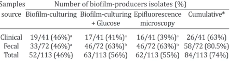
RESULTS
Detection of biofilm formation
The results may be verified in Table 1, briefly, biofilm
formation was observed in 46% (52/113), 56% (63/113)
Fig.1. Biofilm formation by Rhodococcus equi isolates. The bacteria were grown on a glass slide during 24 h, stained with
DAPI and examined with an epifluorescence microscopy (100x). (a) Aggregate of R. equi cells without EPS matrix.
(b) Aggregate of R. equi cells surrounded by EPS matrix (clouding effect surrounding the bacteria due the biofilm
formation). Scale bar = 20μm.
Table 1. Total number and relative frequency of Rhodococcus equi biofilm-producers according the sample source and
assay employed
Samples Number of biofilm-producers isolates (%)
source Biofilm-culturing Biofilm-culturing Epifluorescence Cumulative* + Glucose microscopy
Clinical 19/41 (46%)a 17/41 (41%)a 16/41 (39%)a 26/41 (63%) Fecal 33/72 (46%)a 46/72 (63%)b 46/72 (63%)b 58/72 (80.5%) Total 52/113 (46%) 63/113 (56%) 62/113 (55%) 84/113 (74%) * It was classified as biofilm-producer the isolate positive in at least one assay performed.
and 55% (62/113) of
R. equi
isolates using BC, BCG and
EM assay, respectively. There is no statistically significant
difference between clinical and fecal samples regarding
the biofilm formation, however, we found significant in
-crease in fecal
R. equi
isolates when glucose was added
(Table 1). Biofilm positive and negative
R. equi
cells
ac-cessed by EM are demonstrated in Figure 1a and 1b, res
-pectively.
Correspondence among assays
The biofilm-positive
Rhodococcus equi
in at least one
of the tests performed were considered biofilm-produ
-cer. Therefore, biofilm formation was verified in 74.3%
(84/113) of
R. equi
isolates analyzed, with 63.4% (26/41)
and 80.5% (58/72) of clinical and fecal isolates, respective
-ly. Eight clinical
R. equi
isolates recovered as biofilm-posi
-tive by BC became biofilm-nega-tive in BCG, and six isolates
recovered as biofilm-negative by BC had a biofilm-positive
phenotype in BCG. On the other hand, in fecal isolates, 18
biofilm-negative strains in BC were observed as biofilm
--positive in BCG. All biofilm--positive fecal isolates in BC re
-mained biofilm-positive in BCG.
For clinical isolates, a correlation index of 0.56 was ob
-served among BC, BCG and EM (
P
=0.0001) using the Spear
-man analysis. On the other hand, fecal
R. equi
isolates
sho-wed an increase in the biofilm-formation frequency when
used BCG and EM tests, which had the same sensibility to
detect biofilm formation, showing a strong correlation in
-dex of 0.52 (P = 0.0001).
Antimicrobial activity
Antimicrobial susceptibility testing of planktonic
R.
equi
suspensions.
We evaluated the
in vitro
antimicrobial
susceptibility of eight planktonic suspensions of
R. equi
(four from clinical samples and four from fecal samples) to
AZT, CLAR and ERY. All isolates analyzed were susceptible
to AZT and CLAR. To ERY, four isolates showed
intermedia-te susceptibility and four were susceptible. The results may
be observed in Table 2.
Antimicrobial susceptibility testing of
R. equi
in es
-tablished biofilms.
We used the antimicrobials at
concen-trations at least 4 times their respective MICs to evaluate
their effects on established biofilms. At 24 h incubation
there was significant (
P
<0.05) decrease in
R. equi
concen-tration in presence of AZT, CLAR and ERY. We observed a
nearly 3-log order decrease in
R. equi
concentration
rela-tive to control levels (untreated cells) (Table 3). However,
none of the antimicrobials used were able to eradicate
R.
equi
in established biofilms (Table 3).
DISCUSSION
The biofilm formation by bacterial pathogens of veterinary
importance has received relatively little attention (Jacques
et al. 2010). According to Kaplan (2010) the microorganis
-ms detached from biofil-ms have an important role in the
bacterial dissemination of environmental reservoirs to
hu-man or animal, as well as the microorganism transmission
among hosts. It is well known that biofilm provides resis
-tance to antimicrobials and play an important virulence
factor in chronic bacterial infections in animals, such as
en-dometritis in horses (Ferris et al. 2014). The ability of
Rho-dococcus equi
to form biofilm can be highly favorable for it
survival in the environment as well as in the host. This is
the first study evaluating the biofilm formation by
R. equi
with significant number of isolates.
Several conditions can influence the biofilm forma
-tion/detection, such as the method employed, nutritional
requirements and metabolic pathways (e.g. glucose, iron
Table 3. Azithromycin, clarithromycin and erythromycin activity on
Rhodococcus equi established biofilm
Strain (n = 8) Mean cell density (CFU/ml) after 24 h1
Biofilm untreated Biofilm + AZT Biofilm + CLAR Biofilm + ERY
R. equi 488/00‡ 3.1 x 10⁸ 5.9 x 105* 1 x 105* 3 x 105*
R. equi 27/98‡ 1.5 x 10⁸ 6.9 x 105* 3 x 105* 7.2 x 105*
R. equi 353/93‡ 2.2 x 10⁸ 7.8 x 105* 2.5 x 105* 3.2 x 105*
R. equi 25/03‡ 1.8 x 10⁸ 6.8 x 105* 1.1 x 105* 7 x 105*
R. equi 490/95 DID† 1.9 x 10⁸ 4.5 x 105* 7 x 104* 3.3 x 105*
R. equi 490/95 INB† 1.2 x 10⁸ 6.7 x 105* 1.1 x 105* 5.5 x 105*
R. equi 490/95 TRA† 1.2 x 10⁸ 2.2 x 105* 8 x 104* 1.1 x 105*
R. equi 490/95 IST† 1.9 x 10⁸ 3.2 x 105* 8.5 x 105* 1.7 x 105*
‡ Clinical sample; † Fecal sample.
1Cell density was determined by enumerating CFU by 10-fold serial dilutions plated on TSA; the values are the average of duplicate-independent experiments.
*Indicates statistically significant difference from control by one-way ANOVA and Turkey’s test (P<0.05).
Table 2. Minimal inhibitory concentration of azithromycin, clarithromycin and erythromycin against clinical and fecal
Rhodococcus equi isolates
Strain (n = 8) MIC (µg/mL)
Azithromycin Clarithromycin Erythromycin
R. equi 488/00‡ 1.0a 0.25a 0.5a
R. equi 27/98‡ 1.0a 0.125a 2.0b
R. equi 353/93‡ 0.125a 0.25a 0.5a
R. equi 25/03‡ 1.0a 0.0625a 1.0b
R. equi 490/95 DID† 1.0a 0.125a 1.0b
R. equi 490/95 INB† 0.5a 0.0625a 0.5a
R. equi 490/95 TRA† 1.0a 0.0625a 2.0b
R. equi 490/95 IST† 0.5a 0.0625a 0.5a
‡ Clinical sample; † Fecal sample. a Susceptible; b Intermediate susceptibility.
and phosphate concentration) (Jacques et al. 2010). Con
-sidering this, we aimed verify the
R. equi
biofilm formation
using two different methods and one nutritional variable.
For isolates from the same source, all approaches showed
high concordance (
P
=0.0001). Thus we classified as bio
-film-producer the
R. equi
isolate positive in at least one of
the tests performed. Although some isolates were found
biofilm-positive in one method and negative in another,
this was not unexpected. Similar findings was observed by
Knobloch et al. (2002) studying biofilm formation by clini
-cal and commensal
Staphylococcus aureus
.
Glucose seems as an important substrate required for
biofilm-formation by fecal-isolates of
R. equi
(Table 1). Stu
-dies involving several microorganisms also observed an
in-crease in the ability to form biofilm when used glucose me
-dium supplemented (reviewed by Stepanovic et al. 2007).
In contrast, the presence of glucose did not influence the
biofilm formation by clinical isolates (Table 1). We believe
that different regulatory mechanisms or conditions could
be active in biofilm expression between pathogenic and en
-vironmental
R. equi
. According to Coelho et al. (2008) the
biofilm formation is complex and probably multifactorial,
with different substrates and expression autoinducers,
es-pecially among isolates from different source.
Among the fecal-isolates of
R. equi
, 80.5% (58/72) were
classified as biofilm-producers. The protection ensured by
the EPS matrix during stress conditions, including
exposi-tion to UV radiaexposi-tion (Espeland et al. 2001) and metal toxi
-city (Teitzel & Parsek 2003) may be highly advantageous
during the saprophytic life of
R. equi
. In this way, Tripathi
et al. (2012) showed a mechanism by which the virulence
plasmid can move among
R. equi
in the soil. These authors
highlight a possible relationship between conjugation and
biofilm formation by environmental
R. equi
.
We found 63% (26/41) of clinical-isolates of
R. equi
as biofilm-producers. Usually, bacterial biofilms are asso
-ciated with prolonged-treatment infections (Kulka et al.
2012), as well as with its recurrence or persistence (Hall
--Stoodley et al. 2004). In general,
R. equi
infection has a
long-term antimicrobial therapy, around 4-9 weeks, and,
besides, these infections become chronic (Prescott 2004).
To the best of our knowledge, there are three studies
regar-ding
R. equi
biofilm formation that together evaluated only
ten isolates, six clinical (associated to chronic infection in
humans) (Akhrass et al. 2012, Remuzgo-Martínez et al.
2013) and four from soil (Mart`yanov et al. 2014). Akhrass
et al. (2012) found three strains positive for biofilm forma
-tion, while Remuzgo-Martínez et al. (2013) verified only
one.
None isolate analyzed here showed resistant profile to
antimicrobials (ATM) tested, as presented in Table 2. To
access the susceptibility of
R. equi
in established biofilms
it was used 8µg/mL of each ATM, 8 to 64xMIC verified for
AZT, 32 to 128xMIC for CLAR and 4 to 8xMIC for ERY. A pre
-vious study demonstrated that the average of 4 to 8 times
the MIC could eradicate biofilm of Gram-positive bacteria
(Raja et al. 2011). In the present study,
R. equi
in
establi-shed biofilms were recovered even after exposure to high
ATM concentrations, however, CFU amount reduction was
statically significant (Table 3). Two studies concerning an
-timicrobial susceptibility of clinical and environmental
R.
equi
producing biofilm were performed by Akhrass et al.
(2012) and Mart`yanov et al. (2014). The first study found
R. equi
biofilm reduced completely or partially by antimi
-crobial solutions. Mart`yanov et al. (2014) found a unex
-pected pattern,
R. equi
biofilm persistence was observed in
the treatments with high AZT concentration (15 to 50µg/
mL), while at 8µg/mL the planktonic and biofilm growth
were similar.
In the present study, we observed a nearly 3-log order
decrease in
R. equi
concentration relative to control levels
(untreated cells) (Table 3). Similar results were verified by
Ojha et al. (2008) with
Mycobacterium tuberculosis
biofilm
after rifampicin (50µg/mL) treatment. Although the reduc
-tion have been expressive, these authors indicate that
sub-population of cells surviving this treatment were able to
replicate following 7 day of exposure. Even exopolysaccha
-ride matrix does not form an impenetrable barrier to the
diffusion of antimicrobial agents (Mah & O’Toole 2001), the
observation of tolerant subpopulation may mean an
impor-tant way of ATMs resistance.
Oggioni et al. (2006) observed that cells from biofilm
--producing pneumococcal are more effective in inducing
pneumonia than the planktonic cells. In addition,
micro-organisms growing on EPS matrix are able to resist to the
host defenses, i.e., by impairing the phagocytic activity of
neutrophils (Yamanaka et al. 2009) - and act as reservoirs
for antibiotic resistance genes (Jacques et al. 2010). The in
-fluence of biofilm in
R. equi
pneumonia, especially in the
cases unresponsive to treatment, may require some special
attention.
CONCLUSIONS
Both clinical and fecal
Rhodococcus equi
isolates are
able to form biofilm according the methods used. Those
may represent a quick and reliable methodology to study
R. equi
biofilm.
Glucose addiction seems to increase the ability of fecal
isolates to produce biofilms.
None antimicrobials tested was able to eradicate
R.
equi
in biofilm, even at high concentrations.
Our findings indicate that
R. equi
biofilm-producers may
be more resistant to the antimicrobials evaluated. Further
studies are warranted to test this hypothesis.
Conflict of interest statement.- All authors declare there are no perso-nal conflicts of interests that could inappropriately influence or bias the content of the paper.
Acknowledgements.- The authors acknowledge the financial support from CNPq (Conselho Nacional de Desenvolvimento Científico e Tecnoló -gico), specially for Letícia Trevisan Gressler Master of Science Scholarship (process number: 130023/2012-4) and FAPERGS (Fundação de Amparo à Pesquisa do Estado do Rio Grande do Sul). We thank to Dr. Berta Maria Heinzmann for helpful comments to the manuscript.
REFERENCES
Akhrass F.A., Wohoush I.A., Chaftari A.M., Reitzel R., Jiang Y., Ghannoum M., Tarr J., Hachem R. & Raad I. 2012. Rhodococcus bacteremia in cancer pa-tients is mostly catheter related and associated with biofilm formation. Plos One 7:e32945.
Arlotti M., Zoboli G., Moscatelli G.L., Magnani G., Maserati R., Borghi V., An-dreoni M., Libanore M., Bonazzi L. & Piscina A. 1996. Rhodococcus equi infection in HIV-positive subjects: a retrospective analysis of 24 cases. Scand. J. Infect. Dis. 28:463-467.
CLSI 2008. Performance Standards for Antimicrobial Disk and Dilution Susceptibility Tests for Bacteria Isolated from Animals: approved stan-dard. 3rd ed. Clinical and Laboratory Standards Institute Document M31-A3. Clinical and Laboratory Standards Institute, Wayne, PA. CLSI 2009. Performance Standards for Antimicrobial Susceptibility Test
-ing; approved standard, 10th ed. Clinical and Laboratory Standards Institute Document M100-S19. Clinical and Laboratory Standards Insti -tute, Wayne, PA.
CLSI 2013. Performance Standards for Antimicrobial Disk and Dilution Susceptibility Tests for Bacteria Isolated from Animals: approved stan-dard. 4th ed. Clinical and Laboratory Standards Institute Document VET01-A4. Clinical and Laboratory Standards Institute, Wayne, PA. Cerca N., Martins S., Cerca F., Jefferson K.K., Pier G.B., Oliveira R. & Azere
-do J. 2005. Comparative assessment of antibiotic susceptibility of co-agulase-negative staphylococci in biofilm versus planktonic culture as assessed by bacterial enumeration or rapid XTT colorimetry. J. Antimi-crob. Chemother. 56:331-336.
Coelho L.R., Souza R.R., Ferreira F.A., Guimarães M.A., Ferreira-Carvalho B.T. & Figueiredo M.A.S. 2008. agr RNAIII divergently regulates glucose--induced biofilm formation in clinical isolates of Staphylococcus aureus. Microbiology 154:3480-3490.
Costerton J.W., Stewart P.S. & Greenberg E.P. 1999. Bacterial biofilms: a common cause of persistent infections. Science 284:1318-1322. Cucarella C., Tormo M.A., Ubeda C., Trotonda M.P., Monzon M., Peris C.,
Amorena B., Lasa I. & Penades J.R. 2004. Role of biofilm-associated pro -tein Bap in the pathogenesis of bovine Staphylococcus aureus. Infect. Immun. 72:2177-2185.
Donlan R.M. & Costerton J.W. 2002. Biofilms: survival mechanisms of clini -cally relevant microorganims. Clin. Microbiol. Rev. 15:167-193. Espeland E.M. & Wetzel R.G. 2001. Complexation, stabilization, and UV
photolysis of extracellular and surface-bound glucosidase and alkaline phosphatase: implications for biofilm microbiota. Microb. Ecol. 42:572-585.
Feazel L.M., Baumgartner L.B., Peterson K.L., Frank D.N., Harris J.K. & Pace N.R. 2009. Oppotunistic pathogens enriched in showerhead biofilms. Pnas 106:16393-16399.
Fernandes M.C., Takai S., Leite D.S., Pinto J.P.A.N., Brandão P.E., Santarém V.A., Listoni F.J.P., Da Silva A.V. & Ribeiro M.G. 2013. Identification of pathogens and virulence profile of Rhodococcus equi and Escherichia coli strains obtained from sand of parks. Braz. J. Microbiol. 44:485-492. Ferris R.A., Wittstock S.M., McCue P.M. & Borlee B.R. 2014. Evaluation of
biofilms in gram-negative bacteria isolated from the equine uterus. J. Equine Vet. Sci. 34:121.
Giguère S. & Prescott J.F. 1997. Clinical manifestations, diagnosis, treat -ment, and prevention of Rhodococcus equi infections in foals. Vet. Mi-crobiol. 7:313-334.
Giguère S., Lee E., Williams E., Cohen N.D., Chaffin M.K., Halbert N., Mar -tens R.J., Franklin R.P., Clark C.C. & Slovis N.M. 2010. Determination of the prevalence of antimicrobial resistance to macrolide antimicrobials or rifampin in Rhodococcus equi isolates and treatment outcome in foals infected with antimicrobial-resistant isolates of R equi. J. Am. Vet. Med. Assoc. 237:74-81.
Hal-Stoodley L., Costerton J.W. & Stoodley P. 2004. Bacterium biofilm: from the natural environmental to infections disease. Nat. Rev. Microbiol. 2:95-108.
Jacques M., Aragon V. & Tremblay Y.D.N. 2010. Biofilm formation in bacte -rial pathogens of veterinary importance. Anim. Health Res. Rev. 11:97-121.
Kaplan J.B. 2010. Biofilm dispersal: mechanisms, clinical implications, and potential therapeutic uses. J. Dent. Res. 89:205-218.
Knobloch J.K.M., Horstkotte M.A., Rohde H. & Mack D. 2002. Evaluation of different detection methods of biofilm formation in Staphylococcus
aureus. Med. Microbiol. Immunol. 191:101-106.
Kulka K., Hatfull G. & Ojha A.K. 2012. Growth of Mycobacterium tuberculo-sis biofilms. J. Vis. Exp. 60:e3820.
Lemon K.P., Earl A.M., Vlamakis H.C., Aguilar C. & Kolter R. 2008. Biofilm development with an emphasis on Bacillus subtilis. Curr. Top. Microbiol. Immunol. 322:1-16.
Letek M., González P., MacArthur I., Rodríguez H., Freeman T.C., Vale -ro-Rello A., Blanco M., Buckley T., Cherevach I., Fahey R., Hapeshi A., Holdstock J., Leadon D., Navas J., Ocampo A., Quail M.A., Sanders M., Scortti M.M., Prescott J.F., Fogarty U., Meijer W.G., Parkhill J., Bentley S.D. & Vázquez-Boland J.A. 2010. The genome of a pathogenic Rhodococcus: cooptive virulence underpinned by key gene acquisitions. Plos Genet. 6:1001145.
Mah T.F. & O’Toole G.A. 2001 Mechanisms of biofilm resistance to antimi -crobial agents. Trends Microbiol. 9:34-39.
Marques S.C., Rezende J.G.O.S., Alves L.A.F., Silva B.C., Alves E., Abreu L.R. & Piccoli R.H. 2007. Formation of biofilms by Staphylococcus aureus on stainless steel and glass surfaces and its resistance to some selected chemical sanitizers. Braz. J. Microbiol. 38:538-543.
Mathur T., Singhal S., Khan S., Upadhyay D.J., Fatma T. & Rattan A. 2006. Detection of biofilm formation among the clinical isolates of staphylo -cocci: an evaluation of three different screening methods. Indian J. Med. Microbiol. 24:9-25.
Merino N., Toledo-Arana A., Vergara-Irigaray M., Valle J., Solano C., Calvo E., Lopez J.A., Foster T.J., Penadés J.R. & Lasa I. 2009. Protein A-mediated multicellular behavior in Staphylococcus aureus. J. Bacteriol. 191:832-843.
Monego F., Maboni F., Krewer C., Vargas A., Costa M. & Loreto E. 2009. Molecular characterization of Rhodococcus equi from horse-breeding farms by means of multiplex PCR for the vap gene family. Curr. Micro-biol. 58:399-403.
Muscatello G., Anderson G.A., Gilkerson J.R. & Browning G.F. 2006. Asso -ciations between the ecology of virulent Rhodococcus equi and the ep-idemiology of R. equi pneumonia on Australian thoroughbred farms. Appl. Environ. Microbiol. 72:6152-6160.
Muscatello G., Leadon D.P., Klayt M., Ocampo-Sosa A., Lewis D.A., Fogarty U., Buckley T., Gilkerson J.R., Meijer W.G. & Vazquez-Boland J.A. 2007. Rhodococcus equi infection in foals: the science of ‘rattles’. Equine Vet. J. 39:470-478.
Oggioni M.R., Trappetti C., Kadiougiu A., Cassone M., Iannelli F., Ricci S., Andrew P.W. & Pozzi G. 2006. Switch from planktonic to sessile life: a major event in pneumococcal pathogenesis. Mol. Microbiol. 61:1196-1210.
Ojha A.K., Baughn A.D., Sambandan D., Hsu T., Trivelli X., Guerardel Y., Ala -hari A., Kremer L., Jacobs W.R. & Hatfull Jr G.F. 2008. Growth of
Mycobac-terium tuberculosis biofilms containing free mycolic acids and harbour
-ing drug-tolerant bacteria. Mol. Microbiol. 69(1):164-174.
O’Toole G., Kaplan H.B. & Kolter R. 2000. Biofilm formation as microbial development. Annu. Rev. Microbiol. 54:49-79.
Prescott J.F. 1991. Rhodococcus equi: an animal and human pathogen. Clin. Microbiol. Rev. 4:20-34.
Prescott J.F. 2004. Rhodococcus equi, p.87-95. In: Prescott C.L., Thoen C.O., Prescott J.F. & Songer J.G. (Eds), Pathogenesis of Bacterial Infections of Animals. Blackwell Publ., Ames.
Quinn P.J. 1994. Corynebacterium species and Rhodococcus equi, p.137-143. In: Quinn P.J., Carter M.E., Markey B.K. & Carter G.R. (Eds), Clinical Veterinary Microbiology. Wolfe Publ., London.
Raja A.F., Ali F., Khan I.A., Shawl A.S., Arora D.S., Shah B.A. & Taneja S.C. 2011. Anti-staphylococcal and biofilm inhibitory activities of acetyl-11-keto-β-boswellic acid from Boswellia serrate. BMC Microbiol. 11: 1-9.
Ruzicka F. 2007. Quantification of biofilm in microtiter plates: overview of testing conditions and practical recommendations for assessment of biofilm production by staphylococci. APMIS 115:891-899.
Takai S., Ohbushi S., Koike K., Tsubaki S., Oishi H. & Kamada M. 1991. Pre -valence of virulent Rhodococcus equi in isolates from soil and feces of horses from horse-breeding farms with and without endemic infections. J. Clin. Microbiol. 29:2887-2889.
Takai S., Fukunaga N., Ochiai S., Sakai T., Sasaki Y. & Tsubaki S. 1996. Isola -tion of virulent and intermediately virulent Rhodococcus equi from soil and sand on parks and yards in Japan. J. Vet. Med. Sci. 58:669-672. Takai S. 1997. Epidemiology of Rhodococcus equi infections: A review. Vet.
Microbiol. 56:167-176.
Teitzel G.M. & Parsek M.R. 2003. Heavy metal resistance of biofilm and planktonic Pseudomonas aeruginosa. Appl. Environ. Microbiol. 69:2313-2320.
Tripathi V.N., Harding W.C., Willingham-Lane J.M. & Hondalus M.K. 2012. Conjugal transfer of a virulence plasmid in the opportunistic intracellu-lar actinomycete Rhodococcus equi. J. Bacteriol. 194(24):6790-6801. Von Bargen K. & Haas A. 2009. Molecular and infection biology of the hor
-se pathogen Rhodococcus equi. FEMS Microbiol. Rev. 33(5):870-891. Yamanaka T., Furukawa T., Matsumoto-Mashimo C., Yamane K., Sugimori
C., Nambu T., Mori N., Nishikawa H., Walker C.B., Leung K. & Fukushima H. 2009. Gene expression profile and pathogenicity of biofilm-forming